
Rationale and Objectives
Since the introduction of computed tomographic (CT) imaging in the 1970s, the number of examinations has increased steadily. CT imaging is an essential part of routine workup in diagnostic radiology. The great advantage of multidetector computed tomography is the acquisition of a large amount of data in a short time period, thus speeding up diagnostic procedures. To protect patients from unnecessary radiation exposure, different approaches have been developed. In this study, the efficacy of automated exposure control (AEC) software in multidetector CT imaging with a focus on dose reduction in pediatric examinations was assessed.
Materials and Methods
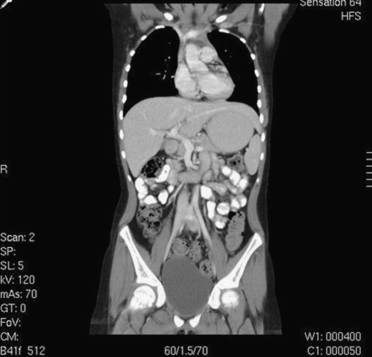
Between August 2004 and September 2005, a total of 71 children (40 male, 31 female; age range, 2–13 years; mean age, 7.2 years) were examined using a multisource CT scanner. Three different regions (chest, upper abdomen, and pelvis) were examined. Overall image quality was assessed with a subjective scale (1 = excellent, 2 = diagnostic, 3 = nondiagnostic). For all examinations, AEC was used. From the scanner’s patient protocol, dose-length product, volume CT dose index, and tube current–time product were calculated for each examination.
Results
With AEC, a mean dose reduction of 30.6% was calculated. Images were rated as excellent ( n = 39) or diagnostic ( n = 32). Nondiagnostic image quality was not seen. Dose-length product and volume CT dose index were reduced by 30.4% and 29.5%, respectively. Overall, a mean dose reduction of 30.1% of the effective dose (5.8 ± 3.1 vs 8.4 ± 4.6 mSv) was achieved ( P < .001).
Conclusions
With AEC software, a mean dose reduction of 30% without any loss in diagnostic image quality is possible.
The number of computed tomographic (CT) examinations, especially in children, is rising steadily. It is known that children are more sensitive to ionizing radiation and face a higher risk for developing malignancy because of their longer life expectancy compared to adults . In general, radiation-free imaging modalities must be considered when imaging children, or whenever indicated if radiation exposure can be lowered or even minimized . Vendors of CT scanners have addressed this issue by developing different dose reduction software to reduce tube current in a sinusoidal way while correlating individual anatomy in real time . Recently, a new dose reduction software (CARE Dose 4D; Siemens Healthcare, Forchheim, Germany) was introduced, in which tube current values are adapted to individual patient anatomy prior to the CT exam. This type of automatic exposure control combines the attenuation-based online tube current modulation presented in earlier studies with an automated evaluation of patient anatomy and setting of exposure parameters for every slice. The purpose of this study was to evaluate this new software in pediatric CT scans, with a special focus on the level of dose reduction and effects on image quality.
Materials and methods
A total of 71 consecutive pediatric patients (41 male, 30 female; age range 2–13 years; mean age, 7.2 years) were included from August 2004 to September 2005. CT scans were performed on a multidetector CT system (Somatom Sensation 64; Siemens Healthcare). Care Dose 4D software uses data from the prescan projection radiogram (topogram) to determine “ideal” tube current values for each z-position by calculating object attenuation. The projection data acquired for the topogram measure the patient’s attenuation for every z-position and allow calculation of the optimal tube current for the projection angle used, anteroposterior or lateral. Evaluation of the attenuation profile’s shape is applied to estimate the attenuation perpendicular to the projection angle and calculate the optimal tube current for this angle. The adequate tube current–time product for this z-position is derived as the average of the two tube current values multiplied by the rotation time. During the scan, the online tube current modulation calculates and applies the optimal tube current for each projection angle. From these data, the software controls the CT scan and adapts tube current values for each z-position. Performed CT scans included three different regions (chest, upper abdomen, and pelvis). From the individual patient protocol information dose-length product, volume CT dose index, and tube current–time product were calculated for each examination. This prospective study was approved by the local ethics committee, and written informed consent was obtained.
Scan Parameters
Get Radiology Tree app to read full this article<
Quantitative Assessment
Get Radiology Tree app to read full this article<
Table 1
Body Weight–Adapted Tube Current–Time Product Values
Body Weight (kg) Chest (mAs) Abdomen/Pelvis (mAs) 0–8.9 40 60 9–17.9 50 70 18–26.9 60 80 27–35.9 70 100 36–44.9 80 120 45–69.9 100 140 >70 100 160
Get Radiology Tree app to read full this article<
Qualitative Assessment
Get Radiology Tree app to read full this article<
Results
Quantitative Assessment
Get Radiology Tree app to read full this article<
Table 2
Comparison of Mean Dose Values with and without AEC and Rate of Mean Dose Reduction for DLP, CTDIvol, and Effective Dose Values
Without AEC With AEC Reduction rate (%)P ∗ DLP (mGy/cm) 457.4 ± 280.8 318.7 ± 184.3 30.4 <.001 CTDIvol 10.9 ± 3.2 7.7 ± 2.1 29.5 <.001 Effective dose (mSv) 8.4 ± 4.6 5.8 ± 3.1 30.1 <.001
AEC, automated exposure control; CTDIvol, volume computed tomography dose index; DLP, dose-length product.
Data are expressed as mean ± standard deviation.
Get Radiology Tree app to read full this article<
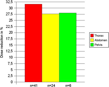
Table 3
Calculated Mean Dose Reduction Values per Exam Region
Region_n_ Mean Dose Reduction (%) Thorax 41 31.6 Upper abdomen 24 27.6 Pelvis 6 28.0
Get Radiology Tree app to read full this article<
Qualitative Assessment
Get Radiology Tree app to read full this article<
Table 4
Qualitative Assessment of Image Quality
Category_n_ % 1 (excellent) 39 54 2 (diagnostic) 32 46 3 (nondiagnostic) 0 0
Get Radiology Tree app to read full this article<
Discussion
Get Radiology Tree app to read full this article<
Get Radiology Tree app to read full this article<
Get Radiology Tree app to read full this article<
Get Radiology Tree app to read full this article<
Conclusions
Get Radiology Tree app to read full this article<
References
1. Brenner D.J., Elliston C.D., Hall E.J., et. al.: Estimated risks of radiation-induced fatal cancer from pediatric CT. AJR Am J Roentgenol 2001; 176: pp. 289-296.
2. Don S.: Radiosensitivity of children potential for overexposure in CR and DR and magnitude of doses in ordinary radiographic examination. Pediatr Radiol 2004; 34: pp. 167-172.
3. Greess H., Lutze J., Nömayr A., et. al.: Dose reduction in subsecond multislice spiral CT examination of children by online tube current modulation. Eur Radiol 2004; 14: pp. 995-999.
4. Kalender W.A.: Computertomographie: Grundlagen, Gerätetechnologie, Bildqualität, Anwendungen.2006.Publicis MCD VerlagMunich, Germany 216
5. Kalender W.A., Wolf H., Suess C., et. al.: Dose reduction in CT by on-line tube current control: principles and validation on phantoms and cadavers. Eur Radiol 1999; 9: pp. 323-328.
6. Stamm G.: Nagel HD. CT-Expo—a novel program for dose evaluation in CT. Fortschr Röntgenstr 2002; 174: pp. 1570-1576.
7. Greess H., Wolf H., Suess C., et. al.: Dosisautomatik bei der Mehrzeilenspiral-CT: Phantommessungen und klinische Ergebnisse. Fortschr Röntgenstr 2004; 176: pp. 862-869.
8. Greess H., Wolf H., Baum U., et. al.: Dose reduction in computed tomography by attenuation-based online modulation of tube current: evaluation of six anatomical regions. Eur Radiol 2000; 10: pp. 391-394.
9. Greess H., Baum U., Wolf H., et. al.: Dosisreduktion bei der Spiral CT: Detektion von Lungenrundherden mit und ohne anatomisch angepasster Röhrenstrommodulation. Fortschr Röntgenstr 2001; 173: pp. 466-470.
10. Das M., Mahnken A.H., Muhlenbruch G., et. al.: Individually adapted examination protocols for reduction of radiation exposure for 16-MDCT chest examinations. AJR Am J Roentgenol 2005; 184: pp. 1437-1443.