
Rationale and Objectives
The aim of this study was to develop a new technique, the use of magnetic resonance (MR) imaging (MRI) to monitor gene/MR–cotransferred stem-progenitor cells (SPCs) recruited to atherosclerosis.
Materials and Methods
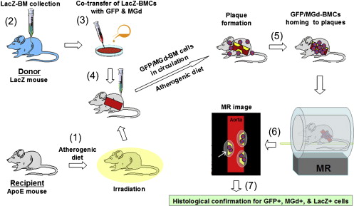
First, a green fluorescent protein (GFP) gene and a T1 MR contrast agent (motexafin gadolinium [MGd]) were cotransferred into neural or bone marrow (BM)–derived SPCs. GFP expression and MGd signal were confirmed by fluorescent microscopy and quantified by flow cytometry. Cell viability and proliferation were then evaluated by trypan blue exclusion and 3-(4,5-dimethylthiazol-2-yl)-5-(3-carboxymethoxyphenyl)-2-(4-sulfophenyl)-2H-tetrazolium assay, and GFP/MGd–transferred cells were imaged using 1.5-T and 9.4-T MR scanners. For in vivo validation, GFP/MGd–cotransferred β-galactosidase–BM SPCs were transplanted to apolipoprotein E–knockout mice, and cell migration to atherosclerotic aortas was monitored using 9.4-T micro-MRI with subsequent histologic correlations.
Results
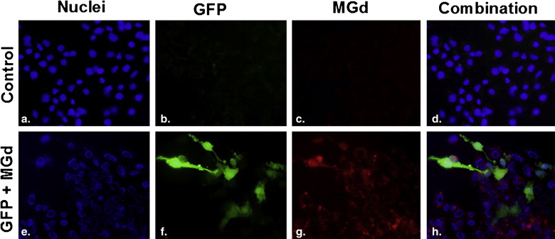
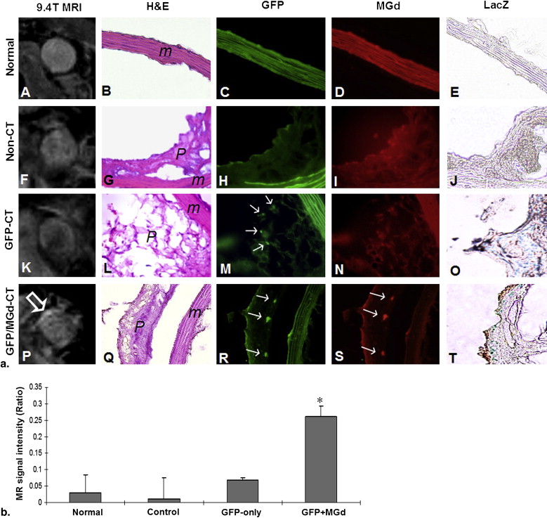
Fluorescent microscopy demonstrated simultaneous GFP expression and MGd signals in cotransferred-cells. Quantitative flow cytometry showed GFP-positive cells at 47 ± 25% and 56 ± 12% and MGd-positive cells at 96 ± 6% and 57 ± 11% for neural stem cells and BM cells, respectively. Cell viability and metabolic rates of cotransferred cells were 86 ± 4% and 84 ± 12%, respectively. In vivo MRI revealed high MR signals of the aortic walls in GFP/MGd–transferred mice, which were confirmed by histologic correlations.
Conclusion
This study has initially proven the new concept of MRI for plaque-specific, cell-mediated gene expression of atherosclerosis.
A characteristic feature of atherosclerotic cardiovascular disease is its diffuse involvement of arteries across the entire human body and the presence of multiple and simultaneous atherosclerotic lesions. Endovascular interventional procedures, such as balloon angioplasty and stenting, are currently used as routine “local” treatments of atherosclerotic arteries. However, these local interventional approaches do not treat multiple diffuse atherosclerosis. Thus, it is essential to seek alternatives to treat all atherosclerotic arteries at once.
Hematopoietic stem-progenitor cells (SPC) can give rise to vascular progenitor cells that migrate or home to atherosclerotic arteries and differentiate into either smooth-muscle cells or endothelial cells . Intravenously transfused hematopoietic SPCs circulate in the blood system, flow through the entire body, and thus home to all atherosclerotic lesions. Gene therapy is an exciting frontier in cardiovascular medicine . The transfer of therapeutic genes into hematopoietic SPCs prior to their transplantation to the body may enable the SPC-mediated plaque-specific delivery of therapeutic genes to diffuse multiple atherosclerotic lesions.
Get Radiology Tree app to read full this article<
Get Radiology Tree app to read full this article<
Materials and methods
Study Design
Get Radiology Tree app to read full this article<
In Vitro Evaluation
Agents
Get Radiology Tree app to read full this article<
Dual Transfer of GFP and MGd Into the Cells
Get Radiology Tree app to read full this article<
Laboratory Conformations
Get Radiology Tree app to read full this article<
Evaluation of Cell Viability and Proliferation
Get Radiology Tree app to read full this article<
In Vitro MRI
Get Radiology Tree app to read full this article<
In Vivo Validation
Extraction of Donor β-galactosidase SPCs
Get Radiology Tree app to read full this article<
Get Radiology Tree app to read full this article<
Cell Transplantation
Get Radiology Tree app to read full this article<
Get Radiology Tree app to read full this article<
In Vivo MRI
Get Radiology Tree app to read full this article<
Histologic Correlation
Get Radiology Tree app to read full this article<
Image Analysis
Get Radiology Tree app to read full this article<
Results
In Vitro Evaluation
Get Radiology Tree app to read full this article<
![Figure 3, (a) Photo taken immediately after centrifuging cells at the bottoms of tubes, showing the success of motexafin gadolinium (MGd)–transferred cells as dark brown color, which is not seen with nontransferred control cells. (b) Representative 1.5-T magnetic resonance (MR) image of cell pellets at bottoms of tubes, demonstrating a brighter signal in transferred cells compared to nontransferred cells. (c) T1-weighted MR imaging of the nontransferred and MGd-transferred cells suspended in 4% gelatin, showing a brighter signal in transferred cells. (d) Comparison of average MR signal intensities (arbitrary units [au]) between nontransferred and transferred bone marrow cells, demonstrating higher average signal intensity in MGd-transferred cells.](https://storage.googleapis.com/dl.dentistrykey.com/clinical/DualTransferofGFPGeneandMGdintoStemProgenitorCells/2_1s20S107663321000111X.jpg)
Get Radiology Tree app to read full this article<
In Vivo Validation
Get Radiology Tree app to read full this article<
Get Radiology Tree app to read full this article<
Discussion
Get Radiology Tree app to read full this article<
Get Radiology Tree app to read full this article<
Get Radiology Tree app to read full this article<
Get Radiology Tree app to read full this article<
Acknowledgment
Get Radiology Tree app to read full this article<
Get Radiology Tree app to read full this article<
References
1. Xu Y., Arai H., Zhuge X., et. al.: Role of bone marrow-derived progenitor cells in cuff-induced vascular injury in mice. Arterioscler Thromb Vasc Biol 2004; 24: pp. 477-482.
2. Orlic D., Kajstura J., Chimenti S., et. al.: Bone marrow stem cells regenerate infarcted myocardium. Pediatr Transplant 2003; 7: pp. 86-88.
3. Krause D., Theise N., Collector M., et. al.: Multi-organ, multi-lineage engraftment by a single bone marrow-derived stem cell. Cell 2001; 105: pp. 369-377.
4. Sata M., Saiura A., Kunisato A., et. al.: Hematopoietic stem cells differentiate into vascular cells that participate in the pathogensis of atherosclerosis. Nat Med 2002; 8: pp. 403-409.
5. Nabel E.: Gene therapy for cardiovascular disease. Circulation 1995; 91: pp. 541-548.
6. Sinnaeve P., Varenne O., Collen D., et. al.: Gene therapy in the cardiovascular system: an update. Cardiovasc Res 1999; 44: pp. 498-506.
7. Yang X.: Imaging of vascular gene therapy. Radiology 2003; 228: pp. 36-49.
8. Bulte J.W., Douglas T., Witwer B., et. al.: Magnetodendrimers allow endosomal magnetic labeling and in vivo tracking of stem cells. Nat Biotechnol 2001; 19: pp. 1141-1147.
9. Qiu B., Yang X.: Molecular MRI of hematopoietic stem-progenitor cells: in vivo monitoring of gene therapy and atherosclerosis. Nat Clin Pract Cardiovasc Med 2008; 5: pp. 396-404.
10. Gao F., Kar S., Zhang J., et. al.: MRI of intravenously injected bone marrow cells homing to the site of injured arteries. NMR Biomed 2007; 20: pp. 673-681.
11. Qiu B., Gao F., Walczak P., et. al.: In vivo MR imaging of bone marrow cells trafficking to atherosclerotic plaques. J Magn Reson Imaging 2007; 26: pp. 339-343.
12. National Institutes of Health. Guide for the care and use of laboratory animals. NIH Publication No 86-23 (revised 1985). Bethesda, MD: National Institutes of Health.
13. Woodburn K., Young S., Fan Q.: Selective uptake of texaphyrins by atheromatous plaque. SPIE 1996; 2671: pp. 62-71.
14. Woodburn K.: Intracellular localization of the radiation enhancer motexafin gadolinium using interferometric Fourier fluorescence microscopy. J Pharmacol Exp Ther 2001; 297: pp. 888-894.
15. Brushett C, Qiu B, Kraitchman D, et al. Comparison of two MR contrast agents for characterization of atherosclerotic plaque (#1649). Presented at: Annual meeting of the International Society of Magnetic Resonance in Medicine; Toronto, Canada; May 10–17, 2003.
16. Evens A.: Motexafin gadolinium: a redox-active tumor selective agent for the treatment of cancer. Curr Opin Oncol 2004; 16: pp. 576-580.
17. Russell J.: Of mice and men, rats and atherosclerosis. Cardiovasc Res 2003; 59: pp. 810-811.
18. Kleemann R., Zadelaar S., Kooistra T.: Cytokines and atherosclerosis: a comprehensive review of studies in mice. Cardiovasc Res 2008; 79: pp. 360-376.