
Rationale and Objectives
Magnetic resonance (MR) imaging has been widely used to detect bone marrow (BM) changes after radiotherapy. However, little information about the dynamic MR appearance of early radiation-induced BM injury is available. This experimental study was designed to determine the MR appearance of irradiated BM during the initial 4 weeks after irradiation.
Materials and Methods
After focal BM irradiation (20 Gy, single dose, x-ray), 12 of 20 rabbits underwent serial MR studies weekly from days 7 to 28; eight rabbits were used for histologic investigation on days 7, 14, 21, and 28 after irradiation.
Results
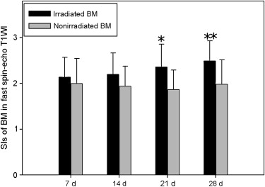
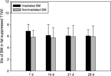
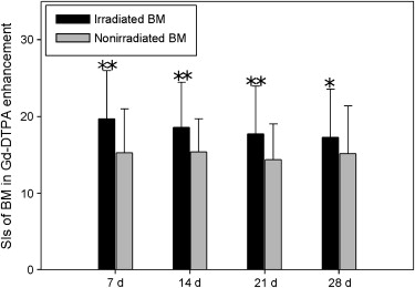
Under microscopy, early BM changes after irradiation consisted of sinusoid dilatation and congestion, followed by a progressive decrease in cellularity and later fat degeneration. All irradiated BM showed relative hyperintensity on short-inversion time inversion recovery (STIR) imaging from days 7 to 21 after irradiation and increased enhancement with gadolinium diethylenetriamine pentaacetic acid (DTPA) administration from days 7 to 28 after irradiation. However, on STIR imaging and gadolinium DTPA enhancement, the relative signal intensity of irradiated BM appeared to decline in a time-dependent way. On fast spin-echo (FSE) T1-weighted imaging, relative hyperintensity was detected in irradiated BM from day 21 after irradiation. On fat-suppressed FSE T1-weighted imaging, a slight increase in signal intensity was shown in some irradiated BM (in five of 12 rabbits) on day 7 after irradiation.
Conclusion
STIR imaging was sensitive to early BM congestion and sinusoidal dilatation, spin-echo T1-weighted imaging was effective in detecting later fatty degeneration in irradiated BM, and gadolinium DTPA enhancement may contribute to the evaluation of BM vascular injury in response to irradiation.
As the main hematogenic organ in the human body, bone marrow (BM) is sensitive to radiation . In medical practice, BM may be exposed to radiation for radiotherapy of malignancies, leading to resultant radiation-induced injury to varying degrees . Generally, BM changes in response to radiation are dose and time dependent, which is characterized in histologic structure by early marrow edema, sinusoids disruption, hypocellularity with vasculature destruction, and late fatty replacement and endosteal fibrosis . Magnetic resonance (MR) imaging (MRI) has been considered an accurate and integral technique in the detection of radiation-induced effects on BM . Studies using short-inversion time inversion recovery (STIR) imaging reported that early BM edema attributable to radiation was presented as hyperintensity . Fatty transformation in irradiated BM appeared as bright signal intensity (SI) in spin-echo (SE) T1-weighted imaging or opposed-phase chemical-shift imaging . A prospectively study by Otake et al described transient enhancement in irradiated BM early after the initiation of radiotherapy.
To our knowledge, the majority of previous MR studies in radiation-induced BM injury detection were performed in patients undergoing radiotherapy for malignancies, who were irradiated to different bones and with varied fractionated doses . Little information about the dynamic MR appearance of early radiation-induced BM injury is available. In this MR study using STIR imaging, routine SE sequences, and gadolinium diethylenetriamine pentaacetic acid (DTPA) enhancement, we prospectively performed a serial evaluation of early radiation-induced BM changes in a rabbit model. Our purposes were to determine the MR appearance of irradiated BM during the initial 4 weeks after irradiation and to reassess the role of different MR techniques in early radiation-induced BM injury detection.
Materials and methods
Animals
Get Radiology Tree app to read full this article<
X-Irradiation
Get Radiology Tree app to read full this article<
MR Investigation
Get Radiology Tree app to read full this article<
Get Radiology Tree app to read full this article<
Get Radiology Tree app to read full this article<
Histologic Examination
Get Radiology Tree app to read full this article<
Results
Get Radiology Tree app to read full this article<
Get Radiology Tree app to read full this article<
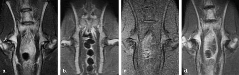
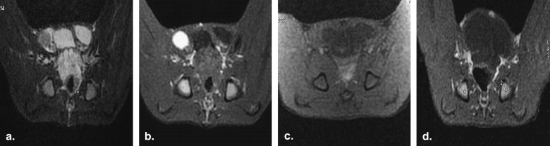
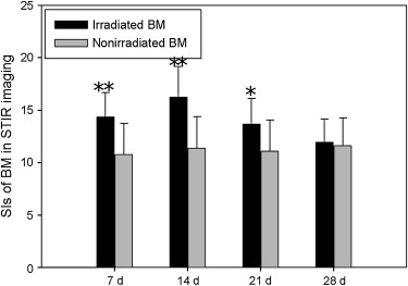
Table 1
Relative Signal Intensity of Irradiated Bone Marrow on Magnetic Resonance Imaging on Days 7, 14, 21, and 28 After Irradiation
Modality Day 7 Day 14 Day 21 Day 28 STIR imaging 58.3 ± 8.7% 57.1 ± 9.0% 37.7 ± 10.3% 21.2 ± 6.6% T1WI 5.1 ± 3.0% 8.1 ± 2.8% 21.8 ± 11.3% 37.2 ± 15.3% FS T1WI 15.5 ± 4.6% 12.0 ± 4.3% 9.5 ± 2.0% 8.4 ± 1.9% CE 36.9 ± 15.4% 32.0 ± 9.6% 24.9 ± 10.8% 16.4 ± 8.3%
CE, gadolinium diethylenetriamine pentaacetic acid enhancement; FS, fat-suppressed; T1WI, T1-weighted imaging.
Data are expressed as mean ± standard error.
Get Radiology Tree app to read full this article<
Get Radiology Tree app to read full this article<
Get Radiology Tree app to read full this article<
Get Radiology Tree app to read full this article<
Get Radiology Tree app to read full this article<
Get Radiology Tree app to read full this article<
Get Radiology Tree app to read full this article<
Get Radiology Tree app to read full this article<
Discussion
Get Radiology Tree app to read full this article<
Get Radiology Tree app to read full this article<
Get Radiology Tree app to read full this article<
Get Radiology Tree app to read full this article<
Get Radiology Tree app to read full this article<
Conclusion
Get Radiology Tree app to read full this article<
References
1. Till J.E., Mcculloch E.A.: A direct measurement of the radiation sensitivity of normal mouse bone marrow cells. Radiat Res 1961; 14: pp. 213-222.
2. Onu M., Savu M., Lungu-Solomonescu C., et. al.: Early MR changes in vertebral BM for patients following radiotherapy. Eur Radiol 2001; 11: pp. 1463-1469.
3. Otake S., Mayr N.A., Ueda T., et. al.: Radiation-induced changes in MR signal intensity and contrast enhancement of lumbosacral vertebrae: do changes occur only inside the radiation therapy field?. Radiology 2002; 222: pp. 179-183.
4. Li X., Wang C., Zhou Y.: MR evaluation: spine changes after radiotherapy. J Tongji Med Univ 1999; 19: pp. 72-76.
5. Park S.W., Lee J.H., Ehara S., et. al.: Single shot fast spin echo diffusion weighted MR imaging of the spine: is it useful in differentiating malignant metastatic tumor infiltration from benign fracture edema?. Clin Imaging 2004; 28: pp. 102-108.
6. Sugimura H., Kisanuki A., Tamura S., et. al.: Magnetic resonance imaging of BM changes after irradiation. Invest Radiol 1994; 29: pp. 35-41.
7. Kauczor H.U., Dietl B., Kreitner K.F., et. al.: BM changes following radiotherapy. Results of MR tomography. Radiology 1992; 32: pp. 516-522.
8. Patt H.M., Maloney M.A.: Bone marrow regeneration after local injury: a review. Exp Hematol 1975; 3: pp. 135-148.
9. Kanberoglu K., Mihmanli I., Kurugoglu S., et. al.: BM changes adjacent to the sacroiliac joints after pelvic radiotherapy mimicking metastases on MRI. Eur Radiol 2001; 11: pp. 1748-1752.
10. Lehar T.J., Kiely J.M., Pease G.L., et. al.: Effect of focal irradiation on human BM. AJR Am J Roentgenol 1966; 96: pp. 183-190.
11. Blomlie V., Rofstad E.K., Skjønsberg A., et. al.: Female pelvic BM: serial MR imaging before, during, and after radiation therapy. Radiology 1995; 194: pp. 537-543.
12. Tong X.Q., Sugimura H., Kisanuki A., et. al.: Multiple fractionated and single-dose irradiation of BM. Evaluation by MR and correlation with histopathological findings. Acta Radiol 1998; 39: pp. 620-624.
13. Stevens S.K., Moore S.G., Kaplan I.D.: Early and late bone-marrow changes after irradiation: MR evaluation. AJR Am J Roentgenol 1990; 154: pp. 745-750.
14. Sacks E.L., Goris M.L., Glatstein E., et. al.: BM regeneration following large field radiation: influence of volume, age, dose, and time. Cancer 1978; 42: pp. 1057-1065.
15. Rubin P., Casarett G.W.: Clinical radiation pathology,Vol II.1968.SaundersPhiladelphia, PA 557–608
16. Knopse W.H., Blom J., Crosby W.H.: Regeneration of locally irradiated bone marrow: dose dependent, long-term changes in the rat, with particular emphasis upon vascular and stromal reaction. Blood 1966; 28: pp. 398-415.
17. Sykes M.P., Chu F.H., Wilkerson W.G.: Local bone-marrow changes secondary to therapeutic irradiation. Radiology 1960; 75: pp. 919-924.
18. Durie B.G., Salmon S.E.: A clinical staging system for multiple myeloma. Correlation of measured myeloma cell mass with presenting clinical features, response to treatment, and survival. Cancer 1975; 36: pp. 842-854.
19. Coller B.S., Chabner B.A., Gralnick H.R.: Frequencies and patterns of bone marrow involvement in non-Hodgkin lymphomas: observations on the value of bilateral biopsies. Am J Hematol 1977; 3: pp. 105-119.
20. Brunning R.D., Bloomfield C.D., McKenna R.W., et. al.: Bilateral trephine bone marrow biopsies in lymphoma and other neoplastic diseases. Ann Intern Med 1975; 82: pp. 365-366.
21. Yankelevitz D.F., Henschke C.I., Knapp P.H., et. al.: Effect of radiation therapy on thoracic and lumbar bone marrow: evaluation with MR imaging. AJR Am J Roentgenol 1991; 157: pp. 87-92.
22. Vande Berg B.C., Lecouvet F.E., et. al.: Normal variants and frequent marrow alterations that simulate BM lesions at MR imaging. Radiol Clin North Am 2005; 43: pp. 761-770.
23. Hwang S., Panicek D.M.: Magnetic resonance imaging of bone marrow in oncology, part 1. Skeletal Radiol 2007; 36: pp. 913-920.
24. Mirowitz S.A., Apicella P., Reinus W.R., et. al.: MR imaging of bone marrow lesions: relative conspicuousness on T1-weighted, fat-suppressed T2-weighted, and STIR images. AJR Am J Roentgenol 1994; 162: pp. 215-221.
25. Montazel J.L., Divine M., Lepage E., et. al.: Normal spinal bone marrow in adults: dynamic gadolinium-enhanced MR imaging. Radiology 2003; 229: pp. 703-709.
26. Chen W., Shih T., Chen R., et. al.: Vertebral bone marrow perfusion evaluated with dynamic contrast-enhanced MR imaging: significance of aging and sex. Radiology 2001; 220: pp. 213-218.
27. Moulopoulos L.A., Maris T.G., Papanikolaou N., et. al.: Detection of malignant BM involvement with dynamic contrast-enhanced magnetic resonance imaging. Ann Oncol 2003; 14: pp. 152-158.
28. Kauczor H.U., Dieti B., Brix G., et. al.: Fatty replacement of bone marrow after radiation therapy for Hodgkin disease: quantification with chemical shift imaging. J Magn Reson Imaging 1993; 3: pp. 575-580.