
Rationale and Objectives
To investigate motion artifacts, image quality, and practical differences in electrocardiographic (ECG)-gated versus non-ECG-gated high-pitch dual-source computed tomography angiography (CTA) of the whole aorta.
Materials and Methods
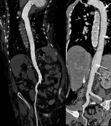
Two groups, each including 40 patients, underwent either ECG-gated or non-ECG-gated high-pitch dual-source CTA of the whole aorta. The aortic annulus, aortic valve, coronary ostia, and the presence of motion artifacts of the thoracic aorta as well as vascular contrast down to the femoral arteries were independently assessed by two readers. Additional objective parameters including image noise and signal-to-noise ratio were analyzed.
Results
Subjective and objective scoring revealed no presence of motional artifacts regardless of whether the ECG-gated or the non-ECG-gated protocol was used ( P > 0.1). Image acquisition parameters (examination length, examination duration, radiation dose) were comparable between the two groups without significant differences. The aortic annulus, aortic valve, and coronary ostia were reliably evaluable in all patients. Vascular contrast was rated excellent in both groups.
Conclusions
High-pitch dual-source CTA of the whole aorta is a robust and dose-efficient examination strategy for the evaluation of aortic pathologies whether or not ECG gating is used.
Introduction
Various improvements in computed tomography (CT) technology are currently commonly used such as wide-detector, single-source, or dual-source systems, and have led to shorter image acquisition durations and less motion artifacts. With the introduction of the latest dual-source CT devices, CT angiography (CTA) of the aorta is feasible in seconds (e.g., a whole body aorta examination within 3 seconds) . Faster image acquisition has become possible because dual-source CT allows pitch values of up to 3.4 with or without electrocardiographic (ECG) synchronization . Other factors influencing image acquisition are the use of fast gantry rotation times, fast table movement, and wide detector systems. One major advantage of high-pitch dual-source CT imaging is its ability to virtually freeze motion for the evaluation of the thoracic aorta, as heart motion can lead to diagnostic difficulties .
Previous studies have been conducted on bolus timing in high-pitch dual-source CT, and there have been feasibilities comparing high-pitch CT to single-source CT techniques . Many of these studies showed advantages such as fast image acquisition, motionless imaging of the thoracic vessels, and the possibility of evaluating the coronary arteries without ECG gating .
Get Radiology Tree app to read full this article<
Get Radiology Tree app to read full this article<
Materials and Methods
Patients and CT Protocols
Get Radiology Tree app to read full this article<
TABLE 1
Study Population and Evaluation of Examination Parameters
Group 1 Group 2P Value: Group 1 vs. Group 2 Patients 40 40 Male 23 29 Female 17 11 Age (years) 63 ± 21.1 (39–82) 64 ± 24.2 (29–88) 0.72 BMI (kg/m 2 ) 27.8 ± 3.9 (18.9–31.5) 28.3 ± 3.8 (18.3–32) 0.24 Scanning range (cm) 71.6 ± 9.3 (64.2–84.1) 71.7 ± 10.5 (40.2–83.9) 0.61 Scanning duration (s) 1.7 ± 0.2 (1.2–2.2) 1.8 ± 0.5 (1.1–2.1) 0.97 CTDIvol (mGy) 3.7 ± 0.5 (2.9–4.2) 3.8 ± 0.6 (3.2–4.3) 0.44
BMI, body mass index; CTDIvol, volume CT dose index.
Values in brackets represent ranges.
Get Radiology Tree app to read full this article<
Get Radiology Tree app to read full this article<
Get Radiology Tree app to read full this article<
TABLE 2
Examination Parameters
Group 1 Group 2 Imaging mode Dual-source Dual-source Machine Definition flash Definition flash Slice × collimation 2 × 128 × 0.6 2 × 128 × 0.6 Pitch 3.0 3.0 ECG gating On Off ROI Descending aorta Descending aorta HU threshold 200 200 Delay (s) 7 7
ECG, electrocardiographic; HU, Hounsfield units; ROI, region of interest.
Get Radiology Tree app to read full this article<
Get Radiology Tree app to read full this article<
Get Radiology Tree app to read full this article<
Get Radiology Tree app to read full this article<
Get Radiology Tree app to read full this article<
Image Analysis
Get Radiology Tree app to read full this article<
TABLE 3
Image Quality Rating Between the Different Groups
Group 1 Group 2P Value Overall image quality 1.0 (1.0–2.0) 1.0 (1.0–2.0) >0.1 Homogeneity of contrast enhancement along patient z-axis 1.5 (1.0–3.0 1.5 (1.0–3.0) >0.1 Evaluation of coronary ostia 1.0 (1.0–2.0) 1.0 (1.0–2.0) >0.1 Motion artifacts 1.0 (1.0–3.0) 1.0 (1.0–3.0) >0.1 Image noise (HU) 10.2 ± 6.1 (5.9–20.3) 9.9 ± 5.9 (6.1–20.1) 0.55 Median overall (HU) 384.2 ± 162.1 (229.1–616.9) 392.1 ± 168.6 (224.1–628.3) 0.43 SNR 38.7 ± 16.8 (18.9–71.8) 40.6 ± 22.9 (20.1–68.3) 0.73
HU, Hounsfield units; SNR, signal-to-noise ratio.
Values in brackets represent ranges.
Get Radiology Tree app to read full this article<
Get Radiology Tree app to read full this article<
Get Radiology Tree app to read full this article<
Get Radiology Tree app to read full this article<
Radiation Exposure
Get Radiology Tree app to read full this article<
Statistical Analysis
Get Radiology Tree app to read full this article<
Results
Get Radiology Tree app to read full this article<
Get Radiology Tree app to read full this article<
Get Radiology Tree app to read full this article<
Get Radiology Tree app to read full this article<
Get Radiology Tree app to read full this article<
Discussion
Get Radiology Tree app to read full this article<
Get Radiology Tree app to read full this article<
Get Radiology Tree app to read full this article<
Get Radiology Tree app to read full this article<
Get Radiology Tree app to read full this article<
Get Radiology Tree app to read full this article<
References
1. Achenbach S., Goroll T., Seltmann M., et. al.: Detection of coronary artery stenoses by low-dose, prospectively ECG-triggered, high-pitch spiral coronary CT angiography. JACC Cardiovascular Imaging 2011; 4: pp. 328-337.
2. Goetti R., Feuchtner G., Stolzmann P., et. al.: High-pitch dual-source CT coronary angiography: systolic data acquisition at high heart rates. Eur Radiol 2010; 20: pp. 2565-2571.
3. Wuest W., Anders K., Schuhbaeck A., et. al.: Dual source multidetector CT-angiography before Transcatheter Aortic Valve Implantation (TAVI) using a high-pitch spiral acquisition mode. Eur Radiol 2012; 22: pp. 51-58.
4. Beeres M., Schell B., Mastragelopoulos A., et. al.: High-pitch dual-source CT angiography of the whole aorta without ECG synchronisation: initial experience. Eur Radiol 2012; 22: pp. 129-137.
5. Bae K.T.: Intravenous contrast medium administration and scan timing at CT: considerations and approaches. Radiology 2010; 256: pp. 32-61.
6. Amacker N.A., Mader C., Alkadhi H., et. al.: Routine chest and abdominal high-pitch CT: an alternative low dose protocol with preserved image quality. Eur J Radiol 2012; 81: pp. e392-e397.
7. Karlo C., Leschka S., Goetti R.P., et. al.: High-pitch dual-source CT angiography of the aortic valve-aortic root complex without ECG-synchronization. Eur Radiol 2011; 21: pp. 205-212.
8. Beeres M., Loch M., Schulz B., et. al.: Bolus timing in high-pitch CT angiography of the aorta. Eur J Radiol 2013; 82: pp. 1028-1033.
9. Goetti R., Baumuller S., Feuchtner G., et. al.: High-pitch dual-source CT angiography of the thoracic and abdominal aorta: is simultaneous coronary artery assessment possible?. AJR Am J Roentgenol 2010; 194: pp. 938-944.
10. Ertel D., Lell M.M., Harig F., et. al.: Cardiac spiral dual-source CT with high pitch: a feasibility study. Eur Radiol 2009; 19: pp. 2357-2362.
11. Bamberg F., Marcus R., Sommer W., et. al.: Diagnostic image quality of a comprehensive high-pitch dual-spiral cardiothoracic CT protocol in patients with undifferentiated acute chest pain. Eur J Radiol 2012; 81: pp. 3697-3702.
12. Qanadli S.D., El Hajjam M., Mesurolle B., et. al.: Motion artifacts of the aorta simulating aortic dissection on spiral CT. J Comput Assist Tomogr 1999; 23: pp. 1-6.
13. Farshad-Amacker N.A., Alkadhi H., Leschka S., et. al.: Effect of high-pitch dual-source CT to compensate motion artifacts: a phantom study. Acad Radiol 2013; 20: pp. 1234-1239.
14. Zheng M., Wu Y., Wei M., et. al.: Low-concentration contrast medium for 128-slice dual-source CT coronary angiography at a very low radiation dose using prospectively ECG-triggered high-pitch spiral acquisition. Acad Radiol 2015; 22: pp. 195-202.
15. Scharf M., Bink R., May M.S., et. al.: High-pitch thoracic CT with simultaneous assessment of coronary arteries: effect of heart rate and heart rate variability on image quality and diagnostic accuracy. JACC Cardiovasc Imaging 2011; 4: pp. 602-609.
16. Zhang L.J., Zhao Y.E., Schoepf U.J., et. al.: Seventy-peak kilovoltage high-pitch thoracic aortic CT angiography without ECG gating: evaluation of image quality and radiation dose. Acad Radiol 2015; 22: pp. 890-897.
17. Wichmann J.L., Varga-Szemes A., Suranyi P., et. al.: Transcatheter aortic valve replacement: imaging techniques for aortic root sizing. J Thorac Imaging 2015; Epub ahead of print
18. Mangold S., Castillo-Sang M., Schoepf U.J., et. al.: Imaging in minimally invasive mitral valve repair. J Thorac Imaging 2015; PMID: 26258600; Epub ahead of print
19. Masson J.B., Kovac J., Schuler G., et. al.: Transcatheter aortic valve implantation: review of the nature, management, and avoidance of procedural complications. JACC Cardiovasc Interv 2009; 2: pp. 811-820.
20. Rixe J., Schuhbaeck A., Liebetrau C., et. al.: Multi-detector computed tomography is equivalent to trans-oesophageal echocardiography for the assessment of the aortic annulus before transcatheter aortic valve implantation. Eur Radiol 2012; 22: pp. 2662-2669.
21. Apfaltrer P., Schymik G., Reimer P., et. al.: Aortoiliac CT angiography for planning transcutaneous aortic valve implantation: aortic root anatomy and frequency of clinically significant incidental findings. AJR Am J Roentgenol 2012; 198: pp. 939-945.
22. Geyer L.L., De Cecco C.N., Schoepf U.J., et. al.: Low-volume contrast medium protocol for comprehensive cardiac and aortoiliac CT assessment in the context of transcatheter aortic valve replacement. Acad Radiol 2015; 22: pp. 1138-1146.
23. Harris B.S., De Cecco C.N., Schoepf U.J., et. al.: Dual-source CT imaging to plan transcatheter aortic valve replacement: accuracy for diagnosis of obstructive coronary artery disease. Radiology 2015; 275: pp. 80-88.
24. Meinel F.G., Canstein C., Schoepf U.J., et. al.: Image quality and radiation dose of low tube voltage 3rd generation dual-source coronary CT angiography in obese patients: a phantom study. Eur Radiol 2014; 24: pp. 1643-1650.
25. Gordic S., Husarik D.B., Desbiolles L., et. al.: High-pitch coronary CT angiography with third generation dual-source CT: limits of heart rate. Int J Cardiovasc Imaging 2014; 30: pp. 1173-1179.
26. Meyer M., Haubenreisser H., Schoepf U.J., et. al.: Closing in on the K edge: coronary CT angiography at 100, 80, and 70 kV-initial comparison of a second- versus a third-generation dual-source CT system. Radiology 2014; 273: pp. 373-382.
27. Haubenreisser H., Meyer M., Sudarski S., et. al.: Unenhanced third-generation dual-source chest CT using a tin filter for spectral shaping at 100 kVp. Eur J Radiol 2015; 84: pp. 1608-1613.