
Rationale and Objectives
In recent years, picture archiving and communication systems and electronic transfer of radiologic images using the digital imaging and communications in medicine file standard has become more widely employed in diagnostic radiology. It seems to be likely that nuclear medicine will be integrated within such systems. On the other hand, many departments possess older nuclear medicine equipment without digital output facilities. There is an increasing tendency to display and archive evaluated images (“save-screens,” printouts) on nondedicated, inexpensive systems using file formats capable of data compression. This was the reason for examining the value of the JPEG format in this pilot study.
Materials and Methods
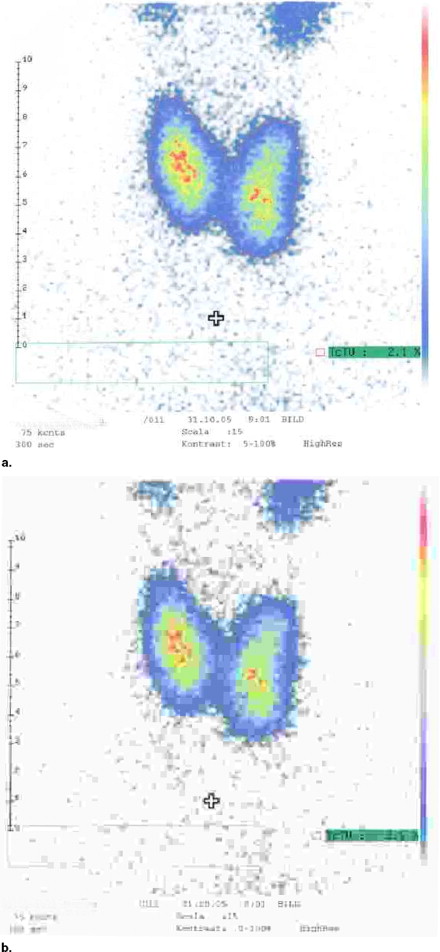
Fifty scanned planar bitmap images of the most frequent scintigraphic examinations (thyroid, bone, myocardium, lungs, and kidneys) were compared with JPEG format at different data compressions by two blinded observers. The visualization of details (eg, pathologic findings) is described for all these images as the visual appearance of the images and the storage capacity required.
Results
Relevant loss of clinical information did not occur up to compression factors of 0.75. A major decrease of subjective image quality was seen at compression factors >0.90. Compared with bitmap files, the use of these factors reduced the storage capacity required by 98% at a (JPEG-related) compression factor of 0.50, and 99% at a compression factor of 0.90. Compared with the GIF format, a reduction by 4.0–5.7 could be achieved.
Conclusions
Use of the JPEG format can therefore be recommended to save costs of image transfer or archiving of standard planar scans for nuclear medical evaluation.
Even for conventional personal computers, there has been a rapid increase of performance (speed of calculation, storage capacity, and access times) and a reduction of costs in recent years. This continuously leads to an ever greater number of users with “normal capacity” personal computers and a growth of networking tasks. Data compression must therefore be used to ensure effective storage management and rapid transfer of data.
Many smaller departments of nuclear medicine may refrain from buying a (DICOM)-related picture archiving and communication systems for reasons of costs. Furthermore, they will continue to use older equipment without digital or DICOM-based output or archiving facilities. This pilot study examines the use of a simple method for storing the output material in a conventional personal computer to fulfill the statutory requirement to store the results of the examination for evaluation purposes to be used in the patient’s follow-up.
Get Radiology Tree app to read full this article<
Get Radiology Tree app to read full this article<
Table 1
Common Image Formats
Name Extension Comments Compression Bitmap *.bmp Raster None Paintbrush *.pcx Raster Lossless, simple compression Portable network graphics *.png Raster Lossless, low compression Graphic interchange format *.gif Raster Lossless, reduced colors Joint photographic experts group *.jpg Raster Lossy, high compression Tagged image file format *.tif Raster Extensive Drawing interchange file (AutoCAD) *.dxf Picture Vector-related Computer graphics metafile *.cgm Picture Vector-related CorelDRAW *.cdr Picture Vector/bitmap Windows metafile *.wmf Picture Vector/bitmap Enhanced metafile *.emf Picture Vector/bitmap Encapsulated postscript *.eps Picture Vector/bitmap, printer related
Get Radiology Tree app to read full this article<
Get Radiology Tree app to read full this article<
Get Radiology Tree app to read full this article<
Get Radiology Tree app to read full this article<
Get Radiology Tree app to read full this article<
Materials and methods
Images
Get Radiology Tree app to read full this article<
Get Radiology Tree app to read full this article<
Get Radiology Tree app to read full this article<
Get Radiology Tree app to read full this article<
Results
Get Radiology Tree app to read full this article<
Table 2
Effects of Compression Factors CF (n = 50, Mean/SD)
(a) Storage Amount in Bytes (Mean/SD) – for Bitmap File: 2,670,414 Bytes/Image in All Cases CF 0.5 0.75 0.90 0.95 0.99 Thyroid
Bone
Myocardium
Lung
Kidneys
Mean storage: 53,687 31,402 26,565 20,832 17,932 % of bitmap file 2.01 1.18 0.99 0.78 0.67
(b) Image Quality (Mean/SD) CF 0.5 0.75 0.90 0.95 0.99 Thyroid 1.0/0.0 2.0/0.0 3.0/0.0 4.0/0.0 5.1/0.3 Bone 1.0/0.0 1.3/0.5 2.9/0.3 4.0/0.5 6.0/5.6 Myocardium 1.0/0.0 2.0/0.0 3.0/0.0 5.0/0.0 6.0/0.0 Lung 1.0/0.0 2.2/0.4 3.4/0.5 4.6/0.5 6.0/0.0 Kidneys 1.0/0.0 2.0/0.0 3.0/0.0 4.0/0.0 5.0/0.0 Mean 1.0 1.9 3.1 4.3 5.6
(c) Image Quality Regarding Differences in Pathologic Lesions (Mean/SD) CF 0.5 0.75 0.90 0.95 0.99 Thyroid 2.0/0.0 2.0/0.0 4.0/0.0 5.0/0.0 6.0/0.0 Bone 2.0/0.0 3.0/0.0 4.2/0.4 5.0/0.0 6.0/0.0 Myocardium 2.0/0.0 2.0/0.0 4.0/0.0 5.0/0.0 6.0/0.0 Lung 2.0/0.0 3.0/0.0 4.0/0.0 5.0/0.0 6.0/0.0 Kidneys 2.0/0.0 2.0/0.0 4.0/0.0 5.0/0.0 6.0/0.0 Mean 2.0 2.4 4.0 5.0 6.0
CF: compression factor; SD: standard deviation.
Get Radiology Tree app to read full this article<
Get Radiology Tree app to read full this article<
Get Radiology Tree app to read full this article<
Get Radiology Tree app to read full this article<
Get Radiology Tree app to read full this article<
Get Radiology Tree app to read full this article<
Discussion
Get Radiology Tree app to read full this article<
Get Radiology Tree app to read full this article<
Get Radiology Tree app to read full this article<
Get Radiology Tree app to read full this article<
Get Radiology Tree app to read full this article<
Get Radiology Tree app to read full this article<
Get Radiology Tree app to read full this article<
Get Radiology Tree app to read full this article<
Get Radiology Tree app to read full this article<
Get Radiology Tree app to read full this article<
Get Radiology Tree app to read full this article<
Get Radiology Tree app to read full this article<
Get Radiology Tree app to read full this article<
Conclusion
Get Radiology Tree app to read full this article<
Acknowledgment
Get Radiology Tree app to read full this article<
References
1. Nissen S.E., Goldburgh M., ACC/ACR/NEMA Ad Hoc Group: ACC, ACR and industry develop standard for digital transfer of angiographic images. J Am Coll Cardiol 1995; 25: pp. 800-802.
2. Bidgood W.D., Horii S.C.: Modular extension of the ACR-NEMA DICOM standard to support new diagnostic imaging modalities and services. J Digit Imaging 1996; 9: pp. 67-77.
3. Parisot C.: The DICOM standard. Int J Card Imaging 1995; 11: pp. 171177.
4. Campioni P., Valentine V., Vincenzoni M., et. al.: Standardization of digital radiologic images. Rays 1996; 21: pp. 213-227.
5. Jao C.S., Hier D.B., Brint S.U.: The display of photographic-quality images on the Web: a comparison of two technologies. IEEE Trans Inf Technol Biomed 1999; 3: pp. 70-73.
6. Hart J.C.: Fractal image compression and recurrent iterated function systems. IEEE Comp Graphics Appl 1996; 16: pp. 25-33.
7. Pennebaker W.B., Mitchell J.L.: 1993.Van Nostrand ReinholdNew York
8. Rigolin V.H., Robiolio P.A., Spero L.A., et. al.: Lossy JPEG compression of digital coronary angiograms does not effect visual or quantitative assessment of coronary stenosis severity. Am J Cardiol 1996; 78: pp. 131-135.
9. Baker W.A., Hearne S.E., Spero L.A., et. al.: Lossy (15:1) JPEG compression of digital coronary angiograms does not limit detection of subtle morphological features. Circulation 1997; 96: pp. 1157-1164.
10. Karson T.H., Chandra S., Morehead A.J., et. al.: JPEG compression of digital echocardiographic images: impact on image quality. J Am Soc Echocardiography 1995; 8: pp. 306-308.
11. Sheigh H.R., Bovik A.C.: Image information and visual quality. IEEE Trans Image Process 2006; 15: pp. 430-444.
12. Levine B.A., Mun S.K., Benson H.R., et. al.: Assessment of the integration of a HIS/RIS with a PACS. Proc SPIE 1990; 1234: pp. 391-397.
13. Feingold E., Seshadri S.B., Arenson R.L.: Folder management on a multimodality PACS display station. Proc SPIE 1991; 1446: pp. 211-216.