
Rationale and Objectives
The study goal was to retrospectively evaluate the effectiveness of computed tomography (CT)-guided spinal biopsies.
Material and Methods
Two hundred two CT-guided vertebral biopsies performed between May 1999 and June 2004 in 187 patients were retrospectively analyzed. Patient characteristics (age, sex, antibiotic therapy), technical parameters (depth and number of biopsies, needle approach), lesion features (spinal level, osteolysis, fluid collections, soft tissue tumor), and complications were documented. Furthermore, histopathological and microbiological diagnoses were considered.
Results
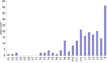
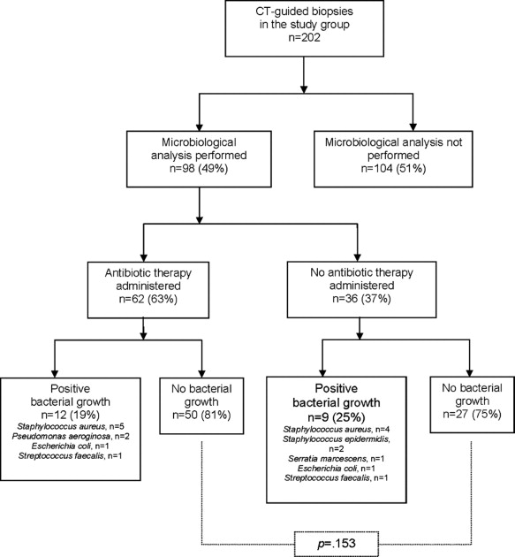
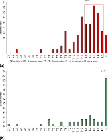
There were two focal hematomas in our study group (complication rate: 1%). Histopathological diagnosis was established in 74% of examinations with spondylitis (41% of cases) being most common. In spinal tumors (27% of cases), malignant lesions were found in 52 of 54 examinations (96%). Osteolysis was diagnosed in 98% of patients with spondylitis and in 87% of patients with tumors ( P < .01). Spinal tumors were most commonly seen in the sacrum (53%, P < .001), whereas spondylitis typically occurred in the lumbar spine (55%, P = .001). Neither patient age nor sex, needle approach, needle depth, or vertebral abnormalities showed a significant impact on diagnostic accuracy. Microbiological tests were performed in 98 patients (49%); 62 of 98 patients (65%) received antibiotic therapy. In 12 of 62 patients (19%) with antibiotic therapy and in 9 of 36 patients (25%) without antibiotic treatment, microbiological tests were positive ( P = .153). Staphylococcus aureus was found in 9 of 21 examinations (43%).
Conclusions
CT-guided vertebral biopsy is a safe and effective procedure to establish final diagnosis in spinal lesions of unclear origin. Patient characteristics, lesions features, and technical considerations did not influence sample quality. In spondylitis, which was commonly caused by Staphylococcus aureus , microbiological yield was low regardless of antibiotic therapy.
Precise histological diagnosis has become a necessary prelude to successful management of diseases of the spine. Although many symptomatic patients with spinal lesions present with osteolysis, spinal masses, or sclerosis on image studies, radiographic findings can be misleading. For years open biopsy has been the reference standard to obtain adequate tissue specimen in spinal lesions. After early attempts of performing biopsies blindly ( ), intensified fluoroscopic guidance became the major imaging device to obtain spinal biopsies. Although early results were promising ( ), this method was not without disadvantages. Conventional radiography does not accurately distinguish between the physical characteristics of a lesion and its surrounding structures ( ). Moreover, poor spatial resolution and superposition of structures further limited its use. Ultrasound has also been used to navigate spinal biopsies ( ), but its inability to assess osseous structures substantially limits its value.
First reports of computed tomography (CT)-guided biopsies of the spine were presented in the late 1970s ( ), after CT had been extensively applied to characterize spinal lesions based on imaging features. Recent advances in imaging technology, including the clinical introduction of multidetector CT, have further improved navigation techniques for spinal biopsies. Although CT guidance is now widely accepted as the standard procedure for image-guided spinal biopsies ( ), little is known about the dependence of its diagnostic accuracy on patient characteristics and on technical considerations of the biopsy procedure. We describe our techniques of CT-guided spinal biopsies and provide an analysis of all examined patients considering final histopathological and microbiological diagnoses.
Materials and methods
Patient Selection
Get Radiology Tree app to read full this article<
Get Radiology Tree app to read full this article<
Biopsy Procedure
Get Radiology Tree app to read full this article<
Get Radiology Tree app to read full this article<
Get Radiology Tree app to read full this article<
Get Radiology Tree app to read full this article<
Get Radiology Tree app to read full this article<

Get Radiology Tree app to read full this article<
Get Radiology Tree app to read full this article<
Statistical Analyses
Get Radiology Tree app to read full this article<
Results
Patient Group
Get Radiology Tree app to read full this article<
Get Radiology Tree app to read full this article<
Biopsy Procedure
Get Radiology Tree app to read full this article<
Histopathological Results
Get Radiology Tree app to read full this article<
Table 1
Histopathological Diagnoses in Spinal Tumor Lesions Established by CT-Guided Percutaneous Biopsy
Histopathological Diagnosis Incidence ( N = 58) Plasmocytoma_n_ = 9 (16%) Metastasis of undifferentiated malignant tumor_n_ = 7 (12%) Metastasis of small cell lung cancer_n_ = 5 (9%) Metastasis of adenocarcinoma_n_ = 5 Metastasis of prostate cancer_n_ = 4 (8%) Metastasis of colon carcinoma_n_ = 3 (5%) Metastasis of renal cell carcinoma_n_ = 2 (4%) Metastasis of urothelial carcinoma_n_ = 2 Primary hemangioma/lymphangioma_n_ = 2 Non-Hodgkin’s lymphoma_n_ = 2 Chondrosarcoma_n_ = 2 Metastasis of spinocellular carcinoma_n_ = 1 (2%) Metastasis of carcinoid carcinoma_n_ = 1 Metastasis of squamous cell carcinoma_n_ = 1 Metastasis of thyroid carcinoma_n_ = 1 Metastasis of breast cancer_n_ = 1 Angiosarcoma_n_ = 1 Leukemia with osseous infiltration_n_ = 1 Metastasis of malignant melanoma_n_ = 1 Lymphangiosis carcinomatosa_n_ = 1 Osteoblastic osteosarcoma_n_ = 1
Get Radiology Tree app to read full this article<
Microbiological Results
Get Radiology Tree app to read full this article<
Get Radiology Tree app to read full this article<
Get Radiology Tree app to read full this article<
Morphology and Level of Vertebral Lesions
Get Radiology Tree app to read full this article<
Get Radiology Tree app to read full this article<
Table 2
Comparison of Level of Spinal Findings and Major Histopathological Diagnoses
Cervical Spine Thoracic Spine Lumbar Spine Sacral Spine Final Histopathological Diagnosis ( n = 4) ( n = 69) ( n = 83) ( n = 40) Tumor 2 of 4 (50%) 16 of 69 (23%) 21 of 40 (53%)n = 54 of 196 (28%)P = .305P = .201 15 of 83 (18%)☆ P = .008P <.001 Spondylitis 1 of 4 (25%) 30 of 69 (43%) 5 of 40 (13%)n = 82 of 196 (42%)P = .443P = .423 46 of 83 (55%) P = .001P < .001 No histopathological diagnosis established 1 of 4 (25%) 20 of 68 (29%) 17 of 83 (20%) 14 of 40 (35%)n = 52 of 196 (27%)P = .711P = .319P = .064P = .129
Get Radiology Tree app to read full this article<
Inconclusive Histopathological/Microbiological Results
Get Radiology Tree app to read full this article<
Table 3
Comparison of Patients With and Without Final Histopathological Diagnosis With Respect to Patient Characteristics, Technical Considerations of the Biopsy Procedure, and Vertebral Abnormalities
Patients With Final Histopathological Diagnosis ( n = 143) Patients Without Final Histopathological Diagnosis (normal findings or insufficient specimen quality) ( n = 52)P value Patient characteristics .034 Age (63 ± 14 y) 63 ± 12 y 59 ± 16 y .374 ⁎ Sex (117 of 195 male, 60%) 90 of 143 male (63%) 27 of 52 male (52%) .111 Biopsy procedure No. of biopsies performed (1.2 ±.47) 1.2 ± 0.44 1.2 ± 0.55 .103 Patient position during biopsy (67% POS) 100 of 143 POS (70%) 21 of 52 POS (40%) .119 Biopsy approach (47% TPA) 67 of 143 TPA (47%) 25 of 52 TPA (48%) .504 Final biopsy depth (7.5 ± 1.8 cm) 7.6 ± 1.7 cm 7.0 ± 1.5 cm .561 Vertebral abnormalities Vertebral osteolysis ( n = 180, 92%) 135 of 143 (94%) 45 of 52 (87%) .069 Spinal stenosis ( n = 53, 27%) 41 of 143 (29%) 12 of 52 (23%) .279 Paravertebral soft tissue mass ( n = 107, 55%) 79 of 143 (55%) 28 of 52 (57%) .495 Paravertebral fluid collection ( n = 36, 18%) 27 of 143 (19%) 9 of 52 (17%) .492
POS, position on the side; TPA, transpedicular approach.
Get Radiology Tree app to read full this article<
Get Radiology Tree app to read full this article<
Get Radiology Tree app to read full this article<
Discussion
Get Radiology Tree app to read full this article<
Get Radiology Tree app to read full this article<
Get Radiology Tree app to read full this article<
Get Radiology Tree app to read full this article<
Get Radiology Tree app to read full this article<
Get Radiology Tree app to read full this article<
Get Radiology Tree app to read full this article<
Get Radiology Tree app to read full this article<
Get Radiology Tree app to read full this article<
Get Radiology Tree app to read full this article<
Get Radiology Tree app to read full this article<
References
1. Robertson R.: Destructive spinal lesions. J Bone Joint Surg 1935; 17: pp. 749-758.
2. Craig F.: Vertebral body biopsy. J Bone Joint Surg 1956; 38: pp. 93-102.
3. Deeley T.: The drill biopsy of bone lesions. Clin Radiol 1972; 23: pp. 536-540.
4. Mazer R., Cozen L.: The diagnostic value of vertebral body needle biopsy. Am J Surg 1952; 135: pp. 245-252.
5. Adapon B.D., Legada B.D., Lim E.V., Silao J.V., Dalmacio-Cruz A.: CT-guided closed biopsy of the spine. J Comput Assist Tomogr 1981; 5: pp. 73-78.
6. Gupta S., Takhtani D., Gulati M., et. al.: Sonographically guided fine-needle aspiration biopsy of lytic lesions of the spine: Technique and indications. J Clin Ultrasound 1999; 27: pp. 123-129.
7. Haaga J., Alfidi R.: Precise biopsy localization by computed tomography. Radiology 1976; 118: pp. 603-607.
8. Adapon B.D., Legada B.D., Lim E.V., Silao J.V., Dalmacio-Cruz A.: CT-guided closed biopsy of the spine. J Comput Assist Tomogr 1981; 5: pp. 73-78.
9. Babu N.V., Titus V.T., Chittaranjan S., Abraham G., Prem H., Korula R.J.: Computed tomographically guided biopsy of the spine. Spine 1994; 19: pp. 2436-2442.
10. Brugieres P., Revel M.P., Dumas J.L., Heran F., Voisin M.C., Gaston A.: CT-guided vertebral biopsy. J Neuroradiol 1991; 18: pp. 351-359.
11. Cleary K., Clifford M., Stoianovici D., Freedman M., Mun S.K., Watson V.: Technology improvements for image-guided and minimally invasive spine procedures. IEEE Trans Inform Technol Biomed 2002; 6: pp. 249-261.
12. Dupuy D.E., Rosenberg A.E., Punyaratabandhu T., Tan M.H., Mankin H.J.: Accuracy of CT-guided needle biopsy of musculoskeletal neoplasms. AJR Am J Roentgenol 1998; 171: pp. 759-762.
13. Kang M., Gupta S., Khandelwal N., Shankar S., Gulati M., Suri S.: CT-guided fine-needle aspiration biopsy of spinal lesions. Acta Radiol 1999; 40: pp. 474-478.
14. Kattapuram S.V., Rosenthal D.I.: Percutaneous biopsy of the cervical spine using CT guidance. AJR Am J Roentgenol 1987; 149: pp. 539-541.
15. Omarini L.P., Garcia J.: Ponctions-biopsies percutanees du rachis sous guidage CT. Schweiz Med Wochenschr 1993; 123: pp. 2191-2197.
16. Sucu H.K., Cicek C., Rezanko T., et. al.: Percutaneous computed-tomography-guided biopsy of the spine: 229 Procedures. Joint Bone Spine Rev Rheumat 2006; 73: pp. 532-537.
17. Omary R.A., Bettmann M.A., Cardella J.F., et. al.: Quality improvement guidelines for the reporting and archiving of interventional radiology procedures. J Vasc Intervent Radiol 2002; 13: pp. 879-881.
18. Valls J., Ottolenghi C.E., Shajowicz F.: Aspiration biopsy in diagnosis of lesions of vertebral bodies. JAMA 1948; 136: pp. 376-382.
19. Puri A., Shingade V.U., Agarwal M.G., et. al.: CT-guided percutaneous core needle biopsy in deep seated musculoskeletal lesions: A prospective study of 128 cases. Skeletal Radiol 2006; 35: pp. 138-143.
20. Kornblum M.B., Wesolowski D.P., Fischgrund J.S., Herkowitz H.N.: Computed tomography-guided biopsy of the spine. Spine 1998; 23: pp. 81-85.
21. Kattapuram S.V., Khurana J.S., Rosenthal D.I.: Percutaneous needle biopsy of the spine. Spine 1992; 17: pp. 561-564.
22. Lis E., Bilsky M.H., Pisinski L., Boland P., Healey J.H., O’Malley B., Krol G.: Percutaneous CT-guided biopsy of osseous lesion of the spine in patients with known or suspected malignancy. AJNR Am J Neuroradiol 2004; 25: pp. 1583-1588.
23. Hau A., Kim I., Kattapuram S., et. al.: Accuracy of CT-guided biopsies in 359 patients with musculoskeletal lesions. Skeletal Radiol 2002; 31: pp. 349-353.
24. Ghelman B., Lospinuso M.F., Levine D.B., O’Leary P.F., Burke S.W.: Percutaneous computed-tomography-guided biopsy of the thoracic and lumbar spine. Spine 1991; 16: pp. 736-739.
25. Stoker D.J., Kissin C.M.: Percutaneous vertebral biopsy: A review of 135 cases. Clin Radiol 1985; 36: pp. 569-577.
26. Ackermann W.: Vertebral trephine biopsy. Ann Surg 1956; 143: pp. 373-385.
27. Ottolenghi C.E.: Aspiration biopsy of the spine. J Bone Joint Surg (Am) 1969; 51: pp. 1531-1544.
28. Siffert R.S., Arkin A.M.: trephine bone biopsy with special reference to the lumbar vertebral bodies. J Bone Joint Surg (Am) 1949; 31: pp. 146-149.
29. Frager D.H., Goldman M.J., Seimon L.P., et. al.: Computed tomography guidance for skeletal biopsy. Skeletal Radiol 1987; 16: pp. 644-646.
30. Brugieres P., Gaston A., Heran F., Voisin M.C., Marsault C.: percutaneous biopsies of the thoracic spine under CT guidance: Transcostovertebral approach. J Comput Assist Tomogr 1990; 14: pp. 446-448.
31. Renfrew D.L., Whitten C.G., Wiese J.A., el-Khoury G.Y., Harris K.G.: CT-guided percutaneous transpedicular biopsy of the spine. Radiology 1991; 180: pp. 574-576.
32. Jelinek J.S., Kransdorf M.J., Gray R., Aboulafia A.J., Malawer M.M.: Percutaneous transpedicular biopsy of vertebral body lesions. Spine 1996; 21: pp. 2035-2040.
33. Michel S.C., Pfirrmann C.W., Boos N., Hodler J.: CT-guided core biopsy of subchondral bone and intervertebral space in suspected spondylodiskitis. AJR Am J Roentgenol 2006; 186: pp. 977-980.
34. Brugieres P., Gaston A., Voisin M.C., Ricolfi F., Chakir N.: CT-guided percutaneous biopsy of the cervical spine: A series of 12 cases. Neuroradiology 1992; 34: pp. 358-360.
35. Gupta R.K., Cheung Y.K., Al Ansari A.G., Naran S., Lallu S., Fauck R.: Diagnostic value of image-guided needle aspiration cytology in the assessment of vertebral and intervertebral lesions. Diagn Cytopathol 2002; 27: pp. 191-196.
36. Minart D., Vallee J.N., Cormier E., Chiras J.: Percutaneous coaxial transpedicular biopsy of vertebral body lesions during vertebroplasty. Neuroradiology 2001; 43: pp. 409-412.
37. Avva R., Vanhemert R.L., Barlogie B., Munshi N., Angtuaco E.J.: CT-guided biopsy of focal lesions in patients with multiple myeloma may reveal new and more aggressive cytogenetic abnormalities. AJNR Am J Neuroradiol 2001; 22: pp. 781-785.
38. Lewin J.S., Petersilge C.A., Hatem S.F., et. al.: Interactive mr imaging-guided biopsy and aspiration with a modified clinical C-arm system. AJR Am J Roentgenol 1998; 170: pp. 1593-1601.