
Rationale and Objectives
To evaluate the effects of iodine contrast agent on diffusion signal intensity and apparent diffusion coefficient (ADC) in diffusion-weighted imaging (DWI) studies in magnetic resonance imaging (MRI) examination just after computed tomography (CT) contrast imaging.
Materials and Methods
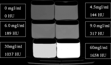
On a 1.5 T MRI scanner, ADC was calculated from the signal intensity of DWI (b = 0 and 1000) using phantoms filled with contrast agent (0, 4.5, 6.0, 9.0, 30, and 60 mgI/mL). We evaluated the signal intensities of DWI and ADC in 10 patients (3 women, 7 men, 35–68 years old) examined by MRI study less than 40 minutes after injection of 100 mL of iopamidol (300 mgI/mL) for CT study.
Results
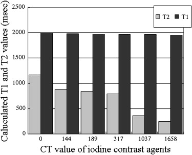
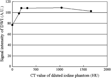
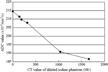
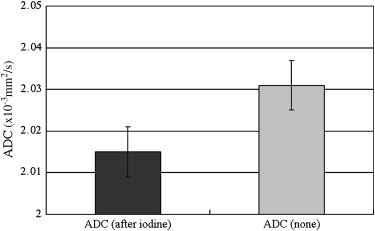
The DWI signal increased until a CT value of 190 HU, but showed no changes above this value. The ADC decreased with increases in CT value. Less than 40 minutes after injection of iopamidol (300 mgI/mL) for CT scan, the signal intensity of DWI was significantly increased and ADC was significantly decreased.
Conclusions
It is necessary to recognize the rate of decrease of ADC, because it is dependent on the density of iodine contrast agents.
Various examination modalities are often used in the same patient for diagnosis, and both computed tomography (CT) and magnetic resonance imaging (MRI) examinations may be performed consecutively on the same day.
Several studies have indicated that contrast agents used for CT and MRI may adversely influence the results of the other imaging modality . In these reports, it was suggested that iodinated contrast agents shorten T1 and T2 on MRI examination, and cause high signal intensity on T1-weighted imaging and low signal intensity on T2-weighted imaging .
Get Radiology Tree app to read full this article<
Get Radiology Tree app to read full this article<
Materials and methods
Get Radiology Tree app to read full this article<
Get Radiology Tree app to read full this article<
Get Radiology Tree app to read full this article<
Get Radiology Tree app to read full this article<
Get Radiology Tree app to read full this article<
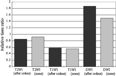
Relativetissueratio=Signalvalueofkidney/Signalvalueoffat(1) Relative
tissue
ratio
=
Signal
value
of
kidney
/
Signal
value
of
fat
(
1
)
Get Radiology Tree app to read full this article<
Get Radiology Tree app to read full this article<
Get Radiology Tree app to read full this article<
Results
Get Radiology Tree app to read full this article<
Table 1
Average Computed Tomography Values and Standard Deviation of Brain Meningiomas and Kidney Cortex and of the Bladder 2 and 15 Minutes after Injection of Contrast Agent, Respectively, in 20 Patients
Averaged CT Value (HU) SD Meningioma after injected two minutes 204.2 15.2 Kidney cortex after injected 2 minutes 182.3 4.11 Bladder after injected 15 minutes 488.2 25.3
Computed tomography values ranged from 180 to 500 HU.
Get Radiology Tree app to read full this article<
Get Radiology Tree app to read full this article<
Get Radiology Tree app to read full this article<
![Figure 3, Diffusion-weighted imaging (repetition time 4000/echo time 107 ms/SENSE factor 2 with EPI [echo planar imaging]) of diluted iodine contrast phantom.](https://storage.googleapis.com/dl.dentistrykey.com/clinical/EffectsofIodinatedContrastAgentonDiffusionWeightedMagneticResonanceImaging/2_1s20S1076633209002566.jpg)

Get Radiology Tree app to read full this article<
Get Radiology Tree app to read full this article<
Get Radiology Tree app to read full this article<
Discussion
Get Radiology Tree app to read full this article<
Get Radiology Tree app to read full this article<
Get Radiology Tree app to read full this article<
Get Radiology Tree app to read full this article<
Get Radiology Tree app to read full this article<
Get Radiology Tree app to read full this article<
Get Radiology Tree app to read full this article<
Conclusions
Get Radiology Tree app to read full this article<
Get Radiology Tree app to read full this article<
References
1. Hergan K., Doringer W., Langle M., et. al.: Effect of iodinated contrast agents in MR imaging. Eur J Radiol 1995; 21: pp. 11-17.
2. Jinkins J.R., Robinson J.W., Sisk L., et. al.: Proton relaxation associated with iodinated contrast agents in MR imaging of the CNS. AJNR 1992; 13: pp. 19-27.
3. Hammer F.D., Goffette P.P., Malaise J., et. al.: Gadolinium dimeglumine: an alternative contrast agent for digital subtraction angiography. Eur Radiol 1999; 9: pp. 128-136.
4. Bloem J.L., Wondergem J.: Gd-DTPA as a contrast agent in CT. Radiology 1989; 171: pp. 578-579.
5. Spinosa D., Angle J.F., Hartwell G., et. al.: Gadolinium-based contrast agents in angiography and interventional radiology. Radiol Clin N Am 2002; 40: pp. 693-710.
6. Arat A., Cekirge H.S., Saatci I.: Gadodiamide as an alternative contrast medium in cerebral angiography in a patient with sensitivity to iodinated contrast medium. Neuroradiology 2000; 42: pp. 34-37.
7. Ogura A., Hayakawa K., Miyati T., et. al.: The effect of susceptibility of gadolinium contrast media on diffusion-weighted imaging and the apparent diffusion coefficient. Acad Radiol 2007; 15: pp. 867-872.
8. Yoshikawa Y., Kawamitsu H., Mitchell D.G., et. al.: ADC measurement of abdominal organs and lesions using parallel imaging technique. AJR Am J Roentegenol 2006; 187L: pp. 1522-1530.
9. Dose M.D., Zhong J., Gore J.C.: In vivo measurement of ADC change due to intravascular susceptibility variation. Magn Reson Med 1999; 41: pp. 236-240.
10. Yamada K., Kubita H., Kizu O., et. al.: Effect of Intravenous gadolinium-DTPA on diffusion weighted images. Stroke 2002; 33: pp. 1799-1802.
11. Chen G., Jespersen S.N., Pederson M., et. al.: Intravenous administration of Gd-DTPA prior to DWI dose not affect the apparent diffusion constant. Magn Reson Imaging 2005; 23: pp. 685-689.
12. Krause W., Miklautz H., Kollenkirchen U., et. al.: Physicochemical parameters of X-ray contrast media. Invest Radiol 1994; 29: pp. 72-80.
13. Gallotti A., Uggeri F., Favilla A., et. al.: The chemistry of iomeprol and physic-chemical properties of its aqueous solutions and pharmaceutical formulations. Eur J Radiol 1994; 18: pp. S1-S12.
14. Pugh N.D.: Haemodynamic and rheological effects of contrast media: the role of viscosity and osmolality. Eur Radiol 1996; 6: pp. 13-15.
15. Bettmann M.A.: Contrast media: safety, viscosity, and volume. Eur Radiol 2005; 15: pp. 62-64.
16. Bihan D.L., Breton E., Lalleman D., et. al.: Separation of diffusion and perfusion in intravoxel incoherent motion MR imaging. Radiology 1988; 168: pp. 497-505.
17. Higano S., Yun Xia, Kumabe T., et. al.: Malignant astrocytic tumors: clinical importance of apparent diffusion coefficient in prediction of grade and prognosis. Radiology 2006; 241: pp. 839-846.
18. Yoshikawa T., Kawamitu H., Mitchell D.G., et. al.: ADC measurement of abdominal organs and lesions using parallel imaging technique. AJR Am J Roentgenol 2006; 187: pp. 1521-1530.
19. Sumi M., Cauteren M.V., Nakamura T.: MR microimaging of benign and malignant nodes in the neck. AJR Am J Roentgenol 2006; 186: pp. 749-757.
20. Woodhams R., Matsubara K., Kan A., et. al.: ADC mapping of benign and malignant breast tumors. MRMS 2005; 4: pp. 35-42.
21. Nakayama T., Yoshimitsu K., Irie H., et. al.: Usefulness of calculated apparent diffusion coefficient value in the differential diagnosis of retroperitoneal masses. JMRI 2004; 20: pp. 735-742.
22. Nakayama T., Yoshimitsu K., Irie H., et. al.: Diffusion-weighted echo-planar MR imaging and ADC mapping in the differential diagnosis of ovarian cystic masses. JMRI 2004; 22: pp. 271-278.
23. Kim C.K., Park B.K., Han J.J., et. al.: Diffusion-weighted imaging of the prostate at 3 T for differentiation of malignant and benign tissue in transition and peripheral zones: preliminary results. JCAT 2007; 31: pp. 449-454.
24. Zhang J., Tehrani Y.M., Wang L., et. al.: Renal masses: characterization with diffusion-weighted MR imaging— a preliminary experience. Radiology 2008; 247: pp. 458-464.
25. Humphries P.D., Sebire N.J., Seigel M.J., et. al.: Tumors in pediatric patients at diffusion-weighted MR imaging: apparent diffusion coefficient and tumor cellularity. Radiology 2007; 245: pp. 848-854.