
Rationale and Objectives
The rates of enrollment of volunteers for brain magnetic resonance imaging (MRI) studies vary by demographic and clinical characteristics. We use data from a large MRI study to identify factors associated with differential enrollment and to examine potential biases this may produce in study results.
Materials and Methods
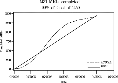
Results from recruitment of 1,431 women into the MRI substudy of the Women’s Health Initiative Memory Study (WHIMS-MRI) are described. A sensitivity analysis was conducted to estimate the degree of bias associated with missing data on estimates of risk factor relationships.
Results
Of 2,345 women contacted from an established cohort of women older than 70 years of age, 72% consented to undergo screening for WHIMS-MRI. Scanning was ultimately completed on 61%. Completion rates varied according to a range of sociodemographic, lifestyle, and clinical characteristics that may be related to study outcomes. Plausible levels of selective enrollment in magnetic resonance imaging studies may produce moderate biases (< ±20%) in characterizations of risk factor relationships. Adverse events, such as claustrophobia, occurred during 1.7% of the attempted scans and, in 0.8% of instances, led to lost data.
Conclusions
Enrollment of older women into brain imaging studies is feasible, although selection biases may limit how well study cohorts reflect more general populations.
Clinical trials and cohort studies increasingly are designed to include magnetic resonance imaging outcomes. Experiences with this new technology have provided successful protocols for standardized measurement, assessment, and safety ( ). Brain magnetic resonance studies conducted in established cohorts have reported consent rates ranging from 71% to 90% and yields of completed imaging in 57%–72% of contacted individuals. However, these rates appear to vary markedly among subgroups based on important demographic and clinical factors ( ). Thus, although it appears that recruitment into brain magnetic resonance imaging (MRI) studies is feasible, differential selection could have the potential to bias research findings.
We used data from the Women’s Health Initiative Memory Study Magnetic Resonance Imaging Study (WHIMS-MRI) to explore potential biases. We report rates of women who consented to undergo screening for the study, as a whole and by various subgroups, to identify potential barriers to recruiting participants. We also report the rates that MRI images were obtained, which are jointly affected by consent, eligibility criteria, and factors that interfere with imaging, such as claustrophobia. A sensitivity analysis was then performed to understand how selection biases inherent in enrollment may ultimately influence estimates of relationships among study outcomes. Additionally, we also describe the rate and nature of serious adverse events that were reported during the scanning procedure, some of which resulted in interruption of scanning and loss of data.
Methods
Study Design
Get Radiology Tree app to read full this article<
Get Radiology Tree app to read full this article<
Get Radiology Tree app to read full this article<
Baseline Data Collection, Variable Definitions, and Data Analysis Plans
Get Radiology Tree app to read full this article<
Get Radiology Tree app to read full this article<
Get Radiology Tree app to read full this article<
Results
Get Radiology Tree app to read full this article<
Get Radiology Tree app to read full this article<
Rates of Consent for WHIMS-MRI Enrollment
Get Radiology Tree app to read full this article<
Table 1
Characteristics of Participants Enrolled in WHIMS Grouped by Whether They Provided at Least Some Level of Consent to Participate in the WHIMS-MRI Study
Characteristic_n_ Consent for Screening MRI Obtained Percent Providing Consent Chi-square P Value Percent MRI Obtained Chi-square P Value Overall 2,345 72% 61% Age 70–75 766 74% 64% 76–81 1,144 73% <.001 63% <.001 82–88 435 64% 50% Education, years <12 (high school) 136 63% 50% High school 574 68% .001 57% <.001 Some college 1,149 72% 61% College diploma 486 77% 67% Race/ethnicity African-American 105 70% 62% American Indian 6 67% 67% Asian 31 81% .594 74% .447 White 2,141 72% 61% Hispanic 31 84% 68% Other/multiple 23 70% 48% Smoking Never 1,324 73% 62% Former 877 71% .123 61% .025 Current 120 64% 49% History of cerebrovascular disease No 2,280 72% .064 61% .364 Yes 65 62% 55% History of heart disease No 2,103 73% .008 62% <.001 Yes 242 64% 48% History of diabetes No 2,193 72% .062 61% .013 Yes 152 65% 51% Alcohol intake None 1,078 71% 58% <1/day 421 73% .684 65% .084 2–3/day 422 73% 64% 4+/day 419 73% 60% Physical activity (METS/wk) <3.5 772 71% 60% 3.6–12.5 801 70% .183 59% .081 12.6+ 772 74% 64% Body mass index (kg/m 2 ) <20.0 62 68% 58% 20.0–24.9 643 71% 60% 25.0–29.9 861 72% .86 62% .612 30.0–34.9 493 73% 62% 35.0+ 275 73% 57% Drop in 3MS score from baseline <8 (<2 SD) 2,138 73% .005 62% .003 8+ (≥2 SD) 207 63% 51% Baseline 3MS score 95–100 1,720 73% 63% 90–94 451 69% .025 57% .005 <90 157 65% 51% Former treatment assignment E+P 719 72% 62% E+P placebo 725 71% .604 63% .334 E-alone 447 74% 58% E-alone placebo 454 70% 59%
METS: metabolic equivalents; SD: standard deviation; 3MS: Modified Mini-Mental state; E = estrogen; P = progestin.
Get Radiology Tree app to read full this article<
Get Radiology Tree app to read full this article<
Table 2
Factors Selected by Backward Stepwise Logistic Regression as being Strongly and Independently Associated with Agreement to Participate in Screening
Characteristic Odds Ratio 95% Confidence Interval_P_ Value Age 70–75 1.48 (1.14–1.92) 76–81 1.47 (1.15–1.87) .004 82–88 1.00 — Education, years <12 (high school) 0.51 (0.34–0.78) High school 0.62 (0.47–0.82) .001 Some college 0.78 (0.61–1.01) College diploma 1.00 — Heart disease No 1.38 (1.04–1.83) .028 Yes 1.00 — Drop in 3MS score from baseline <8 (<2 SD) 1.38 (1.02–1.88) .039 8+ (≥2 SD) 1.00 —
SD: standard deviation; 3MS: Modified Mini-Mental state.
Get Radiology Tree app to read full this article<
Rates of Obtaining MRIs
Get Radiology Tree app to read full this article<
Table 3
Contraindications to Magnetic Resonance Imaging: Frequency and Prevalence Rate and Responding Participants
Report Detailed and/or Summarized Report Contraindication Frequency Percent Pacemakers 40 2.38 Other implantable devices ⁎ 4 0.24 Intracranial aneurysm clip 4 0.24 Cochlear implant 3 0.18 Harrington rods 2 0.12 McGee stapes implant 2 0.12 Defibrillator 1 0.06 Other † 9 0.54 Not specified 26 1.55 Total 91 5.41
Get Radiology Tree app to read full this article<
Get Radiology Tree app to read full this article<
Get Radiology Tree app to read full this article<
Get Radiology Tree app to read full this article<
Get Radiology Tree app to read full this article<
Table 4
Factors Selected by Backward Stepwise Logistic Regression as being Strongly and Independently Associated with Ultimately Obtaining a Study Magnetic Resonance Image
Characteristic Odds Ratio 95% Confidence Interval_P_ Value Age 70–75 1.74 (1.36–2.23) 76–81 1.66 (1.32–2.10) <.001 82–88 1.00 — Education, years <12 (high school) 0.51 (0.34–0.76) High school 0.61 (0.47–0.79) <.001 Some college 0.75 (0.60–0.95) College diploma 1.00 — Heart disease No 1.58 (1.20–2.08) .001 Yes 1.00 — Diabetes No 1.50 (1.07–2.11) .020 Yes 1.00 Smoking Never 1.90 (1.29–2.78) Past 1.78 (1.20–2.62) .005 Current 1.00 — Drop in 3MS score from baseline <8 (<2 SD) 1.36 (1.01–1.83) .423 8+ (≥2 SD) 1.00 —
Get Radiology Tree app to read full this article<
Associations with Depression
Get Radiology Tree app to read full this article<
Rates of Adverse Events
Get Radiology Tree app to read full this article<
Discussion
Get Radiology Tree app to read full this article<
Rates of Consent for Screening and Imaging Yields
Get Radiology Tree app to read full this article<
Table 5
Characteristics Reported to be Associated with Lower Rates of Brain MRI Collection Across the Cohort Targeted for Enrollment
Study Characteristics of Targeted Cohort Consent Rate Image Collection Rate Characteristics Associated with Lower Rates Factors Not Associated with MRI Scan Rates Cardiovascular Health Study ( )n = 5,888 men and women aged ≥69 years 88% 72% Older age, less education, lower family income, history of smoking, cardiovascular disease, cerebrovascular disease, diabetes, hypertension Ethnicity, gender Atherosclerosis Risk in Communities ( )n = 4,474 men and women aged 55–74 years 73% 67% Younger age, less education, greater body weight, lower high density lipoprotein cholesterol Blood pressure, glucose, triglycerides Rotterdam Scan Study ( )n = 1,904 men and women aged 60–90 years 90% 57% Older age, less education, lower cognitive function Cholesterol, body weight, blood pressure Honolulu-Asia Aging Study ( )n = 845 men with mean age 82 years 71% 68% Older age, cerebrovascular disease, dementia, ApoE4 genotype Framingham Scan Study ( )n = 4,100 men and women aged 32–103 years 80% ⁎ 71% ⁎ Older age, higher blood pressure, hypertension, greater body weight, diabetes, higher glucose, cardiovascular disease, cerebrovascular disease Gender, cholesterol, high density lipoprotein-cholesterol, smoking, alcohol intake WHIMS-MRI_n_ = 2,345 women aged 70–88 years 72% 61% Older age, lower education, history of smoking, cardiovascular disease, diabetes, cognitive function, cognitive decline, depression Ethnicity, history of cerebrovascular disease, alcohol intake, physical activity, body weight
MRI: Magnetic resonance imaging.
Get Radiology Tree app to read full this article<
Get Radiology Tree app to read full this article<
Factors Associated with Missing Data
Get Radiology Tree app to read full this article<
Table 6
Expected Relative Bias Associated with Missing MRI Scans When Rates of Missing Data Depend on a Confounding Factor. The Prevalence of the Risk Factor, MRI Outcome, and Confounding Factor were Each Assumed to be 50%
Risk Factor Expected Relative Bias in Estimated Odds Ratio Relating Risk Factor to Outcome Absent Present Confounding Factor Confounding Factor Absent Present Absent Present 20% 20% 20% 20% 0% 40% −9% 40% 20% 9% 40% −1% 40% 20% 20% 9% 40% −1% 40% 20% 19% 40% 8% 40% 20% 20% 20% −9% 40% −17% 40% 20% 0% 40% −9% 40% 20% 20% −1% 40% −10% 40% 20% 9% 40% 0%
Get Radiology Tree app to read full this article<
Get Radiology Tree app to read full this article<
Get Radiology Tree app to read full this article<
Get Radiology Tree app to read full this article<
Potential Impact of Nonconsent on Findings
Get Radiology Tree app to read full this article<
Get Radiology Tree app to read full this article<
Get Radiology Tree app to read full this article<
Get Radiology Tree app to read full this article<
Adverse Events
Get Radiology Tree app to read full this article<
Limitations
Get Radiology Tree app to read full this article<
Conclusions
Get Radiology Tree app to read full this article<
Appendix A
Roster of WHIMS-MRI Sites and Staff
Clinical Centers
Get Radiology Tree app to read full this article<
Clinical Coordinating Center
Get Radiology Tree app to read full this article<
WHIMS-MRI Quality Control Center
Get Radiology Tree app to read full this article<
U.S. National Institutes of Health
Get Radiology Tree app to read full this article<
U.S. National Institutes of Health: National Heart, Lung, and Blood Institute, Bethesda, MD
Get Radiology Tree app to read full this article<
Get Radiology Tree app to read full this article<
References
1. Bryan R.N., Manolio T.A., Schertz L.D., et. al.: A method for using MR to evaluate the effects of cardiovascular disease on the brain—The Cardiovascular Health Study. Am J Neuroradiol 1994; 15: pp. 1625-1633.
2. Manolio T.A., Kronmal R.A., Burke G.L., et. al.: Magnetic-resonance abnormalities and cardiovascular disease in older adults: The Cardiovascular Health Study. Stroke 1994; 25: pp. 318-327.
3. Bryan R.N., Cai J., Burke G., et. al.: Prevalence and anatomic characteristics of infarct-like lesions on MR images of middle-aged adults: the Atherosclerosis Risk in Communities Study. Am J Neuroradiol 1999; 20: pp. 1273-1280.
4. Liao D., Cooper L., Cai J., et. al.: The prevalence and severity of white matter lesions and hypertension, its treatment, and its control. Stroke 1996; 27: pp. 2262-2270.
5. Illes J., Kirschen M.P., Edwards E., et. al.: Incidental findings in brain research. Science 2006; 311: pp. 783-784.
6. Longstreth W.T., Manolio T.A., Arnold A., et. al.: Clinical correlates of white matter findings on cranial magnetic resonance imaging of 3301 elderly people. Stroke 1996; 27: pp. 1274-1282.
7. De Groot J.C., de Leeuw F.-E., Oudkerk M., et. al.: Cerebral white matter lesions and depressive symptoms in elderly adults. Arch Gen Psychiatry 2000; 57: pp. 1071-1076.
8. Havlik R.J., Foley D.J., Sayer B., et. al.: Variability in midlife systolic blood pressure is related to late-life brain white matter lesions: the Honolulu-Asia Aging Study. Stroke 2002; 33: pp. 26-30.
9. DeCarli C., Massaro J., Harvey D., et. al.: Measures of brain morphology and infarction in the Framingham Heart Study: establishing what is normal. Neurobiol Aging 2005; 26: pp. 491-510.
10. Shumaker S.A., Reboussin B.A., Espeland M.A., et. al.: The Women’s Health Initiative Memory Study (WHIMS): a trial of the effect of estrogen therapy in preventing and slowing the progression of dementia. Controlled Clin Trials 1998; 19: pp. 604-621.
11. The Women’s Health Initiative Study Group: Design of the Women’s Health Initiative clinical trial and observational study. Controlled Clin Trials 1998; 19: pp. 61-109.
12. Hays J., Hunt J.R., Hubbell F.A., et. al.: the Women’s Health Initiative recruitment methods and results. Ann Epidemiol 2003; 13: pp. S18-S77.
13. Writing Group for the Women’s Health Initiative Investigators: Risks and benefits of estrogen plus progestin in healthy postmenopausal women: principal results from the Women’s Health Initiative randomized controlled trial. JAMA 2002; 288: pp. 321-333.
14. The Women’s Health Initiative Steering Committee: Effects of conjugated equine estrogens in postmenopausal women with hysterectomy: the Women’s Health Initiative randomized controlled trial. JAMA 2004; 291: pp. 1701-1712.
15. Shumaker S., Legault C., Rapp S., et. al.: The effects of estrogen plus progestin on the incidence of dementia and mild cognitive impairment in postmenopausal women: the Women’s Health Initiative Memory Study. JAMA 2003; 289: 2651–2562
16. Rapp S.R., Espeland M.A., Shumaker S.A., et. al.: Effect of estrogen plus progestin on global cognitive function in postmenopausal women: Women’s Health Initiative Memory Study: a randomized controlled trial. JAMA 2003; 20: pp. 2663-2672.
17. Shumaker S.A., Legault C., Kuller L., et. al.: Conjugated equine estrogen alone, pooled hormone therapy, and incidence of probable dementia and mild cognitive impairment in postmenopausal women: results from the Women’s Health Initiative Memory Study. JAMA 2004; 291: pp. 2947-2958.
18. Espeland M.A., Rapp S.R., Shumaker S.A., et. al.: Conjugated equine estrogens and global cognitive function in postmenopausal women. JAMA 2004; 291: pp. 2959-2968.
19. Teng E.L., Chui H.C.: The Modified Mini-Mental State (3MS) examination. J Clin Psychiatry 1987; 48: pp. 314-318.
20. Resnick S.M., Coker L.H., Maki P.M., et. al.: The Women’s Health Initiative Study of Cognitive Aging (WHISCA): a randomized clinical trial of the effects of hormone therapy on age-associated cognitive decline. Clin Trials 2004; 1: pp. 440-450.
21. Burke W.J., Roccaforte W.H., Wengel S.P.: The short form of the Geriatric Depression Scale: a comparison with the 30-item form. J Geriatr Psychiatry Neurol 1991; 4: pp. 173-178.
22. SAS Institute Inc: 2004.SAS Institute IncCary, NC
23. Katz R.C., Wilson L., Frazer N.: Anxiety and its determinants in patients undergoing magnetic resonance imaging. J Behav Ther Exp Psychiatry 1994; 25: pp. 131-134.
24. Lukins R., Davan I.G., Drummond P.D.: A cognitive behavioural approach to preventing anxiety during magnetic resonance imaging. J Behav Ther Exp Psychiatry 1997; 28: pp. 97-104.
25. Lovato L.C., Hill K., Hertert S., et. al.: Recruitment for controlled clinical trials: literature summary and annotated bibliography. Controlled Clin Trials 1997; 18: pp. 328-357.
26. Stoddart M.L., Jarvis B., Blake B., et. al.: Recruitment of American Indians in epidemiologic research: the Strong Heart Study. Am Indian Alask Native Ment Health Res 2000; 9: pp. 20-37.
27. Shavers V.L., Lynch C.F., Burmeister L.F.: Racial differences in factors that influence the willingness to participate in medical research studies. Ann Epidemiol 2002; 12: pp. 248-256.
28. Wyatt S.B., Diekelmann N., Henderson F., et. al.: A community-driven model of research participation: the Jackson Heart Study Participant Recruitment and Retention Study. Ethn Dis 2003; 13: pp. 438-455.
29. Little R.J.A., Rubin D.B.: 2nd ed.2002.John Wiley and SonsNew York
30. Fitzmaurice G.M., Lipsitz S.R., Molenberghs G., et. al.: Bias in estimating association parameters for longitudinal binary responses with drop-outs. Biometrics 2001; 57: pp. 15-2131.
31. Zhao L.P., Lipsitz S., Lew D.: Regression analysis with missing covariate data using estimating equations. Biometrics 1996; 52: pp. 1165-1182.
32. Cohen J.: 2nd ed.1988.Lawrence Erlbaum AssociatesHillsdale, NJ
33. Stefanick M.L., Cochrane B.B., Hsia J., et. al.: The Women’s Health Initiative postmenopausal hormone trials: overview and baseline characteristics of participants. Ann Epidemiol 2003; 13: pp. 1647-1657.