
Rationale and Objectives
In our earlier studies, we reported an evidence-based computer-assisted decision (CAD) system for location-specific interrogation of mammograms. A content-based image retrieval framework with information theoretic (IT) similarity measures serves as the foundation for this system. Specifically, the normalized mutual information (NMI) was shown to be the most effective similarity measure for reduction of false-positive marks generated by other prescreening mass detection schemes. The objective of this work was to investigate the importance of image filtering as a possible preprocessing step in our IT-CAD system.
Materials and Methods
Different filters were applied, each one aiming to compensate for known limitations of the NMI similarity measure. The study was based on a region-of-interest database that included true masses and false-positive regions from digitized mammograms.
Results
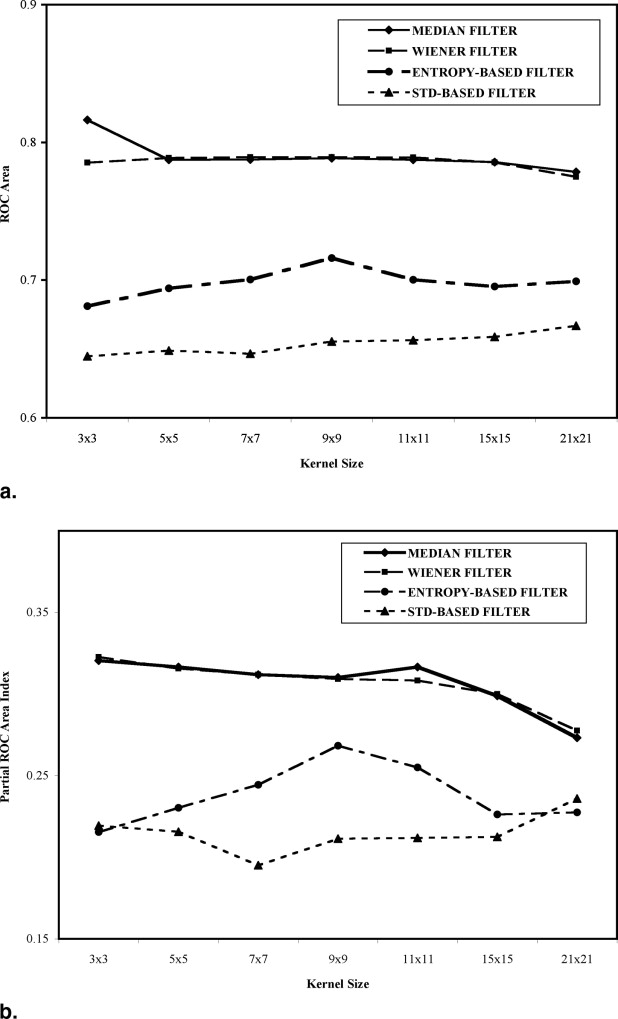
Receiver-operating characteristics (ROC) analysis showed that IT-CAD is affected slightly by image filtering. Modest, yet statistically significant, performance gain was observed with median filtering (overall ROC area index A z improved from 0.78 to 0.82). However, Gabor filtering improved performance for the high-sensitivity portion of the ROC curve where a typical false-positive reduction scheme should operate (partial ROC area index 0.90 A z improved from 0.33 to 0.37). Fusion of IT-CAD decisions from different filtering schemes markedly improved performance (A z = 0.90 and 0.90 A z = 0.55). At 95% sensitivity, the system’s specificity improved by 36.6%.
Conclusions
Additional improvement in false-positive reduction can be achieved by incorporating image filtering as a preprocessing step in our IT-CAD system.
Despite advances in treatment, breast cancer remains the second leading cause of cancer death in women ( ). The role of screening mammography in the battle against breast cancer is well established; women with malignancies detected at an early stage have a significantly better prognosis ( ). However, it is also recognized that the diagnostic interpretation of mammograms continues to be challenging for radiologists with a documented 20% false-negative rate ( ).
The clinical significance of early breast cancer diagnosis and the higher than desired false-negative rate of screening mammography have motivated the development of computer-aided detection (CADe) systems for decision support. These systems typically involve a hierarchical approach, first applying elaborate image preprocessing steps to enhance suspicious structures in the image and then employing morphologic and textural analysis to better classify these structures between true abnormalities and false positives. Detailed reviews of image processing techniques for mammographic image analysis and related CADe systems can be found elsewhere ( ). In addition, several CADe systems are already available commercially for both screen film mammography and full-field digital mammography ( ). Although their true clinical impact is often debated ( ), the scientific community continues to work toward improving the diagnostic performance and clinical integration of CADe technology. Ongoing CADe research efforts focus mainly on the reduction of false-positive computer marks as well as improving the detection rate of breast masses.
Get Radiology Tree app to read full this article<
Get Radiology Tree app to read full this article<
Materials and methods
Materials
Database
Get Radiology Tree app to read full this article<
Get Radiology Tree app to read full this article<
Get Radiology Tree app to read full this article<
Get Radiology Tree app to read full this article<
Overview of the IT-CADe system
Get Radiology Tree app to read full this article<
Get Radiology Tree app to read full this article<
Methods
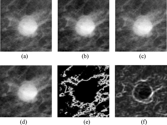
Preprocessing filters
Get Radiology Tree app to read full this article<
Get Radiology Tree app to read full this article<
Get Radiology Tree app to read full this article<
f(x,y)=e{−12[x2σ2x+y2σ2y]}⋅cos(2πμ0(xcos+ysin)) f
(
x
,
y
)
=
e
{
−
1
2
[
x
2
σ
x
2
+
y
2
σ
y
2
]
}
⋅
cos
(
2
π
μ
0
(
x
cos
+
y
sin
)
)
where μ 0 is the frequency of a sinusoidal plane, θ is the orientation, and σ x and σ y are standard deviations (or spatial spread) of the two-dimensional Gaussian envelope ( ). An octave bandwidth of 1 was used in our study because past psychophysical studies have confirmed that an octave bandwidth of 1 is a reasonably good estimate of the human eye when tuned to a frequency ( ). Central frequencies of 0.5, 1, 2, 4, 8, 16, and 32 cycles/degree with orientations at 0°, 45°, 90°, and 135° were used in this study. Get Radiology Tree app to read full this article<
Get Radiology Tree app to read full this article<
Get Radiology Tree app to read full this article<
Get Radiology Tree app to read full this article<
Evaluation Methods
Get Radiology Tree app to read full this article<
Results
Effect of Image Filter
Get Radiology Tree app to read full this article<
Get Radiology Tree app to read full this article<
Get Radiology Tree app to read full this article<
Table 1
Effect of Image Filtering as a Preprocessing Step on the Performance of the IT-CADe System for the Detection of Masses in Screening Mammograms
Preprocessing Filter A z (± σ) 0.90 A z (± σ) Specificity at 95% Sensitivity None 0.778 ± 0.025 0.326 ± 0.055 31.3% (125/399) Median (3 × 3) 0.816 ± 0.025 0.320 ± 0.065 29.6% (118/399) Wiener (3 × 3) 0.785 ± 0.026 0.323 ± 0.057 31.1% (124/399) Gabor 0.783 ± 0.024 0.368 ± 0.053 34.1% (136/399) Entropy (9 × 9) 0.706 ± 0.028 0.268 ± 0.046 27.6% (110/399) Standard deviation (21 × 21) 0.667 ± 0.028 0.236 ± 0.042 23.8% (95/399)
IT-CADe: information theoretic–computer-aided detection.
Get Radiology Tree app to read full this article<
Get Radiology Tree app to read full this article<
Get Radiology Tree app to read full this article<
Get Radiology Tree app to read full this article<
IT-CADe Fusion
Get Radiology Tree app to read full this article<
Get Radiology Tree app to read full this article<
Get Radiology Tree app to read full this article<
Table 2
Performance of Linear Discriminant Analysis Decision Models that Combine the Filter-Specific IT-CADe Outputs
LDA A z (± σ) 0.90 A z (± σ) Specificity at 95% Sensitivity 2 filters: (M, W) 0.884 ± 0.019 0.517 ± 0.067 49.4% (197/399) 3 filters: (M, W, STD) 0.893 ± 0.018 0.523 ± 0.070 47.4% (189/399) 4 filters: (M, W, H, STD) 0.896 ± 0.017 0.535 ± 0.067 48.3% (193/399) 5 filters: (M, W, G, STD, UN) 0.897 ± 0.018 0.549 ± 0.067 49.9% (199/399) ALL: (M, W, G, H, STD, UN) 0.898 ± 0.018 0.548 ± 0.068 50.4% (201/399)
LDA: linear discriminant analysis; IT-CADe: information theoretic–computer-aided detection; UN: unprocessed; M: median; W: adaptive Wiener; G: Gabor; H: entropy-based; STD: standard deviation based.
Different LDA models were built for each possible combination of filtering options. The table shows which combinations emerged as the superior ones depending on the number of inputs allowed in the LDA model.
Get Radiology Tree app to read full this article<
Discussion
Get Radiology Tree app to read full this article<
Get Radiology Tree app to read full this article<
Get Radiology Tree app to read full this article<
Get Radiology Tree app to read full this article<
Get Radiology Tree app to read full this article<
References
1. American Cancer Society: American Cancer Society: cancer facts and figures 2002.2002.American Cancer SocietyAtlanta, GA
2. Tabar L., Vitak B., Chen H.H., et. al.: Beyond randomized controlled trials: organized mammographic screening substantially reduces breast carcinoma mortality. Cancer 2002; 91: pp. 1724-1731.
3. Beam C.A., Conant E.F., Sickles E.A.: Factors affecting radiologist inconsistency in screening mammography. Acad Radiol 2002; 9: pp. 531-540.
4. Bird R.E., Wallace T.W., Yankaskas B.C.: Analysis of cancers missed at screening mammography. Radiology 1992; 184: pp. 613-617.
5. Birdwell R.L., Ikeda D.M., O’Shaughnessy K.F., et. al.: Mammographic characteristics of 115 missed cancers later detected with screening mammography and the potential utility of computer-aided detection. Radiology 2001; 219: pp. 192-202.
6. Yankaskas B.C., Schell M.J., Bird R.E., et. al.: Reassessment of breast cancers missed during routine screening mammography: a community-based study. AJR Am J Roentgenol 2001; 177: pp. 535-541.
7. Sampat M.P., Markey M.K., Bovik A.C.: Computer-aided detection and diagnosis in mammography.Bovik A.C.Handbook of image and video processing.2005.Academic PressNY:pp. 1195-1217.
8. Thangavel K., Karnan M., Sivakumar R., et. al.: Automatic detection of microcalcification in mammograms—a review. ICGST-GVIP J 2005; 5: pp. 31-61.
9. Highnam R.P., Brady J.M.: Mammographic image analysis.1999.Kluwer Academic PublishersDordrecht, The Netherlands
10. Fitzpatrick J.M., Sonka M.: Handbook of medical imaging.2000.SPIE PressBellingham, WA:
11. Burhenne L.J.W., Wood S.A., D’Orsi C.J., et. al.: Potential contribution of computer-aided detection to the sensitivity of screening mammography. Radiology 2000; 215: pp. 554-562.
12. Brem R.F., Baum J., Lechner M., et. al.: Improvement in sensitivity of screening mammography with computer-aided detection: a multiinstitutional trial. AJR Am J Roentgenol 2003; 81: pp. 687-693.
13. Gur D., Sumkin H., Rockette H.E., et. al.: Changes in breast cancer detection and mammography recall rates after the introduction of a computer-aided detection system. J Natl Cancer Inst 2004; 96: pp. 185-190.
14. Taylor P., Given-Wilson R.M.: Evaluation of computer-aided detection (CAD) devices. Br J Radiol 2005; 78: pp. 26-30.
15. Birdwell R.L., Bandodkar P., Ikeda D.M.: Computer-aided detection with screening mammography in a university hospital setting. Radiology 2005; 236: pp. 451-457.
16. Morton M.J., Whaley D.H., Brandt K.R., et. al.: Screening mammograms: Interpretation with computer-aided detection—prospective evaluation. Radiology 2006; 239: pp. 375-383.
17. Krupinski E.A.: Computer-aided detection in clinical environment: benefits and challenges for radiologists. Radiology 2004; 231: pp. 7-9.
18. Fenton J.J., Taplin S.H., Carney P.A., et. al.: Influence of computer-aided detection on performance of screening mammography. N Engl J Med 2007; 356: pp. 1399-1409.
19. Hall F.M.: Breast imaging and computer-aided detection. N Engl J Med 2007; 356: pp. 1464-1466.
20. Tourassi G.D., Vargas-Voracek R., Floyd C.E.: Computer-assisted detection of mammographic masses: a template matching scheme based on mutual information. Med Phys 2003; 30: pp. 2123-2139.
21. Tourassi G.D., Harrawood B., Singh S., et. al.: Evaluation of information-theoretic similarity measures for content-based retrieval and detection of masses in mammograms. Med Phys 2007; 34: pp. 140-150.
22. Tourassi G.D., Harrawood B., Singh S., et. al.: Information-theoretic CAD system in mammography: entropy-based indexing for computational efficiency and robust performance. Med Phys 2007; 34: pp. 3193-3204.
23. Cover T.M., Thomas J.A.: Elements of information theory.1991.John Wiley & SonsNew York
24. Pluim J.P.W., Maintz J.B.A., Viergever M.A.: Mutual-information-based registration of medical images: a survey. IEEE Trans Med Imag 2003; 22: pp. 986-1004.
25. Heath M., Bowyer K., Kopans D., et. al.: Current status of the digital database for screening mammography.1998.Kluwer Academic Publishers http://marathon.csee.usf.edu/Mammography/Database.html Accessed January 7, 2008
26. Catarious D.M., Baydush A.H., Floyd C.E.: A mammographic mass CAD system incorporating features from shape, fractal, and channelized Hotelling observer measurements: preliminary results. SPIE 2003; 5032: pp. 111-119.
27. Catarious D.M., Baydush A.H., Floyd C.E.: Incorporation of an iterative, linear segmentation routine into a mammographic mass CAD system. Med Phys 2004; 31: pp. 1512-1520.
28. Tourassi G.D., Harrawood B., Floyd C.E.: Cross-digitizer robustness of a knowledge-based CAD system for mass detection in screening mammograms. SPIE 2007; 6514: pp. 65141Y1-65474Y8.
29. Hajnal J.V., Hill D.L.G., Hawkes D.J.: Medical image registration.2000.CRC PressBoca Raton, FL
30. Studholme C., Hill D.L.G., Hawkes D.J.: An overlap invariant entropy measure of 3D medical image alignment. Pattern Recognit 1996; 32: pp. 71-86.
31. Dekker N., Ploeger L.S., van Herk M.: Evaluation of cost functions for gray value matching of two-dimensional images in radiotherapy. Med Phys 2003; 30: pp. 778-784.
32. Bovik A.C., Huang S., Manson D.C.: The effect of median filtering on edge estimation and detection. IEEE Trans Pattern Anal Machine Intell 1987; 9: pp. 181-194.
33. Mayo P., Rodenas F., Verdu G.: Comparing methods to denoise mammographic images. IEEE EMBS 2004; 1: pp. 247-250.
34. Gabor D.: Theory of communication. J Inst Elec Eng 1946; 93: pp. 429-459.
35. Zheng Q., Chellappa R.: A computational vision approach to image registration. IEEE Trans Imag Processing 1993; 2: pp. 311-326.
36. Manjunath B.S., Shekhar C., Chellappa R.: A new approach to image feature detection with applications. Pattern Recognit 1996; 29: pp. 627-640.
37. Liu J., Vemuri B.C., Marroquin J.L.: Local frequency representations for robust multimodal imageregistration. IEEE Trans Med Imag 2002; 21: pp. 462-469.
38. Rubner Y., Tomasi C.: Perceptual metrics for image database navigation.2001.SpringerNorwell, MA
39. Hubel D.H., Weisel T.N.: Receptive fields and functional architecture in two nonstriate visual areas (18 and 19) of the cat. J Neurophysiol 1965; 28: pp. 229-289.
40. Campbell F.W., Robson J.G.: Application of Fourier analysis to the visibility of gratings. J Physiol 1968; 197: pp. 551-566.
41. Daugman J.G.: Complete discrete 2D Gabor transforms by neural networks for image analysis and compression. IEEE Trans ASSP 1998; 36: pp. 1169-1179.
42. Chen C.C., Chen C.C.: Gabor transform in texture analysis. SPIE Proc 2003; 2353: pp. 237-245.
43. Watson A.B., Braddick O.J., Sleigh A.C.: Detection and recognition of simple spatial forms.Braddick O.J.Slade A.C.Physical and biological processing of images.1983.Springer-VerlagBerlin, Germany:
44. Gonzalez R.C., Woods R.E., Eddins S.L.: Digital image processing using MATLAB.2003.Prentice HallUpper Saddle River, NJ
45. Obuchowski N.A.: Receiver operating characteristic curves and their use in radiology. Radiology 2003; 229: pp. 3-8.
46. Jiang Y., Metz C.E., Nishikawa R.M.: A receiver operating characteristic partial area index for highly sensitive diagnostic tests. Radiology 1996; 201: pp. 745-750.
47. Tourassi G.D., Jesneck J.A., Mazurowski M.L., et. al.: Stacked generalization in computer-assisted decision systems: empirical comparison of data handling schemes. Proc IJCNN 2007; pp. 1343-1347.
48. Development Core Team: R: a language and environment for statistical computing.2003.R Foundation for Statistical ComputingVienna, Austria
49. The R Project for Statistical Computing: http://www.R-project.org Accessed January 7, 2008
50. Tourassi G.D., Saunders R., Samei E.: Mass detection in full field digital mammograms: validation of an information-theoretic knowledge-based system.2006.RSNA 92nd Scientific AssemblyChicago, IL