
Rationale and Objectives
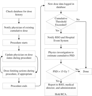
To address the risk of radiation injury during interventional procedures, the Joint Commission has defined prolonged fluoroscopy resulting in a cumulative skin dose of 15 Gy or more to a single field as a reviewable sentinel event. The goal of this work is to present a system for identifying potential fluoroscopic sentinel events (FSE) and describing common case characteristics.
Materials and Methods
Criteria based on fluoroscopic time (FT) > 150 minutes and reference air kerma (RAK) > 6 Gy were used to identify potential sentinel events. Case information including procedure type, number of procedures, and radiation dose parameters was recorded. Peak skin dose (PSD) was calculated by a medical physicist. Values were compared between procedure types and the relationship between FT, RAK, and PSD was evaluated.
Results
Between 2008 and 2011, 183 events exceeding the investigation criteria were identified in three interventional categories: cardiology (54%), neuroradiology (31%), and vascular (16%). The average number of procedures/patient was 1.7 ± 0.1, with the majority (59.6%) having undergone only one procedure. Most cases could be identified using the RAK criterion alone (96.7%). Based on the PSD/RAK ratio, a threshold RAK of 7.5 Gy would effectively identify all cases that would exceed 15 Gy in PSD.
Conclusion
Radiation delivered during interventional cases can place patients at risk of cutaneous radiation injury and potential sentinel events. Using appropriate thresholds to determine which cases require detailed investigation allows efficient utilization of department resources for identifying sentinel events.
As interventional procedures in surgery, cardiology, and radiology have become more prevalent, the risk of radiation-related injuries has become a substantial concern. Prolonged use of radiation during fluoroscopically guided interventional (FGI) procedures can subject patients to a large dose and place them at risk for radiation injury . Effects of high doses of radiation to the skin can range from mild transient erythema to severe necrosis requiring surgical intervention .
To facilitate awareness of radiation overexposure and drive process improvements to enhance patient safety, in 2005 the Joint Commission created a reviewable sentinel event that applied to fluoroscopic procedures. The fluoroscopic sentinel event (FSE) was defined as prolonged fluoroscopy resulting in a cumulative skin dose of 15 Gy or more to a single field. According to guidance from the Joint Commission, this dose may be accumulated either during a single procedure or multiple procedures over 6 months to a year . When a sentinel event occurs, a root cause analysis must be performed to address underlying systems issues that can improve patient safety.
Get Radiology Tree app to read full this article<
Materials and methods
Systematically Monitoring for Potential Sentinel Events
Get Radiology Tree app to read full this article<
Get Radiology Tree app to read full this article<
Data Collection
Get Radiology Tree app to read full this article<
Calculating Patient PSD
Get Radiology Tree app to read full this article<
Group Comparison and Statistical Analysis
Get Radiology Tree app to read full this article<
Results
Number and Type of Cases Identified
Get Radiology Tree app to read full this article<
Get Radiology Tree app to read full this article<
Table 1
Categories of Cases Frequently Identified as Potential Sentinel Events
Potential Sentinel Events Grouped by Category Number of Cases (% of Total Cases)Primary categorySecondary category Cardiologic Coronary angioplasty and stenting 82 (44.8) Electrophysiology ablation 13 (7.10) Embolization - cardiac shunt 1 (0.5) ICD implantation 1 (0.5) SVC angioplasty 1 (0.5)All cardiologic categories98 ( 53.6) Neuroradiologic Embolization - aneurysm 21 (11.5) Embolization - arteriovenous malformation 15 (8.2) Embolization - carotid cavernous fistula 7 (3.8) Embolization - other arteriovenous fistula 7 (3.8) Cerebral stent placement 4 (2.2) Angiography - spinal 1 (0.5) Embolization - spinal tumor 1 (0.5)All neuroradiologic cases56 ( 30.6) Vascular Repair - abdominal aortic aneurysm 5 (2.7) Hepatic transarterial chemoembolization 5 (2.7) Embolization - visceral 5 (2.7) Repair - thoracic aortic aneurysm 3 (1.6) Percutaneous cholangiogram and biliary drain 2 (1.1) Angiography - visceral 2 (1.1) Repair - iliac artery aneurysm 2 (1.1) Embolization - pelvic tumor 1 (0.5) Liver transplant evaluation 1 (0.5) Peripheral revascularization 1 (0.5) Stenting - mesenteric bypass 1 (0.5) Embolization - uterine artery 1 (0.5)All vascular/other cases29 ( 15.8) All 183 (100.0%)
Get Radiology Tree app to read full this article<
Get Radiology Tree app to read full this article<

Get Radiology Tree app to read full this article<
Comparison of Procedure Categories
Get Radiology Tree app to read full this article<
![Figure 3, Features of potential sentinel events when separated by category. (a) Number of procedures per patient, (b) fluoroscopic time (FT [min]), (c) reference air kerma (RAK [Gy]), and (d) peak skin dose (PSD [Gy]) are shown. Values plot the mean with error bars indicating the 95% confidence interval. Statistically significant differences are highlighted by the braces above the graphs with the respective P values.](https://storage.googleapis.com/dl.dentistrykey.com/clinical/EvaluationofFluoroscopicCasesQualifyingasPotentialFluoroscopicSentinelEvents/2_1s20S1076633213000032.jpg)
Get Radiology Tree app to read full this article<
Usefulness of Identification Criteria
Get Radiology Tree app to read full this article<
Table 2
Number of Cases (Percentage) Meeting Identification Criteria by Category
Primary Category Fluoroscopic Time Reference Air Kerma Both Criteria Cardiologic 2 (2.0%) 92 (93.9%) 4 (4.1%) Neuroradiologic 4 (7.1%) 15 (26.8%) 37 (66.1%) Vascular 0 (0.0%) 27 (93.1%) 2 (6.9%)All cases6 ( 3.3%)134 ( 73.2%)43 ( 23.5%)
Get Radiology Tree app to read full this article<
Relationship between Dose Parameters and Peak Skin Dose
Get Radiology Tree app to read full this article<
![Figure 4, Box-and-whisker plots demonstrating (a) ratio of peak skin dose to fluoroscopic time (PSD/FT [Gy/min]) and (b) ratio of peak skin dose to reference air kerma (PSD/RAK) by category. Boxes indicate the 25th, 50th, and 75th percentiles and whiskers indicate the 5th and 95th percentiles. Asterisks denote the maximum value for any case in that category.](https://storage.googleapis.com/dl.dentistrykey.com/clinical/EvaluationofFluoroscopicCasesQualifyingasPotentialFluoroscopicSentinelEvents/3_1s20S1076633213000032.jpg)
Get Radiology Tree app to read full this article<
Discussion
Get Radiology Tree app to read full this article<
Get Radiology Tree app to read full this article<
Get Radiology Tree app to read full this article<
Get Radiology Tree app to read full this article<
Get Radiology Tree app to read full this article<
Get Radiology Tree app to read full this article<
References
1. Wagner L.K., Eifel P.J., Geise R.A.: Potential biological effects following high X-ray dose interventional procedures. J Vasc Interv Radiol 1994; 5: pp. 71-84.
2. Balter S., Hopewell J.W., Miller D.L., et. al.: Fluoroscopically guided interventional procedures: a review of radiation effects on patients’ skin and hair. Radiology 2010; 254: pp. 326-341.
3. The Joint Commission. Radiation overdose as a reviewable sentinel event. Available at: http://www.jointcommission.org/assets/1/18/Radiation_Overdose.pdf . Accessed January 14, 2012.
4. Miller D.L., Balter S., Cole P.E., et. al.: Radiation doses in interventional radiology procedures: the RAD-IR study: part II: skin dose. J Vasc Interv Radiol 2003; 14: pp. 977-990.
5. Miller D.L., Kwon D., Bonavia G.H.: Reference levels for patient radiation doses in interventional radiology: proposed initial values for U.S. practice. Radiology 2009; 253: pp. 753-764.
6. Balter S., Miller D.L.: The new Joint Commission sentinel event pertaining to prolonged fluoroscopy. J Am Coll Radiol 2007; 4: pp. 497-500.
7. Mahesh M.: How to prepare for the Joint Commission’s sentinel event policy pertaining to prolonged fluoroscopy. J Am Coll Radiol 2008; 5: pp. 601-603.
8. den Boer A., de Feijter P.J., Serruys P.W., et. al.: Real-time quantification and display of skin radiation during coronary angiography and intervention. Circulation 2001; 104: pp. 1779-1784.
9. Stecker M.S., Balter S., Towbin R.B., et. al.: Guidelines for patient radiation dose management. J Vasc Interv Radiol 2009; 20: pp. S263-S273.
10. Panuccio G., Greenberg R.K., Wunderle K., et. al.: Comparison of indirect radiation dose estimates with directly measured radiation dose for patients and operators during complex endovascular procedures. J Vasc Surg 2011; 53: pp. 885-894.e1. discussion 94
11. Food and Drug Administration. Avoidance of serious x-ray-induced skin injuries to patients during fluoroscopically-guided procedures. Available at: http://www.fda.gov/downloads/Radiation-EmittingProducts/RadiationEmittingProductsandProcedures/MedicalImaging/MedicalX-Rays/ucm116677.pdf . Accessed January 14, 2012.
12. Rosenthal L.S., Mahesh M., Beck T.J., et. al.: Predictors of fluoroscopy time and estimated radiation exposure during radiofrequency catheter ablation procedures. Am J Cardiol 1998; 82: pp. 451-458.
13. Chida K., Saito H., Otani H., et. al.: Relationship between fluoroscopic time, dose-area product, body weight, and maximum radiation skin dose in cardiac interventional procedures. AJR Am J Roentgenol 2006; 186: pp. 774-778.
14. Fletcher D.W., Miller D.L., Balter S., Taylor M.A.: Comparison of four techniques to estimate radiation dose to skin during angiographic and interventional radiology procedures. J Vasc Interv Radiol 2002; 13: pp. 391-397.
15. Miller D.L., Balter S., Wagner L.K., et. al.: Quality improvement guidelines for recording patient radiation dose in the medical record. J Vasc Interv Radiol 2009; 20: pp. S200-S207.
16. Lickfett L., Mahesh M., Vasamreddy C., et. al.: Radiation exposure during catheter ablation of atrial fibrillation. Circulation 2004; 110: pp. 3003-3010.
17. Cohen M.D.: Optimizing the use of pulsed fluoroscopy to reduce radiation exposure to children. J Am Coll Radiol 2008; 5: pp. 205-209.
18. Vetter S., Faulkner K., Strecker E.P., et. al.: Dose reduction and image quality in pulsed fluoroscopy. Radiat Prot Dosim 1998; 80: pp. 299-301.
19. Miller D.L., Balter S., Noonan P.T., et. al.: Minimizing radiation-induced skin injury in interventional radiology procedures. Radiology 2002; 225: pp. 329-336.
20. Marx M.V.: The radiation dose in interventional radiology study: knowledge brings responsibility. J Vasc Interv Radiol 2003; 14: pp. 947-951.