
Rationale and Objective
We aimed to determine the frequency and clinical significance of homogeneous renal masses measuring 21–39 Hounsfield units on contrast-enhanced computed tomography (CT).
Methods
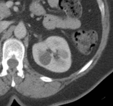
Subjects 40–69 years old undergoing portal-venous-phase contrast-enhanced abdominal CT from January 1, 2006 to December 31, 2010 with slice thickness ≤5 mm and no prior CT or magnetic resonance imaging were identified ( n = 1387) for this institutional review board-approved retrospective cohort study. Images were manually reviewed by three radiologists in consensus to identify all circumscribed homogeneous renal masses (maximum of three per subject) ≥10 mm with a measured attenuation of 21–39 Hounsfield units. Exclusion criteria were known renal cancer or imaging performed for a renal indication. The primary outcome was retrospective characterization as a clinically significant mass, defined as a solid mass, a Bosniak IIF/III/IV mass, or extirpative therapy or metastatic renal cancer within 5 years’ follow-up.
Results
Eligible masses ( n = 74) were found in 5% (63/1387) of subjects. Of those with a reference standard ( n = 42), none (0% [95% CI: 0.0%–8.4%]) were determined to be clinically significant.
Conclusion
Incidental renal masses on contrast-enhanced CT that are homogeneous and display an attenuation of 21–39 Hounsfield units are uncommon in patients 40–69 years of age, unlikely to be clinically significant, and may not need further imaging evaluation. If these results can be replicated in an independent and larger population, the practical definition of a benign cyst on imaging may be able to be expanded.
Introduction
Incidental renal masses are common, with some estimates suggesting that at least half of all patients over the age of 50 have at least one renal mass that may be detected with cross-sectional imaging . When an incidental renal mass is identified at contrast-enhanced computed tomography (CT), a diagnosis of a benign simple cyst can be made if the mass is homogeneous (ie, no wall thickening, septa, calcifications, or visible heterogeneity) and has a measured attenuation between −10 and 20 Hounsfield units (HU) . However, if the mass is heterogeneous, or if the mass is homogeneous and measures greater than 20 HU, it is considered incompletely characterized and in general prompts a recommendation for further imaging .
However, these attenuation criteria may be overly aggressive. For example, it is not possible for a homogeneous renal mass that measures 21–39 HU on contrast-enhanced CT to both fulfill the definition of a nonsimple cyst on unenhanced CT (ie, >20 HU) and meet the threshold for definitive enhancement on contrast-enhanced CT (ie, an increase by 20 HU or more) . In addition, attenuation values of renal masses at contrast-enhanced CT are commonly spuriously high due to pseudoenhancement , an artificial increase in attenuation due to the CT scanner’s overcorrection for beam-hardening . Therefore, an incidentally detected homogeneous renal mass that measures 21–39 HU on contrast-enhanced CT is more likely to be a benign non–enhancing hyperattenuating cyst (ie, Bosniak II cyst), or a simple cyst (ie, Bosniak I) with pseudoenhancement, than it is to be malignant. However, the threshold for definitive enhancement is not always reached for some renal cancers; for example, papillary cancers are known to be hyperattenuating and some may fail to show measurable enhancement on CT .
Get Radiology Tree app to read full this article<
Methods
Get Radiology Tree app to read full this article<
Subjects
Get Radiology Tree app to read full this article<
Get Radiology Tree app to read full this article<
Get Radiology Tree app to read full this article<
Index CT Examinations
Get Radiology Tree app to read full this article<
Reference Standard
Get Radiology Tree app to read full this article<
Data Analysis
Get Radiology Tree app to read full this article<
Results
Get Radiology Tree app to read full this article<
Table 1
Study Population Details
Characteristic Data Subjects 63 Masses 74 Female 44% (28/63) Male 56% (35/63) Mean age (years, range) 58 (40–69) Indication for CT Infection 5% (3/63) Nonrenal malignancy 56% (35/63) Pain 22% (14/63) Trauma 6% (4/63) Other 11% (7/63) Lesion location Exophytic 23% (17/74) Mesophytic 24% (18/74) Endophytic 53% (39/74) Mean lesion size (mm, range) 20 (10–56) Mean lesion attenuation (HU, range) 28 (21–38) Attenuation 21–29 HU 69% (51/74) Attenuation 30–39 HU 31% (23/74) Lesion location Right kidney 41% (30/74) Left kidney 59% (44/74) Reference standard None (ie, not included in primary outcome) 43% (32/74) ≥5 years of clinical follow-up 15% (11/74) Renal protocol CT 16% (12/74) Unenhanced CT 1% (1/74) MRI with and without contrast 8% (6/74) Ultrasound 11% (8/74) More than one of the above 5% (4/74)
CT, computed tomography; HU, Hounsfield unit; MRI, magnetic resonance imaging.
Denominators are 63 for subject-level data and 74 for lesion-level data.
Get Radiology Tree app to read full this article<
Get Radiology Tree app to read full this article<
Discussion
Get Radiology Tree app to read full this article<
Get Radiology Tree app to read full this article<
Get Radiology Tree app to read full this article<
Get Radiology Tree app to read full this article<
Get Radiology Tree app to read full this article<
Compliance with Ethical Standards
Get Radiology Tree app to read full this article<
Get Radiology Tree app to read full this article<
Get Radiology Tree app to read full this article<
Get Radiology Tree app to read full this article<
References
1. Silverman S.G., Israel G.M., Herts B.R., et. al.: Management of the incidental renal mass. Radiology 2008; 249: pp. 16-31.
2. Tada S., Yamagishi J., Kobayashi H., et. al.: The incidence of simple renal cyst by computed tomography. Clin Radiol 1983; 34: pp. 437-439.
3. Kissane J.M.: Congenital malformations.Jenette J.C.Olson J.L.Silva F.G. et. al.Heptinstall’s pathology of the kidney.1998.Lippincott-RavenPhiladelphia:
4. Israel G.M., Bosniak M.A.: How I do it: evaluating renal masses. Radiology 2005; 236: pp. 441-450.
5. Silverman S.G., Israel G.M., Trinh Q.D.: Incompletely characterized incidental renal masses: emerging data support conservative management. Radiology 2015; 275: pp. 28-42.
6. Hollingsworth J.M., Miller D.C., Daignault S., et. al.: Rising incidence of small renal masses: a need to reassess treatment effect. J Natl Cancer Inst 2006; 98: pp. 1331-1334.
7. Bosniak M.A.: The use of the Bosniak classification system for renal cysts and cystic tumors. J Urol 1997; 157: pp. 1852-1853.
8. Maki D.D., Birnbaum B.A., Chakraborty D.P., et. al.: Renal cyst pseudoenhancement: beam-hardening effects on CT numbers. Radiology 1999; 213: pp. 468-472.
9. Birnbaum B.A., Hindman N., Lee J., et. al.: Renal cyst pseudoenhancement: influence of multidetector CT reconstruction algorithm and scanner type in phantom model. Radiology 2007; 244: pp. 767-775.
10. Patel J., Davenport M.S., Khalatbari S., et. al.: In vivo predictors of renal cyst pseudoenhancement at 120 kVp. AJR Am J Roentgenol 2014; 202: pp. 336-342.
11. Dilauro M., Quon M., McInnes M.D., et. al.: Comparison of contrast-enhanced multiphase renal protocol CT versus MRI for diagnosis of papillary renal cell carcinoma. AJR Am J Roentgenol 2016; 206: pp. 319-325.
12. Silverman S.G., Mortele K.J., Tuncali K., et. al.: Hyperattenuating renal masses: etiologies, pathogenesis, and imaging evaluation. Radiographics 2007; 27: pp. 1131-1143.
13. Herts B.R., Coll D.M., Novick A.C., et. al.: Enhancement characteristics of papillary renal neoplasms revealed on triphasic helical CT of the kidneys. AJR Am J Roentgenol 2002; 178: pp. 367-372.
14. Ho V.B., Allen S.J., Hood M.N., et. al.: Renal masses: quantitative assessment of enhancement with dynamic MR imaging. Radiology 2002; 224: pp. 695-700.
15. Hindman N.M., Hecht E.M., Bosniak M.A.: Follow-up of Bosniak category 2F cystic renal lesions. Radiology 2014; 272: pp. 757-766.
16. Smith A.D., Remer E.M., Cox K.L., et. al.: Bosniak category IIF and III cystic renal lesions: outcomes and associations. Radiology 2012; 262: pp. 152-160.
17. O’Malley R.L., Godoy G., Hecht E.M., et. al.: Bosniak category IIF designation and surgery for complex renal cysts. J Urol 2009; 182: pp. 1091-1095.
18. Hwang J.H., Lee C.K., Yu H.S., et. al.: Clinical outcomes of Bosniak category IIF complex renal cysts in Korean patients. Korean J Urol 2012; 53: pp. 386-390.
19. Israel G.M., Bosniak M.A.: Follow-up CT of moderately complex cystic lesions of the kidney (Bosniak category IIF). AJR Am J Roentgenol 2003; 181: pp. 627-633.
20. Frank I., Blute M.L., Cheville J.C., et. al.: Solid renal tumors: an analysis of pathological features related to tumor size. J Urol 2003; 170: pp. 2217-2220.
21. Agochukwu N., Huber S., Spektor M., et. al.: Differentiating renal neoplasms from simple cysts on contrast-enhanced CT on the basis of attenuation and homogeneity. AJR Am J Roentgenol 2017; 208: pp. 801-804.
22. O’Connor S.D., Silverman S.G., Ip I.K., et. al.: Simple cyst-appearing renal masses at unenhanced CT: can they be presumed to be benign?. Radiology 2013; 269: pp. 793-800.
23. Schieda N., Vakili M., Dilauro M., et. al.: Solid renal cell carcinoma measuring water attenuation (−10 to 20 HU) on unenhanced CT. AJR Am J Roentgenol 2015; 205: pp. 1215-1221.