
Purpose
To determine interreader agreement and diagnostic accuracy across varying levels of reader experience using qualitative and quantitative methods of evaluating adrenal nodules using ( 18 F)-fluorodeoxyglucose–positron emission tomography/computed tomography.
Methods
132 adrenal nodules (96 adenomas, 36 metastases) were retrospectively identified in 105 patients (49 men and 56 women, mean age 66 years, age range 45–85 years) with a history of lung cancer who underwent ( 18 F)-fluorodeoxyglucose–positron emission tomography/computed tomography. For each nodule, three readers independently performed one qualitative and two quantitative measurements: visual assessment, standardized uptake value (SUV max ), and standard uptake ratio (SUV ratio ). Interreader agreement was calculated using percent agreement with κ statistic for qualitative analysis and intraclass correlation coefficient (ICC) for quantitative analysis. Accuracy, sensitivity, and specificity for distinguishing benign from malignant adrenal nodules were calculated for each method.
Results
Percent agreement between readers for visual (qualitative) assessment was 92% to 96% and κ statistic was 0.79 to 0.90 (95% confidence limits 0.66–0.99). ICC for SUV max was 92% to 99% (95% CL 0.8–1.0), and ICC for SUV ratio was 89% to 99% (95% CL 0.74–0.99). For diagnosis of malignancy, mean sensitivity and specificity for visual assessment were 80% and 97%, respectively. Mean sensitivity and specificity for SUV max were 91% and 81%, respectively; for SUV ratio , 90% and 80%. Mean diagnostic accuracy was 93%, 83%, and 84% for visual assessment, SUV max , and SUV ratio , respectively.
Conclusion
Excellent interreader agreement is seen for quantitative and qualitative methods of distinguishing benign from malignant adrenal nodules. Qualitative analysis demonstrated higher accuracy but lower sensitivity compared with quantitative analysis.
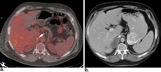
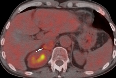
In patients with primary lung malignancy, distinguishing adrenal adenomas from metastases is critical in determining stage of disease, guiding therapy, and predicting prognosis. While the traditional imaging methods of computed tomography (CT) and/or magnetic resonance imaging (MRI) are used to characterize the majority of adrenal nodules, an increasing number of cancer patients are undergoing ( 18 F)-fluorodeoxyglucose–positron emission tomography (FDG-PET) and combined FDG-PET/CT for staging of their primary malignancy. Because of the ability of FDG-PET to detect the increased uptake of radiolabeled glucose associated with adrenal metastases, FDG-PET/CT has emerged as another technique for distinguishing adrenal adenomas from adrenal metastases .
Studies investigating the utility of FDG-PET/CT for characterization of adrenal nodules have accuracies ranging between 75% and 100% . These variable results likely reflect the behavior of some adenomas to demonstrate moderate FDG uptake leading to a false-positive diagnosis of malignancy .
Get Radiology Tree app to read full this article<
Get Radiology Tree app to read full this article<
Get Radiology Tree app to read full this article<
Materials and methods
Get Radiology Tree app to read full this article<
Reference Standard
Get Radiology Tree app to read full this article<
FDG-PET/CT Imaging Protocol
Get Radiology Tree app to read full this article<
Image Analysis
Get Radiology Tree app to read full this article<
Quantitative Adrenal Nodule Assessment
Get Radiology Tree app to read full this article<
Qualitative Adrenal Nodule Assessment
Get Radiology Tree app to read full this article<
Statistical Methods
Qualitative data
Get Radiology Tree app to read full this article<
Quantitative data
Get Radiology Tree app to read full this article<
Get Radiology Tree app to read full this article<
Get Radiology Tree app to read full this article<
Get Radiology Tree app to read full this article<
Results
Descriptive Statistics
Get Radiology Tree app to read full this article<
Get Radiology Tree app to read full this article<
Interreader Agreement
Get Radiology Tree app to read full this article<
Table 1
Three Reader Percent Agreement and κ Statistics for Qualitative Methods
Method Reader Pair ∗ Percent Agreement (%) κ Statistic 95% Confidence Limits Visual assessment 1–2 92 0.79 0.66–0.91 1–3 96 0.90 0.81–0.99 2–3 93 0.81 0.70–0.93
Get Radiology Tree app to read full this article<
Get Radiology Tree app to read full this article<
Get Radiology Tree app to read full this article<
Table 2
Three Reader Intraclass Correlation Coefficients for Quantitative Methods
Method Reader Pair ∗ Intraclass Correlation Coefficient 95% Confidence Limits Standardized uptake value 1–2 0.92 0.88–0.99 1–3 0.99 0.98–1.00 2–3 0.92 0.80–0.98 Standard uptake ratio 1–2 0.89 0.74–0.97 1–3 0.99 0.97–0.99 2–3 0.89 0.73–0.98
Get Radiology Tree app to read full this article<
Get Radiology Tree app to read full this article<
Qualitative Analysis
Get Radiology Tree app to read full this article<
Table 3
Comparison of Three Reader Diagnostic Performance of Qualitative (Visual) and Quantitative (SUV max and SUV ratio ) Methods
Method Parameter Reader 1 (%) Reader 2 (%) Reader 3 (%) Mean (%) 95% Confidence Limits SUV max Sensitivity 94 86 94 91 0.75–1.00 SUV ratio Sensitivity 92 83 94 90 0.69–1.00 Visual Sensitivity 83 72 86 80 0.56–0.97P value ∗ >.08 >.05 >.10 SUV max Specificity 86 77 80 81 0.68–0.93 SUV ratio Specificity 88 74 79 80 0.64–0.95 Visual Specificity 98 96 98 97 0.90–1.00P value ∗ <.01 <.001 <.001 SUV max PPV 72 58 64 65 0.44–0.85 SUV ratio PPV 73 55 63 64 0.40–0.87 Visual PPV 94 87 94 92 0.72–1.00 SUV max NPV 98 94 97 96 0.88–1.00 SUV ratio NPV 97 92 97 95 0.85–1.00 Visual NPV 94 90 95 93 0.83–1.00 SUV max Accuracy 89 77 83 83 0.34–0.86 SUV ratio Accuracy 89 80 84 84 0.41–0.86 Visual Accuracy 94 89 95 93 0.58–0.96P value ∗ >.052 <.02 <.002
Get Radiology Tree app to read full this article<
Get Radiology Tree app to read full this article<
Quantitative Analysis
Get Radiology Tree app to read full this article<
Comparison of Qualitative and Quantitative Diagnostic Performance
Get Radiology Tree app to read full this article<
Integrated FDG-PET/CT Image Analysis
Get Radiology Tree app to read full this article<
Discussion
Get Radiology Tree app to read full this article<
Get Radiology Tree app to read full this article<
Get Radiology Tree app to read full this article<
Get Radiology Tree app to read full this article<
Get Radiology Tree app to read full this article<
Get Radiology Tree app to read full this article<
References
1. Boland G.W.: Adrenal imaging: why, when, what, and how? Part 2. What technique?. Am J Roentgenol 2011; 196: pp. W1-W5.
2. Boland G.W., Dwamena B.A., Jagtiani Sangwaiya M., et. al.: Characterization of adrenal masses by using FDG PET: a systematic review and meta-analysis of diagnostic test performance. Radiology 2011; 259: pp. 117-126.
3. Chong S., Lee K.S., Kim H.Y., et. al.: Integrated PET-CT for the characterization of adrenal gland lesions in cancer patients: diagnostic efficacy and interpretation pitfalls. RadioGraphics 2006; 26: pp. 1811-1824.
4. Boland G.W., Blake M.A., Holalkere N.S., et. al.: PET/CT for the characterization of adrenal masses in patients with cancer: qualitative versus quantitative accuracy in 150 consecutive patients. Am J Roentgenol 2009; 192: pp. 956-962.
5. Brady M.J., Thomas J., Wong T.Z., et. al.: Adrenal nodules at FDG PET/CT in patients known to have or suspected of having lung cancer: a proposal for an efficient diagnostic algorithm. Radiology 2009; 250: pp. 523-530.
6. Caoili E.M., Korobkin M., Brown R.K., et. al.: Differentiating adrenal adenomas from nonadenomas using (18)F-FDG PET/CT: quantitative and qualitative evaluation. Acad Radiol 2007; 14: pp. 468-475.
7. Metser U., Miller E., Lerman H., et. al.: 18F-FDG PET/CT in the evaluation of adrenal masses. J Nucl Med 2006; 47: pp. 32-37.
8. Vikram R., Yeung H.D., Macapinlac H.A., et. al.: Utility of PET/CT in differentiating benign from malignant adrenal nodules in patients with cancer. Am J Roentgenol 2008; 191: pp. 1545-1551.
9. Erasmus J.J., Patz E.F., McAdams H.P., et. al.: Evaluation of adrenal masses in patients with bronchogenic carcinoma using 18F-fluorodeoxyglucose positron emission tomography. Am J Roentgenol 1997; 168: pp. 1357-1360.
10. Yun M., Kim W., Alnafisi N., et. al.: 18F-FDG PET in characterizing adrenal lesions detected on CT or MRI. J Nucl Med 2001; 42: pp. 1795-1799.
11. Gupta N.C., Graeber G.M., Tamim W.J., et. al.: Clinical utility of PET-FDG imaging in differentiation of benign from malignant adrenal masses in lung cancer. Clin Lung Cancer 2001; 3: pp. 59-64.
12. Kumar R., Xiu Y., Yu J.Q., et. al.: 18F-FDG PET in evaluation of adrenal lesions in patients with lung cancer. J Nucl Med 2004; 45: pp. 2058-2062.
13. Blake M.A., Slattery J.M., Kalra M.K., et. al.: Adrenal lesions: characterization with fused PET/CT image in patients with proved or suspected malignancy–initial experience. Radiology 2006; 238: pp. 970-977.
14. Jana S., Zhang T., Milstein D.M., et. al.: FDG-PET and CT characterization of adrenal lesions in cancer patients. Eur J Nucl Med Mol Imaging 2006; 33: pp. 29-35.
15. Sung Y.M., Lee K.S., Kim B.T., et. al.: (18)F-FDG PET versus (18)F-FDG PET/CT for adrenal gland lesion characterization: a comparison of diagnostic efficacy in lung cancer patients. Korean J Radiol 2008; 9: pp. 19-28.
16. Wong T.Z., Paulson E.K., Nelson R.C., et. al.: Practical approach to diagnostic CT combined with PET. Am J Roentgenol 2007; 188: pp. 622-629.
17. Yang Z., Sun X., Hardin J.W.: A note on the tests for clustered matched-pair binary data. Biometrical J 2010; 52: pp. 638-652.
18. Keyes J.W.: SUV: standard uptake or silly useless value?. J Nucl Med 1995; 36: pp. 1836-1839.
19. Boellaard R., Krak N.C., Hoekstra O.S., et. al.: Effects of noise, image resolution, and ROI definition on the accuracy of standard uptake values: a simulation study. J Nucl Med 2004; 45: pp. 1519-1527.