
Rationale and Objectives
To investigate the feasibility of low-volume injections of contrast material with a body weight-adapted iodine-dose protocol in computed tomography coronary angiography (CTCA) using a 320-detector row scanner.
Materials and Methods
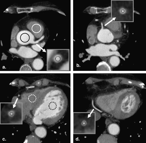
Ninety-eight patients who underwent CTCA in a single heartbeat with electrocardiogram-gating were divided into two groups, receiving 0.8 mL/kg of contrast material injected at a fixed duration of 12 seconds (A; n = 48) or 0.7 mL/kg of contrast material injected at a fixed duration of 10 seconds (B; n = 50); all patients then received 20 mL of saline. Contrast densities were assessed for the ascending aorta, left ventricle, right coronary artery (RCA), and left main coronary artery (LMA).
Results
The mean flow rate was 4.00 ± 0.56 mL/second in group A and 4.06 ± 0.57 mL/second in group B ( P = .51). There were no significant differences in the mean enhancement values of the ascending aorta, LMA and proximal RCA between the two groups. Also, there was no significant difference between the mean enhancement values at the three different levels of the RCA (proximal, middle, and distal segments) (group A; P = .27, group B; P = .07).
Conclusion
The use of 0.7 mL/kg of contrast material injected at a fixed duration of 10 seconds was feasible for CTCA using 320-detector row CT, with a sufficient and reliable contrast enhancement in the ascending aorta and coronary artery.
Computed tomography coronary angiography (CTCA) has become a standard in the noninvasive assessment of coronary arteries in the past few years . In CTCA, high and consistent vascular enhancement is a prerequisite for sufficient evaluation . Recently, it has been reported that a patient weight–adapted iodine-dose protocol with fixed injection duration yielded significantly better image quality than the fixed-dose protocol in 64-detector row CTCA . An injection volume of 1.0 mL/kg body weight of contrast material (370 mgI/mL) is necessary to achieve a sufficient contrast enhancement, and injection duration of 15 seconds is recommended in 64-detector row CTCA .
Very recently, 320-detector row CT scanner has been developed with a z-coverage value of 160 mm; thus, the entire heart can be scanned in a single rotation and within a single heartbeat with a minimum temporal resolution of 175 ms . The data acquisition time for 320-detector row CTCA is less than 3 seconds, which is quite short compared with 64-detector row CTCA at less than 10 seconds. Because this shorter scan time permits a decrease in the contrast dose or the injection duration at CTCA , contrast material protocols must be adjusted and optimized as CT technology evolves.
Get Radiology Tree app to read full this article<
Materials and methods
Patients
Get Radiology Tree app to read full this article<
CT Scanning
Get Radiology Tree app to read full this article<
Get Radiology Tree app to read full this article<
Get Radiology Tree app to read full this article<
Get Radiology Tree app to read full this article<
Methods of Evaluation
Get Radiology Tree app to read full this article<
Get Radiology Tree app to read full this article<
Statistical Analyses
Get Radiology Tree app to read full this article<
Results
Get Radiology Tree app to read full this article<
Get Radiology Tree app to read full this article<
Get Radiology Tree app to read full this article<
Table 1
Patient Characteristics and contrast Dose at Each Group
Group A Group B_P_ Age (y) 69.8 ± 9.8 68.7 ± 9.0 .39 Weight (kg) 59.3 ± 8.4 58.0 ± 8.1 .34 Heart rate (beats/min) 57.1 ± 9.7 58.8 ± 6.4 .14 Flow rate (mL/sec) 4.00 ± 0.56 4.06 ± 0.57 .51Iodine rate (gI/sec) 1.40 ± 0.20 1.42 ± 0.20 .50
Data are the mean ± SD.
Table 2
The Mean Enhancement Values in Each Portion
Group A Group B_P_ Ascending aorta 452.9 ± 50.4 450.7 ± 49.1 .83 Pulmonary artery trunk 188.7 ± 63.7 130.8 ± 31.6 <.01 Right ventricle 146.0 ± 45.4 106.2 ± 27.3 <.01 Left ventricle 414.9 ± 46.4 402.9 ± 49.5 .21 Left main coronary artery 410.6 ± 47.9 416.8 ± 52.6 .52 Proximal right coronary artery 374.6 ± 43.7 384.2 ± 49.8 .38 Middle right coronary artery 361.6 ± 47.0 364.4 ± 48.8 .98 Distal right coronary artery 357.8 ± 51.6 361.2 ± 46.1 .79
Data are the mean ± SD.
Get Radiology Tree app to read full this article<
Discussion
Get Radiology Tree app to read full this article<
Get Radiology Tree app to read full this article<
Get Radiology Tree app to read full this article<
Get Radiology Tree app to read full this article<
Get Radiology Tree app to read full this article<
Get Radiology Tree app to read full this article<
Get Radiology Tree app to read full this article<
Get Radiology Tree app to read full this article<
Get Radiology Tree app to read full this article<
References
1. Achenbach S., Giesler T., Ropers D., et. al.: Detection of coronary artery stenoses by contrast-enhanced, retrospectively electrocardiographically-gated, multislice spiral computed tomography. Circulation 2001; 103: pp. 2535-2538.
2. Nieman K., Oudkerk M., Rensing B.J., et. al.: Coronary angiography with multi-slice computed tomography. Lancet 2001; 357: pp. 599-603.
3. Beck T., Burgstahler C., Kuettner A., et. al.: Clinical use of multislice spiral computed tomography in 210 highly preselected patients: experience with 4 and 16 slice technology. Heart 2005; 91: pp. 1423-1427.
4. Bley T.A., Ghanem N.A., Foell D., et. al.: Computed tomography coronary angiography with 370-millisecond gantry rotation time: evaluation of the best image reconstruction interval. J Comput Assist Tomogr 2005; 29: pp. 1-5.
5. Cademartiri F., Mollet N.R., Lemos P.A., et. al.: Higher intracoronary attenuation improves diagnostic accuracy in MDCT coronary angiography. AJR Am J Roentgenol 2006; 187: pp. W430-W433.
6. Cademartiri F., Mollet N.R., van der Lugt A., et. al.: Intravenous contrast material administration at helical 16-detector row CT coronary angiography: effect of iodine concentration on vascular attenuation. Radiology 2005; 236: pp. 661-665.
7. Schoepf U.J., Zwerner P.L., Savino G., et. al.: Coronary CT angiography. Radiology 2007; 244: pp. 48-63.
8. Yamamuro M., Tadamura E., Kanao S., et. al.: Coronary angiography by 64-detector row computed tomography using low dose of contrast material with saline chaser: influence of total injection volume on vessel attenuation. J Comput Assist Tomogr 2007; 31: pp. 272-280.
9. Nakaura T., Awai K., Yauaga Y., et. al.: Contrast injection protocols for coronary computed tomography angiography using a 64-detector scanner: comparison between patient weight-adjusted- and fixed iodine-dose protocols. Invest Radiol 2008; 43: pp. 512-519.
10. Rybicki F.J., Otero H.J., Steigner M.L., et. al.: Initial evaluation of coronary images from 320-detector row computed tomography. Int J Cardiovasc Imaging 2008; 24: pp. 535-546.
11. Steigner M.L., Otero H.J., Cai T., et. al.: Narrowing the phase window width in prospectively ECG-gated single heart beat 320-detector row coronary CT angiography. Int J Cardiovasc Imaging 2009; 25: pp. 85-90.
12. Einstein A.J., Moser K.W., Thompson R.C., et. al.: Radiation dose to patients from cardiac diagnostic imaging. Circulation 2007; 116: pp. 1290-1305.
13. Bae K.T., Seeck B.A., Hildebolt C.F., et. al.: Contrast enhancement in cardiovascular MDCT: effect of body weight, height, body surface area, body mass index, and obesity. AJR Am J Roentgenol 2008; 190: pp. 777-784.
14. Tatsugami F., Husmann L., Herzog B.A., et. al.: Evaluation of a body mass index-adapted protocol for low-dose 64-MDCT coronary angiography with prospective ECG triggering. Am J Roentgenol 2009; 192: pp. 635-638.
15. Yanaga Y., Awai K., Nakaura T., et. al.: Effect of contrast injection protocols with dose adjusted to the estimated lean patient body weight on aortic enhancement at CT angiography. AJR Am J Roentgenol 2009; 192: pp. 1071-1078.
16. Awai K., Hiraishi K., Hori S.: Effect of contrast material injection duration and rate on aortic peak time and peak enhancement at dynamic CT involving injection protocol with dose tailored to patient weight. Radiology 2004; 230: pp. 142-150.
17. Ichikawa T., Erturk S.M., Araki T.: Multiphasic contrast-enhanced multidetector-row CT of liver: contrast-enhancement theory and practical scan protocol with a combination of fixed injection duration and patients’ body-weight-tailored dose of contrast material. Eur J Radiol 2006; 58: pp. 165-176.
18. Parfrey P.S., Griffiths S.M., Barrett B.J., et. al.: Contrast material-induced renal failure in patients with diabetes mellitus, renal insufficiency, or both. A prospective controlled study. N Engl J Med 1989; 320: pp. 143-149.
19. Solomon R.: Contrast media nephropathy—how to diagnose and how to prevent?. Nephrol Dial Transplant 2007; 22: pp. 1812-1815.
20. Donnino R., Jacobs J.E., Doshi J.V., et. al.: Dual-source versus single-source cardiac CT angiography: comparison of diagnostic image quality. Am J Roentgenol 2009; 192: pp. 1051-1056.
21. Matsumoto M., Kodama N., Endo Y., et. al.: Dynamic 3D-CT angiography. Am J Neuroradiol 2007; 28: pp. 299-304.
22. Schueller G., Schima W., Schueller-Weidekamm C., et. al.: Multidetector CT of pancreas: effects of contrast material flow rate and individualized scan delay on enhancement of pancreas and tumor contrast. Radiology 2006; 241: pp. 441-448.