
Rationale and Objectives
To investigate the expression levels of green fluorescence protein (GFP) into retinal ganglion cells (RGCs) in vitro by ultrasound-mediated microbubble destruction (UMMD) and assess the effect of bcl-xl gene on N-methyl-D-aspartate (NMDA)-induced apoptosis in the cultured RGCs by UMMD.
Materials and Methods
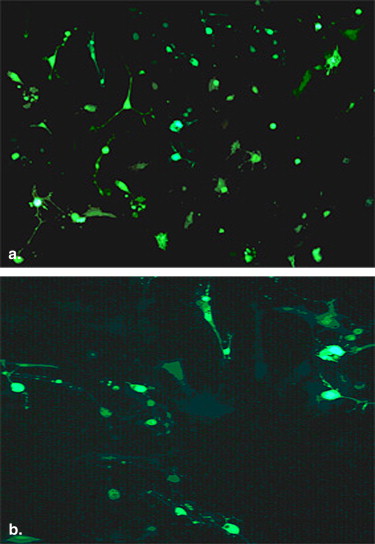
pEGFP-N1 was transfected to RGCs in vitro by UMMD and liposome was used as the control. The transfection effect was detected using microscope and flow cytometry qualitatively and quantitatively. Monotetrazolium was adopted to measure the cell vitality. NMDA was used to induce apoptosis in the cultured RGCs, and the bcl-xl gene was transfected into RGCs by UMMD before NMDA-induced apoptosis. The expression of bcl-xl protein in RGCs was assessed by immunohistochemistry assay. The amorphous character of RGCs was revealed by acridine orange and ethidium bromide staining. DNA fragment was detected by agarose gel electrophoresis.
Results
Ultrasound combined with microbubbles enhanced gene transfection to the cultured cells in some condition. The average transfection rate of pEGFP-N1 with UMMD was 25%. Both ultrasound and microbubble had no effect on cell viability. The expression of bcl-xl protein in transfected and non-transfected RGCs was significantly different. Less apoptotic bodies and no representative DNA fragment were detected in the treatment group.
Conclusions
Microbubble destruction can enhance the reporter gene transfection and expression and have a good target. Transfection of bcl-xl gene has an anti-apoptosis effect on the cultured RGCs induced by NMDA with UMMD.
In recent years, numerous studies have indicated that ultrasonic irradiation itself not only promotes gene transfection and expression in vitro and in vivo and ultrasound-mediated microbubble destruction (UMMD) can further enhance gene transfection efficiency . It has become the research hotspot because it is safe, reliable, and low in cost . This study explored the gene transfection efficiency mediated by UMMD in retinal ganglion cells (RGCs) and the related condition in vitro. With the in-depth research in pathogenesis of glaucoma, it is recognized that to reduce intraocular pressure alone is not enough. Other ways to further protect the optic nerve and prevent or delay damage to RGCs need to be considered . Thus the new concept of optic nerve protection treatment has become the glaucoma research direction and focus in 21st century . Because RGC apoptosis is the key link to glaucoma, we established an RGC excitotoxicity injury model and used ultrasound microbubble–mediated anti-apoptotic gene bcl-xl and determined the optimum parameters for transfecting RGCs with the bcl-xl gene and subsequently investigated its effects on RGC apoptosis. We hope we can protect the optic nerve with new ideas.
Materials and methods
Preparation of Plasmid Enhanced Green Fluorescence Protein N1
Bacillus coli DH5 (Invitrogen, Carlsbad, CA) was resuscitated by the conventional method and plasmid enhanced green fluorescence protein N1 (pEGFP-N 1 ) was inverted into bacteria by classical calcium chloride method. We extracted plasmid according to Kit (EndoFree Plasmid Maxi Kit; Qiagen, Germany) requirements. A small amount of plasmid was electrophoresis, and plasmid concentration was adjusted to 1 μg/μL by spectrophotometry.
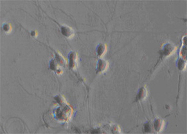
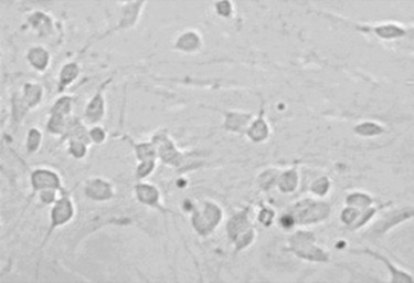
Culture and Identification of Retinal Ganglion Cells
Get Radiology Tree app to read full this article<
Experimental Groups and Transfection of EGFP Gene
Get Radiology Tree app to read full this article<
Get Radiology Tree app to read full this article<
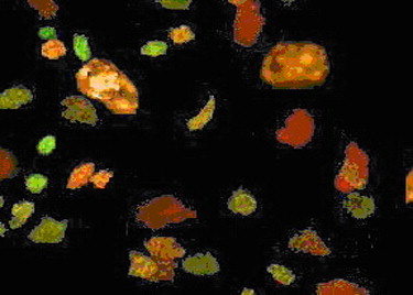
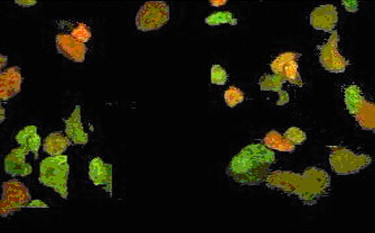
Cell Transfection Analysis
Get Radiology Tree app to read full this article<
Flow Cytometric Analysis of Transfected Quantitative Gene Expression
Get Radiology Tree app to read full this article<
Methyl Thiazolyl Tetrazolium Assay
Get Radiology Tree app to read full this article<
Anti-apoptosis Effect of bcl-xl on RGCs by UMMD
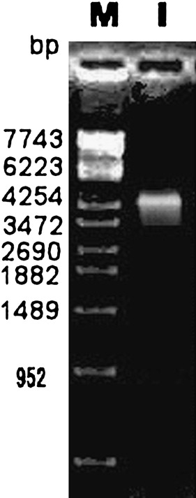
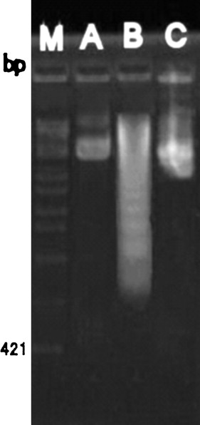
Preparation and identification of the plasmid SFFV.neo-bcl-xl
Get Radiology Tree app to read full this article<
Cultivated RGCs were identified by immunocytochemical method
Get Radiology Tree app to read full this article<
Ultrasound-mediated Microbubble Transfection Cell Model
Get Radiology Tree app to read full this article<
Immunohistochemical Detection
Get Radiology Tree app to read full this article<
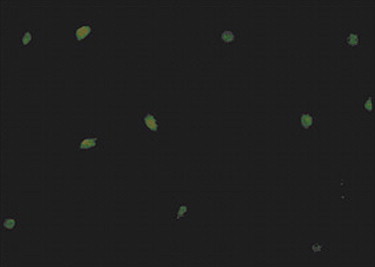
The Morphology Detection of Apoptosis
Get Radiology Tree app to read full this article<
Agarose Electrophoresis Detection
Get Radiology Tree app to read full this article<
Statistical Methods
Get Radiology Tree app to read full this article<
Results
UMMD Parameter Selection
Get Radiology Tree app to read full this article<
Get Radiology Tree app to read full this article<
The Best Transfection Efficiency Parameters of UMMD
Get Radiology Tree app to read full this article<
Get Radiology Tree app to read full this article<
Table 1
The Role of Different Ultrasound Intensity, Exposure Time and Plasmid Concentration to the Transfection Efficiency in the RGC and the Activity of the RGC
Groups Examples n Transfected cells rate AbsorbanceUltrasound Intensity Control group 8 0.62±0.04 0.245±0.032 0.25 8 17.3±4.21 0.246±0.029 0.50 8 19.6±3.22 0.237±0.041 0.75 8 22.8±5.63 0.221±0.034 1.00 8 23.1±4.32 0.185±0.017▴ 1.25 8 20.2±4.25 0.172±0.009Exposure time Control group 8 0.59±0.02 0.241±0.025 30s 8 16.4±3.16 0.238±0.019 60s 8 22.5±4.23 0.226±0.031 90s 8 24.1±3.12 0.195±0.023▴ 120s 8 21.5±2.58 0.181±0.012Plasmid concentration Control group 8 0 0.252±0.034 0.8μg/100μl 8 14.5±2.82 0.243±0.015 2μg/100μl 8 21.6±3.26 0.241±0.021 4μg/100μl 8 25.8±3.83 0.237±0.023 6μg/100μl 8 19.3±3.10 0.185±0.017▴
The role of different machinery index, exposure time and plasmid concentration to the transfection efficiency in the RGC and the activity of the RGC (X±S) note compared with group ahead P<0.05. ▴P<0.05.
Get Radiology Tree app to read full this article<
Cytoactive Determination
Get Radiology Tree app to read full this article<
Comparative Analysis of the Cells Transfection Positive Rate and Expression Level between UMMD and Liposome
Get Radiology Tree app to read full this article<
Get Radiology Tree app to read full this article<
Identify SFFV.neo-bcl-xl Plasmid by Enzyme Digestion
Get Radiology Tree app to read full this article<
Get Radiology Tree app to read full this article<
RGCs Morphology Observation and Identification
Get Radiology Tree app to read full this article<

Get Radiology Tree app to read full this article<
Immunohistochemical Detection
Get Radiology Tree app to read full this article<

Get Radiology Tree app to read full this article<
The Morphology Detection of Apoptosis
Get Radiology Tree app to read full this article<
Get Radiology Tree app to read full this article<
Discussion
Get Radiology Tree app to read full this article<
Get Radiology Tree app to read full this article<
Get Radiology Tree app to read full this article<
Get Radiology Tree app to read full this article<
Get Radiology Tree app to read full this article<
Get Radiology Tree app to read full this article<
Get Radiology Tree app to read full this article<
Get Radiology Tree app to read full this article<
References
1. Wang Z., Ling Z., Ran H., et. al.: Ultrasound-mediated microbubble destruction enhances VEGF gene delivery to the infracted myocardium in rats. Clin Imaging 2004; 28: pp. 395-398.
2. Zhang Q., Wang Z., Ran H., et. al.: Enhanced gene delivery into skeletal muscles with ultrasound and microbubble techniques. Acad Radiol 2006; 13: pp. 363-367.
3. Ren J.L., Wang Z.G., Zhang Y., et. al.: Transfection efficiency of TDL compound in HUVEC enhanced by ultrasound-targeted microbubble destruction. Ultrasound Med Biol 2008; 34: 1857–1567
4. XingSheng L., ZhiGang W., Hai Tao R., et. al.: Experimental research on therapeutic angiogenesis induced by hepatocyte growth factor directed by ultrasound-targeted microbubble destruction in rats. Ultrasound Med 2008; 27: pp. 439-446.
5. Bautista R.D.: Glaucomatous neurodegeneration and the concept of neuroprotection. Int Ophthalmol Clin 1999; 39: pp. 57-70.
6. Weinreb R.N., Levin L.A.: Is neuroprotection a viable therapy for glaucoma?. Arch Ophthalmol 1999; 117: pp. 1540-1544.
7. Zhang J.B.: Cell and tissue culture technique [M].2002.the people health publishing houseBeijing 14
8. Herrmann M., Lorenz H.M., Voll R., et. al.: A Rapid and simple method for the isolation of apoptotic DNA fragments. Nucleic Acid Res 1994; 22: pp. 5506-5507.
9. EI-Aneed A.: An overview of current delivery systems in cancer gene therapy. J Control Release 2004; 94: pp. 1-14.
10. Unger E.C., Hersh E., Vannan M., et. al.: Local drug and gene delivery through microbubles. Prog Cardiovasc Dis 2001; 44: pp. 45-54.
11. Skyba D.M., Price R.J., Linka A.Z., et. al.: Direct in vivo visualization of intravascular destruction of microbubbles by ultra-sound and its local effects on tissue. Circulation 1998; 98: pp. 290-293.
12. Taniyama Y., Tachibana K., Hiraoka K., et. al.: Local delivery of plasmid DNA into rat carotid artery using ultrasound. Circulation 2002; 105: pp. 1233-1239.
13. Taniyama Y., Tachibana K., Hiraoka K., et. al.: Development of safe and efficient novel nonviral gene transfer using ultrasound: enhancement of transfection efficiency of naked plasmid DNA in skeletal muscle. Gene Ther 2002; 9: pp. 372-380.
14. Lawrie A., Brisken A.F., Francis S.E., et. al.: Ultrasound enhances reporter gene expression after transfection of vascular cells in vitro. Circulation 1999; 99: pp. 2617-2620.
15. Shohet R.V., Chen S., Zhou Y.T., et. al.: Echocardiographic destruction of albumin microbubbles directs gene delivery to the myocardium. Circulation 2000; 101: pp. 2554-2556.
16. Koch S., Pohl P., Cobet U., et. al.: Ultrasound enhancement of liposome mediated cell transfection is caused by cavitation effects. Ultrasound Med Biol 2000; 26: pp. 897-903.
17. Lawrie A., Brisken A.F., Francis S.E., et. al.: Microbubble-enhanced ultrasound for vascular gene delivery. Gene Therapy 2000; 7: pp. 2023-2027.
18. Banerjee R.: Liposomes: applications in medicine. J Biomater Appl 2001; 16: pp. 3-21.
19. Sucher N.J., Lipton S.A., Drever E.B.: Molecular basis of glutamate toxicity in retinal ganglion cells. Vision Res 1997; 37: pp. 3483-3493.
20. Dreyer E.B., Zurakowski D., Schumer R.A., et. al.: Elevated glutamate levels in the vitreous body of humans and monkeys with glaucoma. Arch Ophthalmol 1996; 114: pp. 299-305.
21. Siliprandi R., Canella R., Carmignoto G., et. al.: N-methyl-D-aspartate induced neurotoxicity in the adult rat retina. Vis Neurosci 1992; 8: pp. 567-573.
22. Li Y., Schlamp C.L., Nickells R.W.: Experimental induction of retinal ganglion cell death in adult mice. Invest Ophthalmol Vis Sci 1999; 40: pp. 1004-1008.
23. Luo X., Heidinger V., Picaud S., et. al.: Selective excitotoxic degeneration of adult pig retinal ganglion cells in vitro. Invest Ophthalmol Vis Sci 2001; 42: pp. 1096-1106.
24. Levin L.A., Schlamp C.L., Spieldoch R.L., et. al.: Identification of the bcl-2 family of genes in the rat retina. Invest Ophthalmol Vis Sci 1997; 38: pp. 2545-2553.