
Rationale and Objectives
The purpose of this study was to demonstrate the application of gold nanoparticles (AuNP) as a contrast agent for a clinical x-ray computed tomography (CT) system using a phantom and juvenile swine.
Materials and Methods
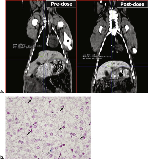
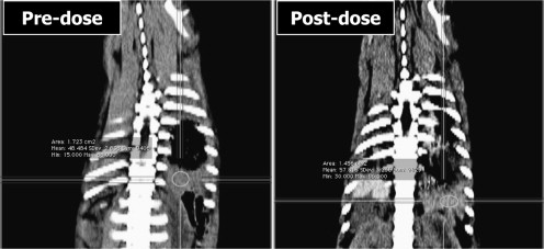
A tissue-mimicking phantom with spherical inclusions containing known concentrations of Au was scanned. Swine were injected with gum Arabic stabilized Au nanoparticles (GA-AuNP), up to 85 mg kg −1 body weight. CT scans were performed before and after the injections. Changes in Hounsfield unit (HU) values between pre- and post- injection scans were evaluated and compared to postmortem determinations of Au uptake. Average uptake of GA-AuNP in the liver of the swine was 380 μg per gram of liver and 680 μg per gram of spleen.
Results
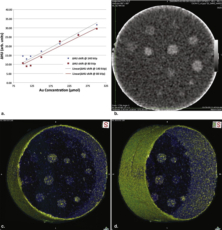
Concentrations of Au in tissues increased the CT numbers in liver by approximately 22 HU per mg Au concentration at 80 kVp and 27 HU per mg Au concentration at 140 kVp. These data were consistent with HU changes observed for similar concentrations in the phantom.
Conclusions
AuNP-based contrast agents may be useful in x-ray based CT. This study provides data for determining concentrations of AuNP in comparison to other contrast materials.
In this work, the authors have used an agarose-based phantom and juvenile swine as a model for evaluating the x-ray contrast properties of gold nanoparticles (AuNP) at low concentrations. This serves to advance our understanding of the effects of these agents and their potential for application to molecular imaging using targeted, hybrid nanoparticle agents.
This work is unique in that we have performed our studies employing the higher energy x-ray spectrum used in clinical computed tomography (CT) equipment. This provides a more realistic challenge for the gold-based nanoparticle agents as a lower photoelectric interaction is in effect at these higher energies than has been reported in earlier published results. This work also is testing the effect of relatively low concentrations of AuNP and provide some framework for expectations of the required uptake and dose to make molecular imaging possible using AuNP with targeting moieties.
Get Radiology Tree app to read full this article<
Get Radiology Tree app to read full this article<
Get Radiology Tree app to read full this article<
Get Radiology Tree app to read full this article<
Get Radiology Tree app to read full this article<
Get Radiology Tree app to read full this article<
Methods and materials
Tissue-mimicking AuNP Phantom
Get Radiology Tree app to read full this article<
Swine Model
Get Radiology Tree app to read full this article<
AuNP Contrast Agent Production
Get Radiology Tree app to read full this article<
Animal Dose Protocol
Get Radiology Tree app to read full this article<
CT Imaging Protocol
Get Radiology Tree app to read full this article<
Get Radiology Tree app to read full this article<
Get Radiology Tree app to read full this article<
Get Radiology Tree app to read full this article<
Get Radiology Tree app to read full this article<
Get Radiology Tree app to read full this article<
Get Radiology Tree app to read full this article<
Get Radiology Tree app to read full this article<
Get Radiology Tree app to read full this article<
Biodistribution of AuNP
Get Radiology Tree app to read full this article<
Results
Get Radiology Tree app to read full this article<
Get Radiology Tree app to read full this article<
Get Radiology Tree app to read full this article<
Get Radiology Tree app to read full this article<
Table 1
Summary of CT Results from Swine Subjects
Liver Spleen Subject Days in Study Dose (mg Au kg-1) AuNP uptake (μmol) ?HU @ 80 kVp ?HU @ 140 kVp AuNP uptake (μmol) ?HU @ 80 kVp ?HU @ 140 kVp A 29.00 86.00 264.21 12.50 13.3 425.26316 9.1 10.9 B 10.00 88.00 148.42 5.70 9.3 297.89474 2.24 2.79 C 10.00 99.00 187.37 7.23 7.87 356.84211 8.5 7
CT, computed tomography; Au, gold; NP, nanoparticles; HU, Hounsfield units.
Mean voxel values for each organ (measured in HU) generally increased in proportion to the Au concentration determined after necropsy. ΔHU values are determined from differences in images acquired at the beginning of the study (Day 0) and at the end of the study (# Days in study).
Get Radiology Tree app to read full this article<
Get Radiology Tree app to read full this article<
Get Radiology Tree app to read full this article<
Discussion
Get Radiology Tree app to read full this article<
Get Radiology Tree app to read full this article<
Get Radiology Tree app to read full this article<
Get Radiology Tree app to read full this article<
Get Radiology Tree app to read full this article<
Get Radiology Tree app to read full this article<
Get Radiology Tree app to read full this article<
Get Radiology Tree app to read full this article<
Get Radiology Tree app to read full this article<
Get Radiology Tree app to read full this article<
Get Radiology Tree app to read full this article<
Get Radiology Tree app to read full this article<
Summary
Get Radiology Tree app to read full this article<
Get Radiology Tree app to read full this article<
References
1. Weissleder R.: Molecular imaging: exploring the next frontier. Radiology 1999; 212: pp. 609-614.
2. Hainfeld J.F., Slatkin D.N., Focella T.M., et. al.: Gold nanoparticles: a new X-ray contrast agent. Br J Radiol 2006; 79: pp. 248-253.
3. Kim D., Park S., Lee J.H., et. al.: Antibiofouling polymer-coated gold nanoparticles as a contrast agent for in vivo X-ray computed tomography imaging. J Am Chem Soc 2007; 129: pp. 7661-7665.
4. Carrascosa P., Capunay C., Bettinotti M., et. al.: Feasibility of gadolinium-diethylene triamine pentaacetic acid enhanced multidetector computed tomography for the evaluation of coronary artery disease. J Cardiovasc Comput Tomogr 2007; 1: pp. 86-94.
5. Alric C., Taleb J., Le Duc G., et. al.: Gadolinium chelate coated gold nanoparticles as contrast agents for both X-ray computed tomography and magnetic resonance imaging. J Am Chem Soc 2008; 130: pp. 5908-5915.
6. Popovtzer R., Agrawal A., Kotov N.A., et. al.: Targeted gold nanoparticles enable molecular CT imaging of cancer. Nano Lett 2008; 8: pp. 4593-4596.
7. Fent G.M., Casteel S.W., Kim D.Y., et. al.: Biodistribution of maltose and gum arabic hybrid gold nanoparticles after intravenous injection in juvenile swine. Nanomedicine 2009; 5: pp. 128-135.
8. Kattumuri V., Katti K., Bhaskaran S., et. al.: Gum arabic as a phytochemical construct for the stabilization of gold nanoparticles: in vivo pharmacokinetics and X-ray-contrast-imaging studies. Small 2007; 3: pp. 333-341.
9. Kannan R., Rahing V., Cutler C., et. al.: Nanocompatible chemistry toward fabrication of target-specific gold nanoparticles. J Am Chem Soc 2006; 128: pp. 11342-11343.
10. Kattumuri V., Chandrasekhar M., Guha S., et. al.: Agarose-stabilized gold nanoparticles for surface-enhanced Raman spectroscopic detection of DNA nucleosides. Appl Phys Lett 2006; 88: pp. 153114-153123.
11. Shukla R., Nune S.K., Chanda N., et. al.: Soybeans as a phytochemical reservoir for the production and stabilization of biocompatible gold nanoparticles. Small 2008; 4: pp. 1425-1436.
12. Katti K., Chanda N., Shukla R., et. al.: Green nanotechnology from cumin phytochemicals: generation of biocompatible gold nanoparticles. Int J Green Nanotechn Biomed 2009; 1: pp. 39-52.
13. D’Souza W.D., Madsen E.L., Unal O., et. al.: Tissue mimicking materials for a multi-imaging modality prostate phantom. Med Phys 2001; 28: pp. 688-700.
14. Wisner E.R., Katzberg R.W., Koblik P.D., et. al.: Iodinated nanoparticles for indirect computed tomography lymphography of the craniocervical and thoracic lymph nodes in normal dogs. Acad Radiol 1994; 1: pp. 377-384.
15. Geso M.: Gold nanoparticles: a new X-ray contrast agent. Br J Radiol 2007; 80: pp. 64-65. author reply 65
16. Jackson P.A., Rahman W.N., Wong C.J., et. al.: Potential dependent superiority of gold nanoparticles in comparison to iodinated contrast agents. Eur J Radiol 2009; ; in Press, doi: 10.1016/j.ejrad.2009.03.057
17. Gore J.C., Yankeelov T.E., Peterson T.E., et. al.: Molecular imaging without radiopharmaceuticals?. J Nucl Med 2009; 50: pp. 999-1007.
18. Roessl E., Proksa R.: K-edge imaging in x-ray computed tomography using multi-bin photon counting detectors. Phys Med Biol 2007; 52: pp. 4679-4696.
19. Chanda N., Kan P., Watkinson L.D., et. al.: Radioactive gold nanoparticles in cancer therapy: therapeutic efficacy studies of (198)AuNP-GA nanoconstruct in prostate tumor-bearing mice. Nanomedicine 2009; in Press, doi: 10.1016/j.nano.2009.11.002
20. Roessl E., Proksa R.: K-edge imaging in x-ray computed tomography using multi-bin photon counting detectors. Phys Med Biol 2007; 52: pp. 4679-4696.
21. Chanda N., Shukla R., Katti K.V., et. al.: Gastrin releasing protein receptor specific gold nanorods: breast and prostate tumor avid nanovectors for molecular imaging. Nano Lett 2009; 9: pp. 1798-1805.