
Rationale and Objectives
The American Board of Radiology Core Examination integrates assessment of physics knowledge into its overall testing of clinical radiology, with an emphasis on understanding image quality and artifacts, radiation dose, and patient safety for each modality or subspecialty organ system. Accordingly, achieving a holistic approach to physics education of radiology residents is a huge challenge. The traditional teaching of radiological physics—simply through didactic lectures—was not designed for such a holistic approach. Admittedly, time constraints and clinical demands can make incorporation of physics teaching into clinical practice problematic. We created and implemented a week-long, intensive physics rotation for fledgling radiology residents and evaluated its effectiveness.
Materials and Methods
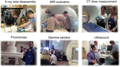
The dedicated physics rotation is held for 1 week during the first month of radiology residency. It comprises three components: introductory lectures, hands-on practical clinical physics operations, and observation of clinical image production. A brief introduction of the physics pertinent to each modality is given at the beginning of each session. Hands-on experimental demonstrations are emphasized, receiving the greatest allotment of time. The residents perform experiments such as measuring radiation dose, studying the relationship between patient dose and clinical practice (eg, fluoroscopy technique), investigating the influence of acquisition parameters (kV, mAs) on radiographs, and evaluating image quality using computed tomography, magnetic resonance imaging, ultrasound, and gamma camera/single-photon emission computed tomography/positron emission tomography phantoms. Quantitative assessment of the effectiveness of the rotation is based on an examination that tests the residents’ grasp of basic medical physics concepts along with written course evaluations provided by each resident.
Results
The pre- and post-rotation tests show that after the physics rotation, the average correct score of 25 questions improved from 13.6 ± 2.4 to 19 ± 1.2. The survey shows that the physics rotation during the first week of residency is favored by all residents and that 1 week’s duration is appropriate. All residents are of the opinion that the intensive workshop would benefit them in upcoming clinical rotations. Residents acknowledge becoming more comfortable regarding the use of radiation and providing counsel regarding radiation during pregnancy.
Conclusions
An immersive, short-duration, clinically oriented physics rotation is well received by new or less experienced radiology trainees, correlates basic physics concepts with their relevance to clinical imaging, and more closely parallels expectations of the American Board of Radiology Core Examination.
Introduction
In 2013, the American Board of Radiology (ABR) replaced the separate written, physics, and oral examinations in diagnostic radiology with the new ABR Core and Certifying Examinations. The ABR Core Examination is intended to validate the candidate’s basic knowledge, skills, and understanding of the entire field of diagnostic radiology, including physics, whereas the certifying examination is intended to validate if the candidate has acquired and is able to apply the requisite knowledge, skills, and understanding for practice .
The ABR Core Examination weaves physics into its sections dealing with imaging modalities and subspecialty organ systems, and shifts the focus from rote knowledge of physics concepts to purposeful application of physics knowledge to solve clinical problems. The content stresses medical physics of practical use to the radiologist. With the integration of clinically relevant physics into the new ABR Core Examination, radiology residency programs are faced with the challenge of how radiological physics should best be taught. In the new ABR Core Examination, the items avoid esoteric minutiae, but instead target practical problems such as radiation dose reduction and image artifact mitigation. Traditional physics teaching to radiology residents through dedicated lectures may not meet these requirements. The Radiological Society of North America/American Association of Physicists in Medicine (RSNA/AAPM) Physics Modules, with their sets of animations and simulations, mitigate this problem to some extent, but do not go far enough in giving the resident an appreciation of the technology they will use in their career.
Get Radiology Tree app to read full this article<
Rationale for the Program
Get Radiology Tree app to read full this article<
Get Radiology Tree app to read full this article<
Get Radiology Tree app to read full this article<
Implementation of the Program
Curriculum
Get Radiology Tree app to read full this article<
TABLE 1
Curriculum and Schedule for Hands-on Physics Education of Residents in Radiology
X-ray tubes (day 1: 8:00 AM to 12:00 noon) X-ray tube disassembly and assembly
Effects of mAs and kV on X-ray quantity and quality
Half value layer
Heel effect and its clinical concerns X-ray projection imaging (day 1: 1:00 PM to 5:00 PM) Effect of kV and mAs on image quality and radiation exposure
Effect of field size and grid on image quality and radiation exposure
Effect of focal-spot size and object-to-image detector distance on image blur and magnification
Automatic exposure control (AEC) and its appropriate use
Mammography and tomosynthesis Fluoroscopy (day 2: 8:00 AM to 12:00 noon) Effect of acquisition modes on image quality and radiation exposure
Fluoroscopy dose metrics and measurement
Strategies to reduce patient and personnel dose Computed tomography (day 2: 1:00 PM to 5:00 PM) CT dose index and dose-length product
Selection of CT acquisition parameters
Tube current modulation and automatic kV adjustment
Evaluation of CT image reconstruction algorithms and image quality Magnetic resonance imaging (day 3: 8:00 AM to 5:00 PM) Components of the MR imager: magnet, gradients, RF coils, and room shielding
MR safety
Effects of Gd-based contrast agents on MR image
Effects of TE, TR on image contrast Gamma camera/SPECT/PET (day 4: 8:00 AM to 5:00 PM) Survey meters: calibration and application
Dose calibrator
Testing parameters for gamma camera
Evaluation of SPECT system using phantom
Evaluation of PET system using phantom Ultrasound (day 5: 8:00 AM to 12:00 noon) Ultrasound image creation, effect of frequency, focal depth on image appearance
Doppler ultrasound: basic principle, angular dependence, artifacts
Shear wave elastography, basic physics, measuring tissue stiffness Radiation safety/wrap up (day 5: 1:00 PM to 5:00 PM)
CT, computed tomography; Gd, gadolinium; MR, magnetic resonance; PET, positron emission tomography; SPECT, single-photon emission computed tomography.
Get Radiology Tree app to read full this article<
Get Radiology Tree app to read full this article<
TABLE 2
Example of Hands-on Laboratory Operations
Laboratory Operation: X-ray Tube Disassembling and Assembling Objectives: The X-ray tube is the very heart of radiography; however, it is rarely seen by operators. The protective housing renders it inaccessible. This experiment is designed to illustrate the fundamental components and function of an X-ray tube. The objectives are as follows:
1. To become familiar with external components that house and protect X-ray tube
2. To identify purpose of glass or metal enclosure
3. To identify and define cathode and filaments
4. To identify and define anode and induction motor
5. To define line-focus principle and heel effect Discussion X-ray consists of two principal elements, cathode and anode, and additional components such as tube envelope, tube housing, rotor, induction stator, window, cooling oil, and expansion bellows. Cathode and anode are enclosed within a glass envelope and encased in a protective housing. The glass or metal enclosure surrounds the two electrodes of a vacuum tube, the cathode and the anode. The cathode contains the tungsten filament, which is the source of electrons. The rotating anode is the tungsten-rhenium disk, which serves as a target for the electrons accelerated from the cathode. The protective housing covers the X-ray tube and serves three functions: (1) limiting radiation leakage to 100 mR/hr at 1 meter; (2) providing mechanical support while protecting the tube from damage; and (3) acting as a heat conduit from the X-ray tube target. Materials required 1. X-ray tube
2. Tools Procedure 1. Drain fluid from X-ray tube (if not already drained)
2. Disassemble X-ray tube from one side and place parts in order
3. Disassemble X-ray from another side and place parts in order
4. Take insert out
5. Re-assemble X-ray tube Results analysis Visually observe X-ray tube, recognize its parts, and be able to explain their functions Review questions 1. What is leakage radiation?
2. How does the anode heel effect affect radiation intensity?
3. What are the filament sizes and their effects on image quality?
4. What is the line-focus principle and how does it affect image quality?
5. How is off-focus radiation produced, and what are the possible effects on image quality?
Get Radiology Tree app to read full this article<
Get Radiology Tree app to read full this article<
Implementation
Get Radiology Tree app to read full this article<
Get Radiology Tree app to read full this article<
Evaluation
Get Radiology Tree app to read full this article<
Results
Get Radiology Tree app to read full this article<
Get Radiology Tree app to read full this article<
Get Radiology Tree app to read full this article<
Discussions and Conclusions
Get Radiology Tree app to read full this article<
Get Radiology Tree app to read full this article<
Get Radiology Tree app to read full this article<
Get Radiology Tree app to read full this article<
Get Radiology Tree app to read full this article<
Get Radiology Tree app to read full this article<
Acknowledgment
Get Radiology Tree app to read full this article<
Get Radiology Tree app to read full this article<
References
1. Initial Certification https://www.theabr.org/ic-dr-core-exam accessed Nov. 8
2. Andrews J.A.: Online videos: a new tool for medical education. Univ B C Med J 2012; 4: pp. 26-27.
3. Redish E.F., Steinberg R.N.: Teaching physics: figuring out what works. Phys Today 1999; 52: pp. 24-30.
4. Wieman C., Perkins K.: Transforming physics education. Phys Today 2005; 58: pp. 36-41.
5. Sutcliffe R.G., Cogdell B., Hansell M.H., et. al.: Active learning in a large first year biology class: a collaborative resource-based study project on “AIDS in Science and Society. Innov Educ Training Int 1999; 36: pp. 53-64.
6. Aguilar L., Walton G., Wieman C.: Psychological insights for improved physics teaching. Phys Today 2014; 67: pp. 43-49.
7. Haury D.L., Rillero P.: Perspectives of hands-on science teaching.1994.ERIC Clearinghouse for Science, Mathematics, and Environmental EducationColumbus, OH
8. Kontra C., Lyons D.J., Fischer S.M., et. al.: Physical experience enhances science learning. Psychol Sci 2015; 26: pp. 737-749.