
Rationale and Objectives
Hyperpolarized 3 He magnetic resonance imaging ventilation defects have been observed in subjects with respiratory disorders. We quantified 3 He ventilation defects in elderly and middle-aged subjects who had no history of smoking, respiratory, or cardiovascular disorders.
Materials and Methods
Hyperpolarized 3 He magnetic resonance imaging ventilation defect volume (VDV) and ventilation defect score (VDS) were assessed in eight elderly healthy volunteers (mean 67 ± 6 years) scanned twice within 7 ± 2 minutes and again 7 ± 2 days later. A younger cohort of 24 subjects (mean 44 ± 10 years) was also scanned for direct comparison. Four observers blinded to scan timepoint and subject identity scored VDS and manually segmented VDV in all center coronal slices.
Results
Center coronal slice ventilation defects were observed in six of eight elderly subjects (ages 63–74 years, 5 males) in all scans acquired and in no middle-aged subjects. At the scan timepoint, mean VDS was 2.7 (mean VDV 52 ± 34 cm 3 ), whereas for same-day rescan, mean VDS was 2.5 (mean VDV 53 ± 35 cm 3 ) and at 7-day rescan, mean VDS was 3.6 (mean VDV 48 ± 39 cm 3 ). Interscan coefficients of variation (COV) for mean VDV was 1.8% (same-day rescan) and 5.3% (7-day rescan) and interobserver COV ranged from 10–12%.
Conclusion
Elderly subjects have ventilation defects that are reproducible in same-day scanning and 7-day scanning visits. The observation of reproducible pulmonary ventilation defects in otherwise healthy elderly volunteers suggests caution must be used in interpreting results from 3 He studies of elderly subjects.
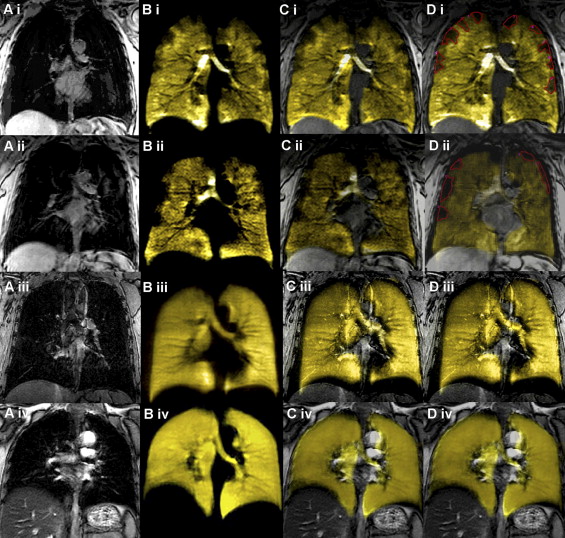
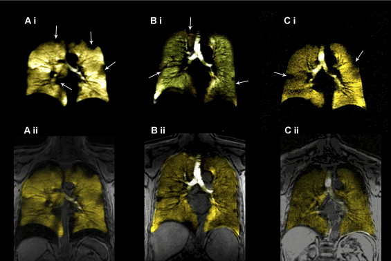
New methods of pulmonary magnetic resonance imaging (MRI) with inhaled hyperpolarized helium-3 ( 3 He) have been shown to provide regional pulmonary 3 He ventilation maps and the location and size of ventilation changes within the lung in asthma ( ), cystic fibrosis ( ) and chronic obstructive pulmonary disease (COPD) ( ). In patients with respiratory disease, areas of decreased ventilation from airflow changes are observed as “ventilation defects” that are visualized as decreased (and/or absence of) 3 He intensity in 3 He MRI spin density images.
Previously, both the size and number of these ventilation defects have been shown to correlate with severity in asthma ( ), and in addition, exercise and methacholine challenge has been shown to alter the size, location, and number of defects that occur in asthma ( ). We have also previously described a preliminary analysis of ventilation defect score (VDS) and ventilation defect volume (VDV) in 3 subjects: one with mild-moderate COPD, one with severe COPD, and a single healthy age-matched control ( ). We noted in this study that significant ventilation defects were observed even in the healthy elderly subject. The finding of numerous center slice ventilation defects in an older healthy individual who was not a smoker, did not have asthma, or cardiovascular disease, and who had normal pulmonary function tests ( ) was surprising and had not been reported previously in other studies of younger healthy volunteers. This result challenged us to explain the physiologic mechanisms related to ventilation defects in healthy lungs as well as the prevalence of ventilation defects in older and middle-aged individuals. To try to address some of these issues, the goal of this study was to examine and compare, using 3 He MRI, ventilation defects in elderly and middle-aged subjects who had no history of smoking, respiratory, or cardiovascular disease.
Materials and methods
Study Population
Get Radiology Tree app to read full this article<
Spirometry
Get Radiology Tree app to read full this article<
Magnetic Resonance Imaging
Get Radiology Tree app to read full this article<
Get Radiology Tree app to read full this article<
Get Radiology Tree app to read full this article<
Image Analysis
Get Radiology Tree app to read full this article<
Get Radiology Tree app to read full this article<
Get Radiology Tree app to read full this article<
Results
Study Subjects
Get Radiology Tree app to read full this article<
Table 1
Study Subject Demographic Characteristics
Elderly Healthy Volunteers (n = 8) Middle-aged Healthy Volunteers (n = 24) Male (n) 5 14 Age (± SD) [range] (yrs) 67 (6) 44 (10) [58–74] [23–57] Weight (± SD) kg 76 (17) 76 (13) Body mass index (± SD) [range] 27 (4) 25 (3) [24–35] [20–34] FEV 1 % predicted (± SD) 106 (19) 101 (11) FEV 1 /FVC % (± SD) 77 (5) 80 (8) Subjects with ventilation defects (n) (male) 6 (3) 0 (0) Total ventilation defects (n) Scan 16 0 Same-day rescan 15 0 7-day Rescan 18 0 Mean VDV cm 3 (± SD) Scan 52 (34) 0 Same-day rescan 53 (35) 0 7-day rescan 48 (39) 0
SD, subgroup standard deviation for eight healthy volunteers; FEV, forced expiratory volume in 1 second; FVC, forced vital capacity.
Get Radiology Tree app to read full this article<
Ventilation Defects and Ventilation Defect Volume
Get Radiology Tree app to read full this article<
Table 2
Ventilation Defects in Elderly Healthy Volunteers
Ventilation Defects in Center Slice Scan Same-day Rescan 7-day Rescan Subject 1001 (n) 0 0 0 Subject 1002 (n) 3 3 ND Subject 1003 (n) 3 3 5 Subject 1004 (n) 2 2 4 Subject 1005 (n) 3 2 3 Subject 1006 (n) 0 0 0 Subject 1007 (n) 2 2 3 Subject 1008 (n) 3 3 3 Total sum defects 16 15 18 Mean defect score 2.7 2.5 3.6
ND, not done (patient withdrew from study due to claustrophobia)
Get Radiology Tree app to read full this article<
Ventilation Defect Volume Interscan and Interobserver Reproducibility
Get Radiology Tree app to read full this article<
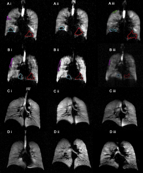
Table 3
Elderly Volunteer Ventilation Defect Volume Interobserver Reproduciblity
Mean VDV cm 3 (± SD) Scan Same-day Rescan 7-day Rescan Observer 1 52 (34) 53 (35) 48 (39) Observer 2 72 (55) 67 (47) 60 (48) Observer 3 67 (43) 64 (42) 53 (40) Observer 4 70 (63) 65 (61) 46 (44) Mean Ob1-4 66 (8) ⁎ 62 (6) ⁎ 52 (6) ⁎ COV (%) 12 10 12
SD, subgroup standard deviation for eight healthy volunteers, except for *SD is observer standard deviation; COV,
Get Radiology Tree app to read full this article<
Get Radiology Tree app to read full this article<
Discussion
Get Radiology Tree app to read full this article<
Get Radiology Tree app to read full this article<
Get Radiology Tree app to read full this article<
Get Radiology Tree app to read full this article<
Get Radiology Tree app to read full this article<
Get Radiology Tree app to read full this article<
Conclusions
Get Radiology Tree app to read full this article<
Acknowledgements
Get Radiology Tree app to read full this article<
Get Radiology Tree app to read full this article<
References
1. Altes T.A., Powers P.L., Knight-Scott J., et. al.: Hyperpolarized 3 He MR lung ventilation imaging in asthmatics: preliminary findings. J Magn Reson Imaging 2001; 13: pp. 378-384.
2. de Lange E.E., Altes T.A., Patrie J.T., et. al.: The variability of regional airflow obstruction within the lungs of patients with asthma: assessment with hyperpolarized helium-3 magnetic resonance imaging. J Allergy Clin Immunol 2007; 119: pp. 1072-1078.
3. Samee S., Altes T., Powers P., et. al.: Imaging the lungs in asthmatic patients by using hyperpolarized helium-3 magnetic resonance: assessment of response to methacholine and exercise challenge. J Allergy Clin Immunol 2003; 111: pp. 1205-1211.
4. de Lange E.E., Altes T.A., Patrie J.T., et. al.: Evaluation of asthma with hyperpolarized helium-3 MRI: correlation with clinical severity and spirometry. Chest 2006; 130: pp. 1055-1062.
5. Altes T.A., de Lange E.E.: Applications of hyperpolarized helium-3 gas magnetic resonance imaging in pediatric lung disease. Top Magn Reson Imaging 2003; 14: pp. 231-236.
6. Altes T.A., Eichinger M., Puderbach M.: Magnetic resonance imaging of the lung in cystic fibrosis. Proc Am Thorac Soc 2007; 4: pp. 321-327.
7. Donnelly L.F., MacFall J.R., McAdams H.P., et. al.: Cystic fibrosis: combined hyperpolarized 3 He-enhanced and conventional proton MR imaging in the lung-—preliminary observations. Radiology 1999; 212: pp. 885-889.
8. Koumellis P., Van Beek E.J., Woodhouse N., et. al.: Quantitative analysis of regional airways obstruction using dynamic hyperpolarized 3 He MRI-preliminary results in children with cystic fibrosis. J Magn Reson Imaging 2005; 22: pp. 420-426.
9. Mentore K., Froh D.K., de Lange E.E., et. al.: Hyperpolarized HHe 3 MRI of the lung in cystic fibrosis: assessment at baseline and after bronchodilator and airway clearance treatment. Acad Radiol 2005; 12: pp. 1423-1429.
10. Salerno M., Altes T.A., Brookeman J.R., et. al.: Dynamic spiral MRI of pulmonary gas flow using hyperpolarized (3)He: preliminary studies in healthy and diseased lungs. Magn Reson Med 2001; 46: pp. 667-677.
11. Ebert M., Grossmann T., Heil W., et. al.: Nuclear magnetic resonance imaging with hyperpolarised helium-3. Lancet 1996; 347: pp. 1297-1299.
12. Fain S.B., Panth S.R., Evans M.D., et. al.: Early emphysematous changes in asymptomatic smokers: detection with 3 He MR imaging. Radiology 2006; 239: pp. 875-883.
13. Kauczor H.U., Hofmann D., Kreitner K.F., et. al.: Normal and abnormal pulmonary ventilation: visualization at hyperpolarized He-3 MR imaging. Radiology 1996; 201: pp. 564-568.
14. Ley S., Zaporozhan J., Morbach A., et. al.: Functional evaluation of emphysema using diffusion-weighted 3Helium-magnetic resonance imaging, high-resolution computed tomography, and lung function tests. Invest Radiol 2004; 39: pp. 427-434.
15. MacFall J.R., Charles H.C., Black R.D., et. al.: Human lung air spaces: potential for MR imaging with hyperpolarized He-3. Radiology 1996; 200: pp. 553-558.
16. Morbach A.E., Gast K.K., Schmiedeskamp J., et. al.: Diffusion-weighted MRI of the lung with hyperpolarized helium-3: a study of reproducibility. J Magn Reson Imaging 2005; 21: pp. 765-774.
17. Parraga G., Ouriadov A., Evans A., et. al.: Hyperpolarized 3 He ventilation defects and apparent diffusion coefficients in chronic obstructive pulmonary disease: preliminary results at 3.0 Tesla. Invest Radiol 2007; 42: pp. 384-391.
18. Swift A.J., Wild J.M., Fichele S., et. al.: Emphysematous changes and normal variation in smokers and COPD patients using diffusion 3 He MRI. Eur J Radiol 2005; 54: pp. 352-358.
19. Woodhouse N., Wild J.M., Paley M.N., et. al.: Combined helium-3/proton magnetic resonance imaging measurement of ventilated lung volumes in smokers compared to never-smokers. J Magn Reson Imaging 2005; 21: pp. 365-369.
20. Pauwels R.A., Buist A.S., Calverley P.M., et. al.: Global strategy for the diagnosis, management, and prevention of chronic obstructive pulmonary disease. Am J Respir Crit Care Med 2001; 163: pp. 1256-1276.
21. Landry A., Spence J.D., Fenster A.: Measurement of carotid plaque volume by 3-dimensional ultrasound. Stroke 2004; 35: pp. 864-869.
22. Landry A., Fenster A.: Theoretical and experimental quantification of carotid plaque volume measurements made by three-dimensional ultrasound using test phantoms. Med Phys 2002; 29: pp. 2319-2327.
23. Landry A., Spence J.D., Fenster A.: Quantification of carotid plaque volume measurements using 3D ultrasound imaging. Ultrasound Med Biol 2005; 31: pp. 751-762.
24. Choudhri A, Altes, TA, Stay R, et al. The Occurence of Ventilation Defects in the Lungs of Healthy Subjects as Demonstrated by Hyperpolarized Helium-3 MR Imaging. RSNA SSA21-05. 2007.
25. Ouriadov A, Etemad-Rezai R, Parraga G, Santyr GE. Correction of errors due to RF field inhomogeneities in hyperpolarized 3 He measurement of alveolar oxygen partial pressure in human lung. 2008. Toronto. Proceedings of the 16th Annual Scientific Meeting of the International Society for Magnetic Resonance in Medicine. Toronto; 2008.
26. Fain S.B., Altes T.A., Panth S.R., et. al.: Detection of age-dependent changes in healthy adult lungs with diffusion-weighted 3 He MRI. Acad Radiol 2005; 12: pp. 1385-1393.