
Purpose
To evaluate the effect of levodopa on apparent diffusion coefficient (ADC) value of the brain parenchyma in patients with idiopathic Parkinson disease (PD).
Material and Methods
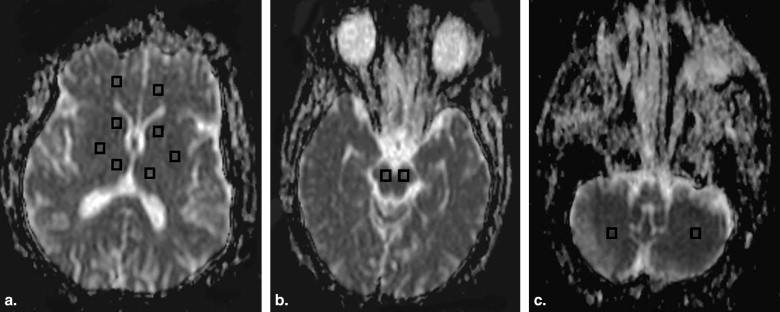
Prospective study was conducted on native PD without treatment ( n = 25) and patients receiving levodopa (L-Dopa) ( n = 25). Diffusion magnetic resonance–weighted imaging was done using a single-shot spin echo type of echo planar imaging. The apparent diffusion coefficient (ADC) value at different regions of the brain on both sides was calculated.
Results
The ADC value of the putamen in patients with native PD was 0.732 ± 0.15 × 10⁻³ mm2/seconds and in patients receiving levodopa was 0.789 ± 0.24 × 10⁻³ mm2/second. There was a statistically significant difference in the ADC value at the putamen ( P = .001) between patients with native PD and patients receiving levodopa. When ADC value of the putamen at 0.745 × 10⁻³ mm2/second was used as a threshold value for differentiating native PD patients and patients receiving L-Dopa, the best results were obtained with an accuracy of 82%, sensitivity of 92%, specificity of 72%, positive predictive value of 77%, negative predictive value of 90%, and area under the curve of 0.955.
Conclusion
ADC value of the putamen is a promising parameter for predication of effect of levodopa on brain parenchyma in patients with PD.
Idiopathic Parkinson disease (PD) is the second most common neurodegenerative disorder. Although different therapy such as surgery, intervention, and gene therapy has been applied for patients with PD, medication with levodopa (L-Dopa) remains the first main most effective line of treatment of patients with of PD. Finding a parameter for the effect of L-Dopa on the brain is important because L-Dopa’s effect on patients’ signs and symptoms dwindles with time and can become toxic . Positron emission tomography scanning has been used to detect effect of L-Dopa on brain parenchyma, but it is limited by relatively poor resolution, lack of chemical specificity, and the need to inject a radioactive agent . Magnetic resonance (MR) spectroscopy shows reduced N Acetyl Aspertate in patients receiving L-Dopa compared to others, but its result overlaps . Voxel-based morphometry at T1 relaxation time was less in patients receiving L-Dopa compared to native PD patients . Also, blood oxygenation level dependent, perfusion, and diffusion MR imaging has been used for study the effect of L-Dopa on brain parenchyma .
The purpose of this work is to evaluate the effect of levodopa on apparent diffusion coefficient (ADC) value at various regions of the brain parenchyma in patients with idiopathic PD.
Materials and methods
Get Radiology Tree app to read full this article<
Table 1
Demographic Data of the Patients
Demographic data Native PD ( n = 25) Levo Dopa ( n = 25) Men/women 15/10 13/12 Mean age (y) 65.44 ± 5.22 66.33 ± 6.22 Mean age at disease onset 58.44 ± 4.32 59.37 ± 7.12 Median hoehn and yahr score 3.2 (1–5) 4 (3–5) Median unified parkinson disease rating scale score 44.25 (22–88) 51.50 (30–95)
Get Radiology Tree app to read full this article<
Get Radiology Tree app to read full this article<
Get Radiology Tree app to read full this article<
Get Radiology Tree app to read full this article<
Get Radiology Tree app to read full this article<
Results
Get Radiology Tree app to read full this article<
Get Radiology Tree app to read full this article<
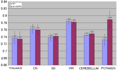
Table 2
The ADC Value at Different Regions of the Brain Parenchyma
ROI Native IPD
( n = 25) Levo Dopa
( n = 25)P value Putamen 0.732 ± 0.15 0.789 ± 0.24 .001 Thalamus 0.737 ± 0.17 0.734 ± 0.21 .52 Caudate nucleus 0.763 ± 0.12 0.760 ± 0.14 .35 Substantia nigra 0.738 ± 0.11 0.742 ± 0.06 .59 White matter 0.784 ± 0.08 0.782 ± 0.05 .32 Cerebellum 0.7452 ± 0.11 0.749 ± 0.08 .17
ADC, apparent diffusion coefficient; IPD, idiopathic Parkinson disease; ROI, region of interest.
Get Radiology Tree app to read full this article<
Discussion
Get Radiology Tree app to read full this article<
Get Radiology Tree app to read full this article<
Get Radiology Tree app to read full this article<
Get Radiology Tree app to read full this article<
Get Radiology Tree app to read full this article<
Get Radiology Tree app to read full this article<
Get Radiology Tree app to read full this article<
Get Radiology Tree app to read full this article<
References
1. Tolosa E., Wenning G., Poewe W.: The diagnosis of Parkinson’s disease. Lancet Neurol 2006; 5: pp. 75-86.
2. Marras C., Lang A.: Invited article: changing concepts in Parkinson disease: moving beyond the decade of the brain. Neurology 2008; 70: pp. 1996-2003.
3. Clarke C.E.: Medical management of Parkinson’s disease. J Neurol Neurosurg Psychiatr 2002; 72: pp. 122-127.
4. Lees A.: Alternatives to levodopa in the initial treatment of early Parkinson’s disease. Drugs Aging 2005; 22: pp. 731-740.
5. Torstenson R., Hartvig P., Lngstrom B., et. al.: Differential effects of levodopa on dopaminergic function in early and advanced Parkinson’s disease. Ann Neurol 1997; 41: pp. 334-340.
6. Muller T., Hefter H., Hueber R., et. al.: Is levodopa toxic?. J Neurol 2004; 251: pp. VI/44-VI/46.
7. Antonini A., Schwarz J., Oertel W., et. al.: [11C] raclopride and positron emission tomography in previously untreated patients with Parkinson’s disease: influence of L-dopa and lisuride therapy on striatal dopamine D2-receptors. Neurology 1994; 44: pp. 1325-1329.
8. Simoes-Ribeiro F., Mendes-Ribeiro J., Soares R., et. al.: Proton magnetic resonance spectroscopy findings in Parkinson’s disease: drug-naive vs levodopa-treated dyskinetic vs levodopa-treated non-dyskinetic patients. Mov Disord 1998; 13: pp. 173-174.
9. Salgado-Pineda P., Delaveau P., Falcon C., et. al.: Brain T1 intensity changes after levodopa administration in healthy subjects: a voxel-based morphometry study. Br J Clin Pharmacol 2006; 62: pp. 546-551.
10. Brusa L., Bassi A., Pierantozzi M., et. al.: Perfusion-weighted dynamic susceptibility (DSC) MRI: basal ganglia hemodynamic changes after apomorphine in Parkinson’s disease. Neurol Sci 2002; 23: pp. S61-S62.
11. Kraft E., Loichinger W., Diepers M., et. al.: Levodopa-induced striatal activation in Parkinson’s disease: A functional MRI study. Parkinsonism Related Disord 2009; 15: pp. 558-563.
12. Degirmenci B., Yaman M., Haktanir A., et. al.: The effects of levodopa use on diffusion coefficients in various brain regions in Parkinson’s disease. Neurosci Lett 2007; 416: pp. 294-298.
13. Hughes A., Daniel S., Kilford L., et. al.: Accuracy of clinical diagnosis of idiopathic Parkinson’s disease: a clinico-pathological study of 100 cases. J Neurol Neurosurg Psychiatry 1992; 55: pp. 181-184.
14. Seppi K., Poewe W.: Brain magnetic resonance imaging techniques in the diagnosis of parkinsonian syndromes. Neuroimaging Clin North Am 2010; 20: pp. 29-55.
15. Sitburana O., Ondo W.: Brain magnetic resonance imaging (MRI) in parkinsonian disorders. Parkinsonism Related Disord 2009; 15: pp. 165-174.
16. Brooks D.: Assessment of Parkinson’s disease with imaging. Parkinsonism Related Disord 2007; 13: pp. S268-S275.
17. Menke R., Scholz J., Miller K., et. al.: MRI characteristics of the substantia nigra in Parkinson’s disease: a combined quantitative T1 and DTI study. NeuroImage 2009; 47: pp. 435-441.
18. Vaillancourt D.E., Spraker M.B., Prodoehl J., et. al.: High-resolution diffusion tensor imaging in the substantia nigra of de novo Parkinson disease. Neurology 2009; 72: pp. 1378-1384.
19. Gattellaro G., Minati L., Grisoli M., et. al.: White matter involvement in idiopathic Parkinson disease: a diffusion tensor imaging study. AJNR Am J Neuroradiol 2009; 30: pp. 1222-1226.
20. Tessa C., Giannelli M., Della N.R., et. al.: A whole-brain analysis in de novo Parkinson disease. AJNR Am J Neuroradiol 2008; 29: pp. 674-680.
21. Kendi A.T., Lehericy S., Luciana M., et. al.: Altered diffusion in the frontal lobe in Parkinson disease. AJNR Am J Neuroradiol 2008; 29: pp. 501-505.
22. Chan L.L., Rumpel H., Yap K., et. al.: Case control study of diffusion tensor imaging in Parkinson’s disease. J Neurol Neurosurg Psychiatry 2007; 78: pp. 1383-1386.
23. Rizzo G., Martinelli P., Manners D., et. al.: Diffusion-weighted brain imaging study of patients with clinical diagnosis of corticobasal degeneration, progressive supranuclear palsy and Parkinson’s disease. Brain 2008; 131: pp. 2690-2700.
24. Paviour D., Thornton J., Lees A., et. al.: Diffusion-weighted magnetic resonance imaging differentiates Parkinsonian variant of multiple system atrophy from progressive supranuclear palsy. Mov Disord 2007; 22: pp. 68-74.
25. Ito M., Watanabe H., Kawai Y., et. al.: Usefulness of combined fractional anisotropy and apparent diffusion coefficient values for detection of involvement in multiple system atrophy. J Neurol Neurosurg Psychiatry 2007; 78: pp. 722-728.
26. Blain C., Barker G., Jarosz J., et. al.: Measuring brain stem and cerebellar damage in parkinsonian syndromes using diffusion tensor MRI. Neurology 2006; 67: pp. 2199-2205.
27. Nicoletti G., Lodi R., Condino F., et. al.: Apparent diffusion coefficient measurements of the middle cerebellar peduncle differentiate the Parkinson variant of MSA from Parkinson’s disease and progressive supranuclear palsy. Brain 2006; 129: pp. 2679-2687.