
Rationale and Objectives
Focal epilepsies potentially can be cured by neurosurgery; other treatment options usually remain symptomatic. High-resolution magnetic resonance (MR) imaging is the central imaging strategy in the evaluation of focal epilepsy. The most common substrate of temporal epilepsies is hippocampal sclerosis (HS), which cannot always be sufficiently characterized with current MR field strengths. Therefore, the purpose of our study was to demonstrate the feasibility of high-resolution MR imaging at 7 Tesla in patients with focal epilepsy resulting from a HS and to improve image resolution at 7 Tesla in patients with HS.
Materials and Methods
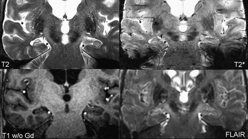
Six patients with known HS were investigated with T1-, T2-, T2 ∗ -, and fluid-attenuated inversion recovery–weighted sequences at 7 Tesla with an eight-channel transmit-receive head coil. Total imaging time did not exceed 90 minutes per patient.
Results
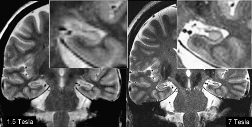
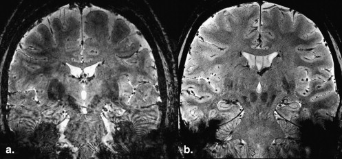
High-resolution imaging at 7 Tesla is feasible and reveals high resolution of intrahippocampal structures in vivo. HS was confirmed in all patients. The maximum non-interpolated in-plane resolution reached 0.2 × 0.2 mm 2 in T2 ∗ -weighted images. The increased susceptibility effects at 7 Tesla revealed identification of intrahippocampal structures in more detail than at 1.5 Tesla, but otherwise led to stronger artifacts. Imaging revealed regional differences in hippocampal atrophy between patients. The scan volume was limited because of specific absorption rate restrictions, scanning time was reasonable.
Conclusions
High-resolution imaging at 7 Tesla is promising in presurgical epilepsy imaging. “New” contrasts may further improve detection of even very small intrahippocampal structural changes. Therefore, further investigations will be necessary to demonstrate the potential benefit for presurgical selection of patients with various lesion patterns in mesial temporal epilepsies resulting from a unilateral HS.
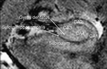
Although the causes of temporal lobe seizures are manifold, the most common finding in patients with severe, medically intractable temporal lobe epilepsy is mesial temporal or hippocampal sclerosis (HS) . HS can occur either as a cause or consequence of focal epileptic seizures . Clinical and neuropathological findings, however, suggest different underlying etiologies and subsequent clinical presentations . Any changes of normal cortical architecture can be associated with severe epileptic syndromes, not restricted to the hippocampus. In general, epileptogenic lesions can be very small and either developmental or acquired.
Epilepsy patients are primarily treated pharmacologically to control seizures. But pharmacological treatment remains symptomatic and fails to sufficiently control seizures in about 20%–25% of patients . To date, the only curative treatment option is the neurosurgical resection of an epileptic lesion . Therefore, the precise presurgical anatomical and functional delineation of epileptogenic lesions by magnetic resonance (MR) imaging as well as by electroencephalographic studies is crucial for diagnosis and mandatory before surgical treatment. In addition, precise delineation of epileptic lesions in relation to functionally important surrounding structures such as eloquent areas, fiber tracts, or vessels may minimize surgical complications.
Get Radiology Tree app to read full this article<
Get Radiology Tree app to read full this article<
Get Radiology Tree app to read full this article<
Get Radiology Tree app to read full this article<
Get Radiology Tree app to read full this article<
Materials and methods
Get Radiology Tree app to read full this article<
Get Radiology Tree app to read full this article<
Table 1
Summary of Basic Patient Characteristics with Respect to Sex, Age, Age at Onset of Seizures, Side of the Hippocampal Sclerosis, and Current Semiology of Seizures
Number Sex Age Age at Onset Side Aura Semiology 1 F 39 4 L Abdominal CPS, GTC 2 F 55 NA R Abdominal CPS 3 F 44 39 R Abdominal CPS, GTC 4 F 60 NA R Abdominal CPS 5 M 26 10 R No CPS 6 F 28 24 L Deja-/jamais-vu CPS, GTC
NA, no data available; CPS, complex partial seizure; GTC, focally induced, secondarily generalized tonic-clonic seizures.
Get Radiology Tree app to read full this article<
Get Radiology Tree app to read full this article<
Get Radiology Tree app to read full this article<
Get Radiology Tree app to read full this article<
Get Radiology Tree app to read full this article<
Get Radiology Tree app to read full this article<
Results
Get Radiology Tree app to read full this article<
Get Radiology Tree app to read full this article<
Get Radiology Tree app to read full this article<
Get Radiology Tree app to read full this article<
Get Radiology Tree app to read full this article<
Get Radiology Tree app to read full this article<
Get Radiology Tree app to read full this article<
Get Radiology Tree app to read full this article<

Get Radiology Tree app to read full this article<
Get Radiology Tree app to read full this article<
Get Radiology Tree app to read full this article<
Discussion
Get Radiology Tree app to read full this article<
Get Radiology Tree app to read full this article<
Get Radiology Tree app to read full this article<
Get Radiology Tree app to read full this article<
Get Radiology Tree app to read full this article<
Get Radiology Tree app to read full this article<
Get Radiology Tree app to read full this article<
Get Radiology Tree app to read full this article<
Conclusion
Get Radiology Tree app to read full this article<
References
1. Koepp M.J., Woermann F.G.: Imaging structure and function in refractory focal epilepsy. Lancet Neurol 2005; 4: pp. 42-53.
2. Jefferys J.G.: Hippocampal sclerosis and temporal lobe epilepsy: cause or consequence?. Brain 1999; 122: pp. 1007-1008.
3. Koepp M.J.: Hippocampal sclerosis: cause or consequence of febrile seizures. J Neurol Neurosurg Psychiatry 2000; 69: pp. 716-717.
4. Cendes F.: Febrile seizures and mesial temporal sclerosis. Curr Opin Neurol 2004; 17: pp. 161-164.
5. Janszky J., Janszky I., Schulz R., et. al.: Temporal lobe epilepsy with hippocampal sclerosis: predictors for long-term surgical outcome. Brain 2005; 128: pp. 395-404.
6. Blumcke I., Pauli E., Clusmann H., et. al.: A new clinico-pathological classification system for mesial temporal sclerosis. Acta Neuropathol 2007; 113: pp. 235-244.
7. Wiebe S., Blume W.T., Girvin J.P., et. al.: A randomized, controlled trial of surgery for temporal-lobe epilepsy. N Engl J Med 2001; 345: pp. 311-318.
8. Elsharkawy A.E., Behne F., Oppel F., et. al.: Long-term outcome of extratemporal epilepsy surgery among 154 adult patients. J Neurosurg 2008; 108: pp. 676-686.
9. Jackson G.D.: The diagnosis of hippocampal sclerosis: other techniques. Magn Reson Imaging 1995; 13: pp. 1081-1093.
10. Woermann F.G., Steiner H., Barker G.J., et. al.: A fast FLAIR dual-echo technique for hippocampal T2 relaxometry: first experiences in patients with temporal lobe epilepsy. J Magn Reson Imaging 2001; 13: pp. 547-552.
11. Bartlett P.A., Symms M.R., Free S.L., et. al.: T2 relaxometry of the hippocampus at 3T. AJNR Am J Neuroradiol 2007; 28: pp. 1095-1098.
12. Duncan J.S.: Imaging and epilepsy. Brain 1997; 120: pp. 339-377.
13. Jackson G.D., Berkovic S.F., Tress B.M., et. al.: Hippocampal sclerosis can be reliably detected by magnetic resonance imaging. Neurology 1990; 40: pp. 1869-1875.
14. Jackson G.D., Connelly A., Duncan J.S., et. al.: Detection of hippocampal pathology in intractable partial epilepsy: increased sensitivity with quantitative magnetic resonance T2 relaxometry. Neurology 1993; 43: pp. 1793-1799.
15. Baldwin G.N., Tsuruda J.S., Maravilla K.R., et. al.: The fornix in patients with seizures caused by unilateral hippocampal sclerosis: detection of unilateral volume loss on MR images. AJR Am J Roentgenol 1994; 162: pp. 1185-1189.
16. Grossman R.I.: Mesial temporal sclerosis.2nd ed.2003.MosbyPhiladelphia, PA
17. Benveniste H., Einstein G., Kim K.R., et. al.: Detection of neuritic plaques in Alzheimer’s disease by magnetic resonance microscopy. Proc Natl Acad Sci U S A 1999; 96: pp. 14079-14084.
18. Chakeres D.W., Whitaker C.D., Dashner R.A., et. al.: High-resolution 8 Tesla imaging of the formalin-fixed normal human hippocampus. Clin Anat 2005; 18: pp. 88-91.
19. Tien R.D., Felsberg G.J., Crain B.: Normal anatomy of the hippocampus and adjacent temporal lobe: high-resolution fast spin-echo MR images in volunteers correlated with cadaveric histologic sections. AJR Am J Roentgenol 1992; 159: pp. 1309-1313.
20. Theysohn J.M., Kraff O., Maderwald S., et. al.: The human hippocampus at 7 T—in vivo MRI. Hippocampus 2009; 19: pp. 1-7.
21. Theysohn J.M., Maderwald S., Kraff O., et. al.: Subjective acceptance of 7 Tesla MRI for human imaging. Magma 2008; 21: pp. 63-72.
22. Naidich T.P., Daniels D.L., Haughton V.M., et. al.: Hippocampal formation and related structures of the limbic lobe: anatomic-MR correlation. Part I. Surface features and coronal sections. Radiology 1987; 162: pp. 747-754.
23. Duvernoy H.: The human hippocampus: functional anatomy, vascularization and serial sections with MRI.3rd ed.2005.SpringerHeidelberg, Germany
24. Kraff O., Theysohn J.M., Maderwald S., et. al.: MRI of the knee at 7.0 Tesla. Rofo 2007; 179: pp. 1231-1235.
25. Yushkevich P.A., Avants B.B., Pluta J., et. al.: A high-resolution computational atlas of the human hippocampus from postmortem magnetic resonance imaging at 9.4 T. Neuroimage 2009; 44: pp. 385-398.
26. Eriksson S.H., Thom M., Bartlett P.A., et. al.: PROPELLER MRI visualizes detailed pathology of hippocampal sclerosis. Epilepsia 2008; 49: pp. 33-39.
27. Sicotte N.L., Kern K.C., Giesser B.S., et. al.: Regional hippocampal atrophy in multiple sclerosis. Brain 2008; 131: pp. 1134-1141.
28. de Lanerolle N.C., Kim J.H., Williamson A., et. al.: A retrospective analysis of hippocampal pathology in human temporal lobe epilepsy: evidence for distinctive patient subcategories. Epilepsia 2003; 44: pp. 677-687.