
Rationale and Objectives
Prospective electrocardiogram (ECG) triggering allows coronary computed tomography angiography (CCTA) scanning with low radiation dose but requires heart rates below 63 beats/min. We assessed the impact of a novel vendor-specific motion-correction algorithm on image quality and interpretability of low-dose CCTA acquired despite insufficient heart rate control.
Materials and Methods
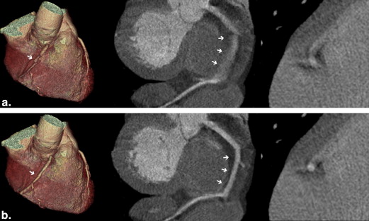
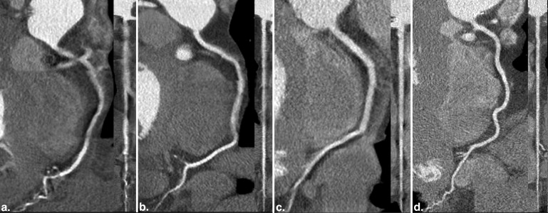
In 40 patients undergoing CCTA for the assessment of known or suspected coronary artery disease who did not reach the target heart rate below 63 beats/min despite β-blockade before prospective low-dose scanning, the temporal acquisition window was increased (80 ms additional padding). The new algorithm detects and integrates vessel path and velocity from adjacent cardiac phases for motion correction. Two blinded observers assessed image quality on a 4-point Likert scale (1, nonevaluative; 2, reduced but evaluative; 3, good; and 4, excellent) and the fraction of interpretable segments (score 2 or more) using motion correction versus standard reconstruction.
Results
Image reconstruction with motion correction resulted in an increased median coronary artery image quality score (excellent interobserver agreement, κ = 0.85) compared to standard reconstruction (3.4 vs. 3.0, P < .001). Consequently, motion-corrected reconstruction significantly improved the overall interpretability of coronary arteries (from 78% to 88%, P < .001). Estimated mean effective radiation dose was 2.3 ± 0.8 mSv.
Conclusions
A novel, vendor-specific, motion-corrected, reconstruction algorithm improves image quality and interpretability of prospectively ECG-triggered low-dose CCTA despite insufficient heart rate control.
Coronary computed tomography angiography (CCTA) is an established noninvasive tool for the evaluation of coronary artery disease (CAD) in daily clinical routine with high accuracy compared to invasive coronary angiography in a large variety of patients .
Despite impressive technical improvements of CT technology over the past years such as increased temporal resolution and growing number of detectors, motion artifacts remain an important limitation of CCTA. Specifically, when using prospective electrocardiogram (ECG)–triggering protocols for achieving low radiation dose CCTA, a low and stable heart rate is crucial as these protocols are prone to motion artifacts because of insufficient heart rate control . Although dual source CT offers a superior temporal resolution, currently scanning with low heart rate is preferred to allow taking full advantage of the high pitch scanning capability . An opening of the padding, that is, adding surrounding X-ray beam time to the middiastolic window or even conventional spiral scanning with retrospective gating has often been suggested to overcome motion-induced image degradation. However, this is associated with a substantial increase in radiation dose exposure to the patient and does not necessarily improve image interpretability . Many studies have shown a negative correlation of image quality score and heart rate or heart rate variability . Therefore, heart rate control for CCTA is essential before the scan and currently recommended by the guidelines of the Society of Cardiovascular Computed Tomography .
Get Radiology Tree app to read full this article<
Get Radiology Tree app to read full this article<
Materials and methods
Study Population
Get Radiology Tree app to read full this article<
CT Acquisition
Get Radiology Tree app to read full this article<
Get Radiology Tree app to read full this article<
Get Radiology Tree app to read full this article<
CT Image Reconstruction
Get Radiology Tree app to read full this article<
CT Image Analysis
Get Radiology Tree app to read full this article<
Get Radiology Tree app to read full this article<
Radiation Dose Estimation
Get Radiology Tree app to read full this article<
Statistical Analysis
Get Radiology Tree app to read full this article<
Results
Patient Characteristics
Get Radiology Tree app to read full this article<
Table 1
Patient Characteristics ( n = 40)
Characteristic Value Age (mean ± SD, y) 60 ± 12 Male (%) 70 BMI (mean ± SD, kg/m 2 ) 27 ± 4 Cardiovascular risk factors (prevalence, %) Hypertension 38 Dyslipidemia 28 Smoking 48 Diabetes 25 Positive family history 33 History of CAD (prevalence, %) Prior PCI 15 Prior CABG 3 Intravenous β-blockade (mean ± SD, mg metoprolol) 21 ± 7 HR during scan (mean ± SD, beats/min) 69 ± 9 HR variability during scan (mean ± SD, beats/min) 3 ± 5 Mean maximum HR during scan (mean ± SD, beats/min) 73 ± 11 Dose–length product (mean ± SD, mGycm) 163 ± 57 Radiation dose (mean ± SD, mSv) 2.3 ± 0.8
BMI, body mass index; CABG, coronary artery bypass grafting; CAD, coronary artery disease; HR, heart rate; PCI, percutaneous coronary intervention; SD, standard deviation.
Get Radiology Tree app to read full this article<
Get Radiology Tree app to read full this article<
Image Quality and Interpretability
Get Radiology Tree app to read full this article<
Table 2
Image Quality and Interpretability: Motion-Corrected Versus Standard Reconstructions
Parameter Motion Correction Standard_P_ Value Overall Image quality Per-artery 3.4 (2.7–4.0) 3.0 (2.4–4.0) <.001 Per-patient 3.3 (2.6–3.6) 3.0 (2.5–3.4) <.001 Overall interpretability Per-segment, % 93 (453/488) 86 (421/488) <.001 Per-artery, % 88 (140/160) 78 (125/160) <.001 Interpretability by artery LM, % 100 (40/40) 100 (40/40) 1.0 LAD, % 83 (33/40) 73 (29/40) .125 LCX, % 83 (33/40) 73 (29/40) .219 RCA, % 85 (34/40) 68 (27/40) .016
LAD, left anterior descending artery; LCX, left circumflex artery; LM, left main artery; RCA, right coronary artery.
Image quality values are given as median and interquartile range. Interpretability is given in percentage, whereby the numbers in brackets indicate the ratio of interpretable divided by all segments and coronaries, respectively.
Get Radiology Tree app to read full this article<
Impact of Mean Heart Rate and Mean Heart Rate Variability on Image Quality
Get Radiology Tree app to read full this article<
Discussion
Get Radiology Tree app to read full this article<
Get Radiology Tree app to read full this article<
Get Radiology Tree app to read full this article<
Get Radiology Tree app to read full this article<
Get Radiology Tree app to read full this article<
Acknowledgments
Get Radiology Tree app to read full this article<
Get Radiology Tree app to read full this article<
Get Radiology Tree app to read full this article<
Get Radiology Tree app to read full this article<
References
1. Schroeder S., Achenbach S., Bengel F., et. al.: Cardiac computed tomography: indications, applications, limitations, and training requirements: report of a Writing Group deployed by the Working Group Nuclear Cardiology and Cardiac CT of the European Society of Cardiology and the European Council of Nuclear Cardiology. Eur Heart J 2008; 29: pp. 531-556.
2. Lee J.H., Chun E.J., Choi S.I., et. al.: Prospective versus retrospective ECG-gated 64-detector coronary CT angiography for evaluation of coronary artery bypass graft patency: comparison of image quality, radiation dose and diagnostic accuracy. Int J Cardiovasc Imaging 2011; 27: pp. 657-667.
3. Pelliccia F., Pasceri V., Evangelista A., et. al.: Diagnostic accuracy of 320-row computed tomography as compared with invasive coronary angiography in unselected, consecutive patients with suspected coronary artery disease. Int J Cardiovasc Imaging 2013; 29: pp. 443-452.
4. Husmann L., Valenta I., Gaemperli O., et. al.: Feasibility of low-dose coronary CT angiography: first experience with prospective ECG-gating. Eur Heart J 2008; 29: pp. 191-197.
5. Achenbach S., Marwan M., Ropers D., et. al.: Coronary computed tomography angiography with a consistent dose below 1 mSv using prospectively electrocardiogram-triggered high-pitch spiral acquisition. Eur Heart J 2010; 31: pp. 340-346.
6. Labounty T.M., Leipsic J., Min J.K., et. al.: Effect of padding duration on radiation dose and image interpretation in prospectively ECG-triggered coronary CT angiography. AJR Am J Roentgenol 2010; 194: pp. 933-937.
7. Wintersperger B.J., Nikolaou K., von Ziegler F., et. al.: Image quality, motion artifacts, and reconstruction timing of 64-slice coronary computed tomography angiography with 0.33-second rotation speed. Invest Radiol 2006; 41: pp. 436-442.
8. Leschka S., Scheffel H., Husmann L., et. al.: Effect of decrease in heart rate variability on the diagnostic accuracy of 64-MDCT coronary angiography. AJR Am J Roentgenol 2008; 190: pp. 1583-1590.
9. Herzog B.A., Husmann L., Burkhard N., et. al.: Low-dose CT coronary angiography using prospective ECG-triggering: impact of mean heart rate and heart rate variability on image quality. Acad Radiol 2009; 16: pp. 15-21.
10. Abbara S., Arbab-Zadeh A., Callister T.Q., et. al.: SCCT guidelines for performance of coronary computed tomographic angiography: a report of the Society of Cardiovascular Computed Tomography Guidelines Committee. J Cardiovasc Comput Tomogr 2009; 3: pp. 190-204.
11. Leipsic J., Labounty T.M., Hague C.J., et. al.: Effect of a novel vendor-specific motion-correction algorithm on image quality and diagnostic accuracy in persons undergoing coronary CT angiography without rate-control medications. J Cardiovasc Comput Tomogr 2012; 6: pp. 164-171.
12. Buechel R.R., Husmann L., Herzog B.A., et. al.: Low-dose computed tomography coronary angiography with prospective electrocardiogram triggering: feasibility in a large population. J Am Coll Cardiol 2011; 57: pp. 332-336.
13. Herzog B.A., Husmann L., Burkhard N., et. al.: Accuracy of low-dose computed tomography coronary angiography using prospective electrocardiogram-triggering: first clinical experience. Eur Heart J 2008; 29: pp. 3037-3042.
14. Husmann L., Herzog B.A., Gaemperli O., et. al.: Diagnostic accuracy of computed tomography coronary angiography and evaluation of stress-only single-photon emission computed tomography/computed tomography hybrid imaging: comparison of prospective electrocardiogram-triggering vs. retrospective gating. Eur Heart J 2009; 30: pp. 600-607.
15. Pazhenkottil A.P., Husmann L., Buechel R.R., et. al.: Validation of a new contrast material protocol adapted to body surface area for optimized low-dose CT coronary angiography with prospective ECG-triggering. Int J Cardiovasc Imaging 2010; 26: pp. 591-597.
16. Kazakauskaite E., Husmann L., Stehli J., et. al.: Image quality in low-dose coronary computed tomography angiography with a new high-definition CT scanner. Int J Cardiovasc Imaging 2013; 29: pp. 471-477.
17. Tatsugami F., Husmann L., Herzog B.A., et. al.: Evaluation of a body mass index-adapted protocol for low-dose 64-MDCT coronary angiography with prospective ECG triggering. AJR Am J Roentgenol 2009; 192: pp. 635-638.
18. Gebhard C., Fiechter M., Fuchs T.A., et. al.: Coronary artery calcium scoring: Influence of adaptive statistical iterative reconstruction using 64-MDCT. Int J Cardiol 2012;
19. Fuchs T.A., Fiechter M., Gebhard C., et. al.: CT coronary angiography: impact of adapted statistical iterative reconstruction (ASIR) on coronary stenosis and plaque composition analysis. Int J Cardiovasc Imaging 2013; 29: pp. 719-724.
20. Bhagalia R., Pack J.D., Miller J.V., et. al.: Nonrigid registration-based coronary artery motion correction for cardiac computed tomography. Med Phys 2012; 39: pp. 4245-4254.
21. Iatrou M., Pack J.D., Bhagalia R., et. al.: Coronary artery motion estimation and compensation: a feasibility study. IEEE Nucl Sci Symp Conf Rec (2010) 2010; pp. 2819-2821.
22. Austen W.G., Edwards J.E., Frye R.L., et. al.: A reporting system on patients evaluated for coronary artery disease. Report of the Ad Hoc Committee for Grading of Coronary Artery Disease, Council on Cardiovascular Surgery, American Heart Association. Circulation 1975; 51: pp. 5-40.
23. Ghadri J.R., Kuest S.M., Goetti R., et. al.: Image quality and radiation dose comparison of prospectively triggered low-dose CCTA: 128-slice dual-source high-pitch spiral versus 64-slice single-source sequential acquisition. Int J Cardiovasc Imaging 2012; 28: pp. 1217-1225.
24. Eltinge J.L., Sribney W.M.: Estimation of means, totals, ratios, and proportions for survey data. Stata Tech Bull 1997; 6:
25. Singer J.D., Willett J.B.: Applied longitudinal data analysis/modeling change and event occurrence.2003.Oxford University PressNew York
26. Landis J.R., Koch G.G.: An application of hierarchical kappa-type statistics in the assessment of majority agreement among multiple observers. Biometrics 1977; 33: pp. 363-374.
27. Kaufmann P.A.: Low-dose computed tomography coronary angiography with prospective triggering: a promise for the future. J Am Coll Cardiol 2008; 52: pp. 1456-1457.
28. Husmann L., Leschka S., Desbiolles L., et. al.: Coronary artery motion and cardiac phases: dependency on heart rate—implications for CT image reconstruction. Radiology 2007; 245: pp. 567-576.
29. Herzog B.A., Wyss C.A., Husmann L., et. al.: First head-to-head comparison of effective radiation dose from low-dose 64-slice CT with prospective ECG-triggering versus invasive coronary angiography. Heart 2009; 95: pp. 1656-1661.
30. Dewey M., Zimmermann E., Deissenrieder F., et. al.: Noninvasive coronary angiography by 320-row computed tomography with lower radiation exposure and maintained diagnostic accuracy: comparison of results with cardiac catheterization in a head-to-head pilot investigation. Circulation 2009; 120: pp. 867-875.