
With growing adoption of digital breast tomosynthesis, an increasing number of imaging abnormalities are being identified only by tomosynthesis. Upright digital breast tomosynthesis-guided stereotactic biopsy is a proven method for sampling these abnormalities as well as abnormalities traditionally evaluated using conventional stereotactic biopsy. In this article, we describe the technique of upright digital breast tomosynthesis-guided stereotactic biopsy and outline a systematic operational approach to implementation of this technique in clinical radiology practices.
Introduction
Core needle biopsy is a widely accepted tissue-sampling method for the diagnosis of nonpalpable image-detected abnormalities. This procedure has been proven to be a safe, accurate, and cost-effective alternative to surgery for biopsy of suspicious image-detected lesions . Core needle biopsy most often is performed with ultrasound, stereotactic, or magnetic resonance imaging guidance.
Digital breast tomosynthesis (DBT) is a useful supplement to full-field digital mammography (FFDM) that addresses some of the limitations of conventional FFDM. Compared to FFDM alone, FFDM plus DBT has been shown to increase the cancer detection rate, detect smaller cancers, and reduce the rate of false-positive findings and associated call-backs; FFDM plus DBT has been particularly beneficial in evaluation of patients with dense breasts . Mass morphology and architectural distortion are often better visualized and characterized by DBT than by FFDM because with DBT there is less obscuration by overlapping breast tissue .
Radiologists need image-guided biopsy techniques to diagnose imaging abnormalities visible only on DBT . In one study of DBT-guided localization of lesions without sonographic or magnetic resonance imaging correlate, although DBT-only lesions comprised 3% of screening and diagnostic cases, 47% of these cases were malignant and 14% were high-risk lesions .
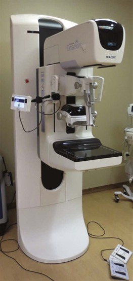
At our facility, noncalcified abnormalities detected by DBT are first evaluated by sonography for further characterization and biopsy planning. For lesions without an ultrasound correlate, we perform DBT-guided biopsy. All of our stereotactic biopsies, including biopsies of calcifications, are performed using DBT guidance as we do not have a separate conventional FFDM stereotactic biopsy unit. We have performed hundreds of successful stereotactic biopsies using DBT-guided biopsy.
Currently, both private-practice and academic radiology practices are pursuing DBT-guided biopsy technology. To assist other radiology practices, we review here the DBT-guided biopsy technique and describe a systematic approach to implementation of upright DBT-guided biopsy in radiology practices.
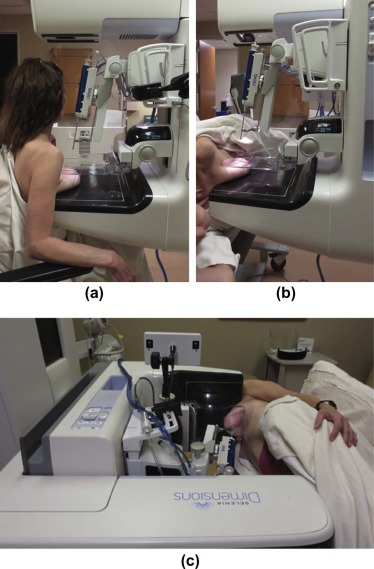
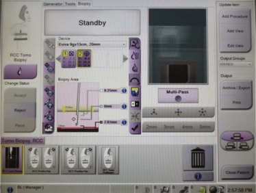
Technique for DBT-Guided Biopsy
Get Radiology Tree app to read full this article<
Get Radiology Tree app to read full this article<
Get Radiology Tree app to read full this article<
Get Radiology Tree app to read full this article<
Get Radiology Tree app to read full this article<
Steps Toward Implementation of DBT-guided Biopsy in a Radiology Practice
Get Radiology Tree app to read full this article<
Evaluation of Current Practice
Get Radiology Tree app to read full this article<
Get Radiology Tree app to read full this article<
Get Radiology Tree app to read full this article<
Product Evaluation and Acquisition
Get Radiology Tree app to read full this article<
Get Radiology Tree app to read full this article<
Get Radiology Tree app to read full this article<
Get Radiology Tree app to read full this article<
Personnel Training
Get Radiology Tree app to read full this article<
Get Radiology Tree app to read full this article<
Integration of DBT-guided Biopsy Into the Existing Workflow
Get Radiology Tree app to read full this article<
Get Radiology Tree app to read full this article<
Patient Selection for DBT-guided Biopsy
Get Radiology Tree app to read full this article<
Get Radiology Tree app to read full this article<
Get Radiology Tree app to read full this article<
Get Radiology Tree app to read full this article<
Get Radiology Tree app to read full this article<
Get Radiology Tree app to read full this article<
Get Radiology Tree app to read full this article<
Conclusions
Get Radiology Tree app to read full this article<
Get Radiology Tree app to read full this article<
References
1. Pettine S., Place R., Babu S., et. al.: Stereotactic breast biopsy is accurate, minimally invasive, and cost effective. Am J Surg 1996; 171: pp. 474-476.
2. Bruening W., Fontanarosa J., Tipton K., et. al.: Systematic review: comparative effectiveness of core-needle and open surgical biopsy to diagnose breast lesions. Ann Intern Med 2010; 152: pp. 238-246.
3. Bassett L., Winchester D.P., Caplan R.B., et. al.: Stereotactic core-needle biopsy of the breast: report of the Joint Task Force of the American College of Radiology, American College of Surgeons, and College of American Pathologists. CA Cancer J Clin 1997; 47: pp. 171-190.
4. Skaane P., Bandos A.I., Gullien R., et. al.: Prospective trial comparing full-field digital mammography (FFDM) versus combined FFDM and tomosynthesis in a population-based screening programme using independent double reading with arbitration. Eur Radiol 2013; 23: pp. 2061-2071.
5. Skaane P., Bandos A.I., Gullien R., et. al.: Comparison of digital mammography alone and digital mammography plus tomosynthesis in a population-based screening program. Radiology 2013; 267: pp. 47-56.
6. Park J.M., Franken E.A., Garg M., et. al.: Breast tomosynthesis: present considerations and future applications. Radiographics 2007; 27: pp. S231-S240.
7. Noroozian M., Hadjiiski L., Rahnama-Moghadam S., et. al.: Digital breast tomosynthesis is comparable to mammographic spot views for mass characterization. Radiology 2012; 262: pp. 61-68.
8. Andersson I., Ikeda D.M., Zackrisson S., et. al.: Breast tomosynthesis and digital mammography: a comparison of breast cancer visibility and BIRADS classification in a population of cancers with subtle mammographic findings. Eur Radiol 2008; 18: pp. 2817-2825.
9. Bernardi D., Ciatto S., Pellegrini M., et. al.: Prospective study of breast tomosynthesis as a triage to assessment in screening. Breast Cancer Res Treat 2012; 133: pp. 267-271.
10. Freer P.E., Niell B., Rafferty E.A.: Preoperative tomosynthesis-guided needle localization of mammographically and sonographically occult breast lesions. Radiology 2015; 275: pp. 377-383.
11. Vedantham S., Karellas A., Vijayaraghavan G.R., et. al.: Digital breast tomosynthesis: state of the art. Radiology 2015; 277: pp. 663-684.
12. Baker J.A., Lo J.Y.: Breast tomosynthesis: state-of-the-art and review of the literature. Acad Radiol 2011; 18: pp. 1298-1310.
13. Schrading S., Distelmaier M., Dirrichs T., et. al.: Digital breast tomosynthesis-guided vacuum-assisted breast biopsy: initial experiences and comparison with prone stereotactic vacuum-assisted biopsy. Radiology 2015; 274: pp. 654-662.
14. U.S. Food and Drug Administration : Frequently asked questions about DBT and MQSA training requirements. Available at http://www.fda.gov/Radiation-EmittingProducts/MammographyQualityStandardsActandProgram/FacilityCertificationandInspection/ucm447869.htm Accessed July 14, 2016
15. Viala J., Gignier P., Perret B., et. al.: Stereotactic vacuum-assisted biopsies on a digital breast 3D-tomosynthesis system. Breast J 2013; 19: pp. 4-9.