
Objectives
A computer-aided diagnosis (CAD) scheme for determining histological classifications of breast masses is expected to be useful for clinicians in making a differential diagnosis. The purpose of this study was to evaluate the usefulness of using the CAD scheme on ultrasonographic images.
Methods
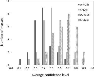
The database consisted of 390 breast ultrasonographic images with masses. Three experienced clinicians independently provided subjective ratings on the likelihood of malignancy for each of the 390 masses. Fifty benign masses (25 cysts and 25 fibroadenomas) and 50 malignant masses (25 noninvasive ductal carcinomas and 25 invasive ductal carcinomas) were selected as unknown cases for an observer study based on a stratified randomization method with the ratings. The likelihood of the histological classification in each unknown case was evaluated by the CAD scheme with image features that clinicians commonly use for describing masses. In the observer study, seven observers provided their confidence levels regarding the malignancy of the unknown case before and after viewing the likelihood of the histological classification. The usefulness of the CAD scheme was evaluated with a multireader multicase receiver operating characteristic (ROC) analysis.
Results
The areas under the ROC curves (AUCs) for all observers were improved by use of the CAD scheme. The average AUC increased from 0.716 without to 0.864 with the CAD scheme ( P = .006).
Conclusion
The presentation of the likelihood of the histological classification evaluated by the CAD scheme improved the clinicians’ performance and therefore would be useful in making a differential diagnosis of masses on ultrasonographic images.
Breast ultrasonography is thought to be more useful than mammography for detecting small breast cancers in dense breasts . However, introducing ultrasonography to breast cancer screening might result in a lower specificity and thereby increase the false positive rate . Tohno et al showed the rate of positive findings by ultrasonography to be 24%, and that it would decrease to 10% if simple cysts would be excluded from the positive findings . Therefore, ultrasonography may be able to achieve more effective breast cancer screening than mammography if clinicians can more accurately make a differential diagnosis on ultrasonographic images.
Computer-aided diagnosis (CAD) is one of the solutions for improving clinicians’ performance . CAD is a diagnostic method in which clinicians use the results analyzed by a computer as a “second opinion.” The usefulness of CAD at mammography has been shown on many studies. Jiang et al conducted observer studies for distinguishing between benign and malignant clustered microcalcifications with and without the computer output indicating the likelihood of malignancy. The average area under the receiver operating characteristic (ROC) curve (AUC) was thus found to increase from 0.61 to 0.75 by the computer aid ( P < .0001) . Timps et al showed the radiologists’ performances to improve significantly ( P < .05) when they used the computer output for the characterization of benign and malignant masses on mammograms using a temporal change analysis . Nakayama et al investigated the effect of presenting similar images on radiologists’ differential diagnosis of clustered microcalcifications on mammograms . The observer study found that the radiologists’ performance significantly increased with the use of similar images ( P = .0009).
Get Radiology Tree app to read full this article<
Materials and methods
Get Radiology Tree app to read full this article<
Case Selection
Get Radiology Tree app to read full this article<
Get Radiology Tree app to read full this article<
Get Radiology Tree app to read full this article<
Get Radiology Tree app to read full this article<
Observer Study
Get Radiology Tree app to read full this article<
Get Radiology Tree app to read full this article<
Get Radiology Tree app to read full this article<
Statistical Analysis
Get Radiology Tree app to read full this article<
Results
Get Radiology Tree app to read full this article<
Get Radiology Tree app to read full this article<
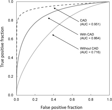
Table 1
AUCs for Clinicians in the Distinction between Benign and Malignant Masses with and without CAD
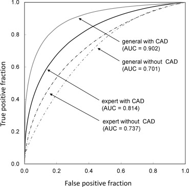
AUC Without CAD With CAD_P_ Expert A 0.787 0.853 .001 B 0.690 0.823 — C 0.733 0.765 .013Mean__0.7370.814.115 Surgeon D 0.625 0.955 — E 0.713 0.922 — F 0.728 0.861 .002 G 0.737 0.870 —Mean__0.7010.902.016 AllMean0.7160.874.002
Get Radiology Tree app to read full this article<
Get Radiology Tree app to read full this article<
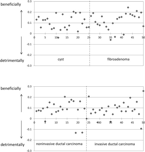
Table 2
Number of Cases in Which CAD had a Beneficial or Detrimental Effect
Beneficially Detrimentally Expert A 17 0 B 25 2 C 5 1Mean__4__2 Surgeon D 65 8 E 64 2 F 32 1 G 26 1Mean__66__3AllMean472
Get Radiology Tree app to read full this article<
Discussion
Get Radiology Tree app to read full this article<
Get Radiology Tree app to read full this article<
Get Radiology Tree app to read full this article<

Get Radiology Tree app to read full this article<
Get Radiology Tree app to read full this article<
Conclusion
Get Radiology Tree app to read full this article<
Appendix
Get Radiology Tree app to read full this article<
References
1. Crystal P., Strano S.D., Shcharynski S., et. al.: Using sonography to screen women with mammographically dense breasts. Am J Roentegenol AJR 2003; 181: pp. 177-182.
2. Tohno E., Ueno E., Watanabe H.: Ultrasound screening of breast cancer. Breast Cancer 2009; 16: pp. 18-22.
3. Berg W.A., Blume J.D., Cormack J.B., et. al.: Combined screening with ultrasound and mammography vs mammography alone in women at elevated risk of breast cancer. JAMA 2008; 299: pp. 2151-2163.
4. Ying X., Lin Y., Xia X., et. al.: A comparison of mammography and ultrasound in women with breast disease: a receiver operating characteristic analysis. Breast J 2012; 18: pp. 130-138. 10.1111/j.1524-4741.2011.01219.x
5. Tohno E., Ide S., Shibata K., et. al.: Occurrence of focal detection abnormalities in breast cancer screening by ultrasound. J Jpn Assoc Breast Cancer Screen 2000; 9: pp. 231-236.
6. Doi K.: Computer-aided diagnosis in medical imaging: historical review, current status and future potential. Comput Med Imaging Graph 2007; 31: pp. 198-211.
7. Jiang Y., Nishikawa R.M., Schmidt R.A., et. al.: Improving breast cancer diagnosis with computer-aided diagnosis. Acad Radiol 1999; 6: pp. 22-33.
8. Timp S., Varela C., Karssemeijer N.: Computer-aided diagnosis with temporal analysis to improve radiologists’ interpretation of mammographic mass lesions. IEEE Trans Inform Technol Biomed 2010; 14: pp. 803-808.
9. Nakayama R., Abe H., Shiraishi J., et. al.: Potential usefulness of similar images in the differential diagnosis of clustered microcalcifications on mammograms. Radiology 2009; 253: pp. 625-631.
10. Hizukuri A, Nakayama R, Kashikura Y, et al. Computer-aided diagnosis scheme for determining histological classification of breast masses using clinical features and texture features. Proc Fourth International Workshop on Regional Innovation Studies - Biomedical Engineering 2012;83–86.
11. Hizukuri A, Nakayama R, Kashikura Y, et al. Computerized quantification method for objective features based on clinicians’ subjective ratings of breast masses on ultrasonographic images. Proc Third International Workshop on Regional Innovation Studies -Biomedical Engineering 2011; 33–36.
12. Dorfman D.D., Berbaum K.S., Metz C.E.: Receiver operating characteristic rating analysis. Generalization to the population of readers and patients with the jackknife method. Invest Radiol 1992; 27: pp. 723-731.
13. Kobayashi T., Xu X.W., MacMahon H., et. al.: Effect of a computer-aided diagnosis scheme on radiologists’ performance in detection of lung nodules on radiographs. Radiology 1996; 199: pp. 843-848.
14. Uozumi T., Nakamura K., Watanabe H., et. al.: ROC analysis of detection of metastatic pulmonary nodules on digital chest radiographs with temporal subtraction. Acad Radiol 2001; 8: pp. 871-878.