
Rationale and Objectives
The aim of this study was to characterize early hepatic lipid changes in an experimental model of liver fibrosis using proton ( 1 H) magnetic resonance spectroscopy (MRS) at high magnetic field in vivo.
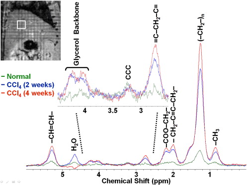
Materials and Methods
Liver fibrosis was induced in 12 Sprague-Dawley rats by twice-weekly carbon tetrachloride (CCl 4 ) administration up to 4 weeks. Eight normal rats were used as controls. Single-voxel 1 H MRS experiments were performed at 7 Tesla to measure signal integrals of various lipid peaks including –CH 3 , (–CH 2 –) n , –CH 2 –C=C–CH 2 –, =C–CH 2 –C= and –CH=CH– at 0.9, 1.3, 2.0, 2.8, and 5.3 ppm, respectively, and peak from choline-containing compounds (CCC) at 3.2 ppm. Total lipid, total saturated fatty acid, total unsaturated fatty acid, total unsaturated bond, polyunsaturated bond, and CCC indices were quantified.
Results
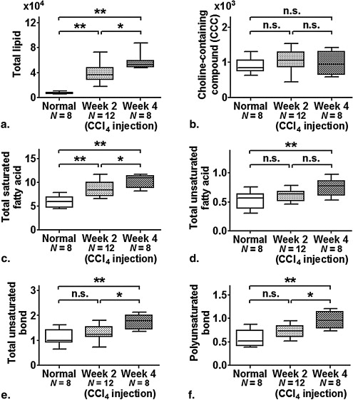
Significant increases ( P < .01) in total lipid and total saturated fatty acid indices were found in animals with CCl 4 -induced fibrosis as compared with normal animals. In addition, total unsaturated bond and polyunsaturated bond indices of animals at 4 weeks after CCl 4 insult were significantly higher than ( P < .01 and P < .05, respectively) those of normal animals and animals at 2 weeks following insult; whereas there was only significant increase ( P < .01) in total unsaturated fatty acid index in animals with 4-week CCl 4 insult as compared with normal animals.
Conclusion
The hepatic lipid changes in CCl 4 -induced experimental fibrosis model were documented in vivo and longitudinally using 1 H MRS at 7 Tesla. The experimental findings suggested that total saturated fatty acid increase contributed mainly to the total lipid increase in animals with CCl 4 insult. This study also demonstrated the potential value of high field MRS to resolve lipid composition and alterations in liver fibrosis.
Liver fibrosis associated with chronic liver injury can progress to cirrhosis and ultimately hepatocellular carcinoma (HCC) . Percutaneous liver biopsy has been considered the standard technique for diagnosis and staging of liver fibrosis. However, liver biopsy is highly invasive and associated with risk of complications, limiting its applicability in longitudinal monitoring of fibrosis progression or regression in response to treatment . Because disease progression to liver cirrhosis can be prevented by early interventions and treatments , there has been a great interest in the development of noninvasive techniques for early diagnosis and characterization of liver fibrosis. Proton magnetic resonance spectroscopy ( 1 H MRS) allows the study of cellular biochemistry and metabolism, and provides a noninvasive means to determine disease abnormalities and progression in vivo and longitudinally. Liver lipid content, which has been suggested to play an important pathogenic role in the development of liver fibrosis and cirrhosis in patients with chronic hepatitis C and nonalcoholic steatohepatitis , can be measured by 1 H MRS noninvasively. Most importantly, specific lipid changes related to saturated and unsaturated fatty acids can be monitored in vivo by high resolution 1 H MRS.
Carbon tetrachloride (CCl 4 ) intoxication is a well-characterized, reproducible and the most commonly used experimental animal model of liver fibrosis. It has been widely studied with respect to the histological, biochemical, cellular, and molecular changes associated with development of fibrosis . By interfering hepatic energy metabolism and protein synthesis, CCl 4 -induced hepatotoxicity can lead to triglyceride accumulation, mitochondrial injury, and necrosis . With the increased availability of high-field (≥3.0 Tesla) magnetic resonance (MR) systems for clinical and preclinical studies, both signal-to-noise ratio (SNR) and spectral resolution of metabolites in the MR spectra can be improved significantly , allowing more accurate metabolite identification and quantification and thus disease characterization. Although MRS can provide insights into liver metabolism noninvasively, detailed in vivo 1 H MRS study of liver fibrosis with high spectral resolution has been limited . The aim of this study was to characterize early hepatic lipid changes in the experimental CCl 4 -induced liver fibrosis model by means of single-voxel 1 H MRS at high magnetic field in vivo.
Materials and methods
Animal Preparation
Get Radiology Tree app to read full this article<
Get Radiology Tree app to read full this article<
In Vivo Liver 1 H MRS Experiments
Get Radiology Tree app to read full this article<
Data and Statistical Analysis
Get Radiology Tree app to read full this article<
Table 1
Peak Area Ratios of Various Metabolite Indices Measured by Proton Magnetic Resonance Spectroscopy ( 1 H MRS)
Index Peak Area Ratio Frequency Total lipid (–CH 2 –) n /noise 1.3 ppm Total saturated fatty acid 3(–CH 2 –)/2(–CH 3 ) 1.3/0.9 ppm Total unsaturated fatty acid 3(–CH 2 –C=C–CH 2 –)/4(–CH 3 ) 2.0/0.9 ppm Total unsaturated bond 3(–CH=CH–)/2(–CH 3 ) 5.3/0.9 ppm Polyunsaturated bond 3(=C–CH 2 –C=)/2(–CH 3 ) 2.8/0.9 ppm Choline-containing compound (CCC) CCC/noise 3.2 ppm
Get Radiology Tree app to read full this article<
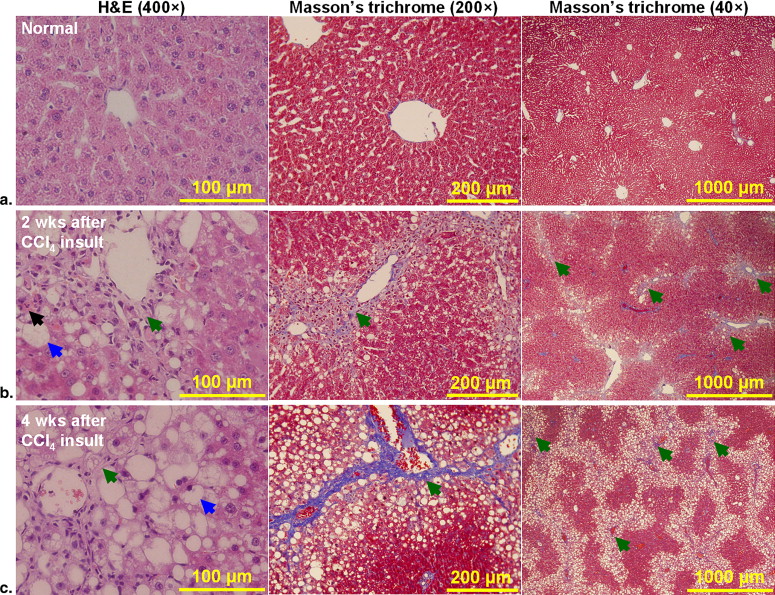
Histology
Get Radiology Tree app to read full this article<
Results
Get Radiology Tree app to read full this article<
Get Radiology Tree app to read full this article<
Get Radiology Tree app to read full this article<
Get Radiology Tree app to read full this article<
Discussion
Get Radiology Tree app to read full this article<
Get Radiology Tree app to read full this article<
Get Radiology Tree app to read full this article<
Get Radiology Tree app to read full this article<
Get Radiology Tree app to read full this article<
Get Radiology Tree app to read full this article<
References
1. Moradpour D., Wands J.R.: The molecular pathogenesis of hepatocellular carcinoma. J Viral Hepat 1994; 1: pp. 17-31.
2. Bataller R., Brenner D.A.: Liver fibrosis. J Clin Invest 2005; 115: pp. 209-218.
3. Manning D.S., Afdhal N.H.: Diagnosis and quantitation of fibrosis. Gastroenterology 2008; 134: pp. 1670-1681.
4. Wanless I.R., Nakashima E., Sherman M.: Regression of human cirrhosis. Morphologic features and the genesis of incomplete septal cirrhosis. Arch Pathol Lab Med 2000; 124: pp. 1599-1607.
5. Arthur M.J.: Reversibility of liver fibrosis and cirrhosis following treatment for hepatitis C. Gastroenterology 2002; 122: pp. 1525-1528.
6. Veldhuijzen I.K., Toy M., Hahne S.J., et. al.: Screening and early treatment of migrants for chronic hepatitis B virus infection is cost-effective. Gastroenterology 2010; 138: pp. 522-530.
7. Hourigan L.F., Macdonald G.A., Purdie D., et. al.: Fibrosis in chronic hepatitis C correlates significantly with body mass index and steatosis. Hepatology 1999; 29: pp. 1215-1219.
8. Castera L., Hezode C., Roudot-Thoraval F., et. al.: Worsening of steatosis is an independent factor of fibrosis progression in untreated patients with chronic hepatitis C and paired liver biopsies. Gut 2003; 52: pp. 288-292.
9. Wang D., Wei Y., Pagliassotti M.J.: Saturated fatty acids promote endoplasmic reticulum stress and liver injury in rats with hepatic steatosis. Endocrinology 2006; 147: pp. 943-951.
10. Gentile C.L., Pagliassotti M.J.: The role of fatty acids in the development and progression of nonalcoholic fatty liver disease. J Nutr Biochem 2008; 19: pp. 567-576.
11. Corbin I.R., Furth E.E., Pickup S., et. al.: In vivo assessment of hepatic triglycerides in murine non-alcoholic fatty liver disease using magnetic resonance spectroscopy. Biochim Biophys Acta 2009; 1791: pp. 757-763.
12. Tsukamoto H., Matsuoka M., French S.W.: Experimental models of hepatic fibrosis: a review. Semin Liver Dis 1990; 10: pp. 56-65.
13. Constandinou C., Henderson N., Iredale J.P.: Modeling liver fibrosis in rodents. Methods Mol Med 2005; 117: pp. 237-250.
14. Recknagel R.O.: Carbon tetrachloride hepatotoxicity. Pharmacol Rev 1967; 19: pp. 145-208.
15. Gruetter R., Weisdorf S.A., Rajanayagan V., et. al.: Resolution improvements in in vivo 1H NMR spectra with increased magnetic field strength. J Magn Reson 1998; 135: pp. 260-264.
16. Cho S.G., Kim M.Y., Kim H.J., et. al.: Chronic hepatitis: in vivo proton MR spectroscopic evaluation of the liver and correlation with histopathologic findings. Radiology 2001; 221: pp. 740-746.
17. Orlacchio A., Bolacchi F., Angelico M., et. al.: In vivo, high-field, 3-Tesla 1H MR spectroscopic assessment of liver fibrosis in HCV-correlated chronic liver disease. Radiol Med 2008; 113: pp. 289-299.
18. Hazle J.D., Narayana P.A., Dunsford H.A.: Chronic carbon tetrachloride and phospholipase D hepatotoxicity in rat: in vivo 1H magnetic resonance, total lipid analysis, and histology. Magn Reson Med 1990; 15: pp. 211-228.
19. Man K., Ng K.T., Lee T.K., et. al.: FTY720 attenuates hepatic ischemia-reperfusion injury in normal and cirrhotic livers. Am J Transplant 2005; 5: pp. 40-49.
20. Proctor E., Chatamra K.: Controlled induction of cirrhosis in the rat. Br J Exp Pathol 1983; 64: pp. 320-330.
21. Armendariz-Borunda J., Seyer J.M., Kang A.H., et. al.: Regulation of TGF beta gene expression in rat liver intoxicated with carbon tetrachloride. FASEB J 1990; 4: pp. 215-221.
22. Zeisberg M., Yang C., Martino M., et. al.: Fibroblasts derive from hepatocytes in liver fibrosis via epithelial to mesenchymal transition. J Biol Chem 2007; 282: pp. 23337-23347.
23. Cheung J.S., Guo H., Leung J.C., et. al.: MRI visualization of rodent liver structure and peritoneal adhesion with dialyzate enhancement. Magn Reson Med 2008; 59: pp. 1170-1174.
24. Cheung J.S., Fan S.J., Chow A.M., et. al.: In vivo DTI assessment of hepatic ischemia reperfusion injury in an experimental rat model. J Magn Reson Imaging 2009; 30: pp. 890-895.
25. Zhong K., Ernst T.: Localized in vivo human 1H MRS at very short echo times. Magn Reson Med 2004; 52: pp. 898-901.
26. Chan K.C., Khong P.L., Lau H.F., et. al.: Late measures of microstructural alterations in severe neonatal hypoxic-ischemic encephalopathy by MR diffusion tensor imaging. Int J Dev Neurosci 2009; 27: pp. 607-615.
27. Chan K.C., So K.F., Wu E.X.: Proton magnetic resonance spectroscopy revealed choline reduction in the visual cortex in an experimental model of chronic glaucoma. Exp Eye Res 2009; 88: pp. 65-70.
28. Hayakawa Y., Yoshioka Y., Yasuda N.: Effects of ligation and reperfusion of hepatic afferent vessels on the composition of liver cell membrane in the rat: 1H- and 31P-magnetic resonance spectroscopic analysis. NMR Biomed 1997; 10: pp. 257-262.
29. Lim A.K., Hamilton G., Patel N., et. al.: 1H MR spectroscopy in the evaluation of the severity of chronic liver disease. Radiology 2003; 226: pp. 288-289.
30. Zancanaro C., Nano R., Marchioro C., et. al.: Magnetic resonance spectroscopy investigations of brown adipose tissue and isolated brown adipocytes. J Lipid Res 1994; 35: pp. 2191-2199.
31. Kim H., Booth C.J., Pinus A.B., et. al.: Induced hepatic fibrosis in rats: hepatic steatosis, macromolecule content, perfusion parameters, and their correlations—preliminary MR imaging in rats. Radiology 2008; 247: pp. 696-705.
32. Wu E.X., Wu Y., Nicholls J.M., et. al.: MR diffusion tensor imaging study of postinfarct myocardium structural remodeling in a porcine model. Magn Reson Med 2007; 58: pp. 687-695.
33. Bruckner J.V., MacKenzie W.F., Muralidhara S., et. al.: Oral toxicity of carbon tetrachloride: acute, subacute, and subchronic studies in rats. Fundam Appl Toxicol 1986; 6: pp. 16-34.
34. Hazle J.D., Narayana P.A., Dunsford H.A.: In vivo NMR, biochemical, and histologic evaluation of alcohol-induced fatty liver in rat and a comparison with CCl4 hepatotoxicity. Magn Reson Med 1991; 19: pp. 124-135.
35. Stark D.D., Bass N.M., Moss A.A., et. al.: Nuclear magnetic resonance imaging of experimentally induced liver disease. Radiology 1983; 148: pp. 743-751.
36. de Vries J.E., Vork M.M., Roemen T.H., et. al.: Saturated but not mono-unsaturated fatty acids induce apoptotic cell death in neonatal rat ventricular myocytes. J Lipid Res 1997; 38: pp. 1384-1394.
37. Foley L.M., Towner R.A., Painter D.M.: In vivo image-guided (1)H-magnetic resonance spectroscopy of the serial development of hepatocarcinogenesis in an experimental animal model. Biochim Biophys Acta 2001; 1526: pp. 230-236.
38. Griffin J.L., Lehtimaki K.K., Valonen P.K., et. al.: Assignment of 1H nuclear magnetic resonance visible polyunsaturated fatty acids in BT4C gliomas undergoing ganciclovir-thymidine kinase gene therapy-induced programmed cell death. Cancer Res 2003; 63: pp. 3195-3201.
39. Ruiz-Cabello J., Cohen J.S.: Phospholipid metabolites as indicators of cancer cell function. NMR Biomed 1992; 5: pp. 226-233.
40. Menon D.K., Sargentoni J., Taylor-Robinson S.D., et. al.: Effect of functional grade and etiology on in vivo hepatic phosphorus-31 magnetic resonance spectroscopy in cirrhosis: biochemical basis of spectral appearances. Hepatology 1995; 21: pp. 417-427.
41. Corbin I.R., Buist R., Peeling J., et. al.: Hepatic 31P MRS in rat models of chronic liver disease: assessing the extent and progression of disease. Gut 2003; 52: pp. 1046-1053.
42. Lim A.K., Patel N., Hamilton G., et. al.: The relationship of in vivo 31P MR spectroscopy to histology in chronic hepatitis C. Hepatology 2003; 37: pp. 788-794.
43. Dezortova M., Taimr P., Skoch A., et. al.: Etiology and functional status of liver cirrhosis by 31P MR spectroscopy. World J Gastroenterol 2005; 11: pp. 6926-6931.
44. Soper R., Himmelreich U., Painter D., et. al.: Pathology of hepatocellular carcinoma and its precursors using proton magnetic resonance spectroscopy and a statistical classification strategy. Pathology 2002; 34: pp. 417-422.
45. Kuo Y.T., Li C.W., Chen C.Y., et. al.: In vivo proton magnetic resonance spectroscopy of large focal hepatic lesions and metabolite change of hepatocellular carcinoma before and after transcatheter arterial chemoembolization using 3.0-T MR scanner. J Magn Reson Imaging 2004; 19: pp. 598-604.
46. Li C.W., Kuo Y.C., Chen C.Y., et. al.: Quantification of choline compounds in human hepatic tumors by proton MR spectroscopy at 3 T. Magn Reson Med 2005; 53: pp. 770-776.
47. Towner R.A., Foley L.M., Painter D.M.: Hepatocarcinogenesis tumor grading correlated with in vivo image-guided 1H-NMR spectroscopy in a rat model. Toxicol Appl Pharmacol 2005; 207: pp. 237-244.
48. Zhao W.D., Guan S., Zhou K.R., et. al.: In vivo detection of metabolic changes by 1H-MRS in the DEN-induced hepatocellular carcinoma in Wistar rat. J Cancer Res Clin Oncol 2005; 131: pp. 597-602.
49. Xu H., Li X., Yang Z.H., et. al.: In vivo 1H MR spectroscopy in the evaluation of the serial development of hepatocarcinogenesis in an experimental rat model. Acad Radiol 2006; 13: pp. 1532-1537.
50. Le Page R.N., Cheeseman K.H., Osman N., et. al.: Lipid peroxidation in purified plasma membrane fractions of rat liver in relation to the hepatoxicity of carbon tetrachloride. Cell Biochem Funct 1988; 6: pp. 87-99.