
Rationale and Objectives
A reliable noninvasive method for in vivo detection of early therapeutic response of non-Hodgkin’s lymphoma (NHL) patients would be of great clinical value. This study evaluates the feasibility of 1 H and 31 P magnetic resonance spectroscopy (MRS) for in vivo detection of response to combination chemotherapy of human diffuse large B-cell lymphoma (DLCL2) xenografts in severe combined immunodeficient (SCID) mice.
Materials and Methods
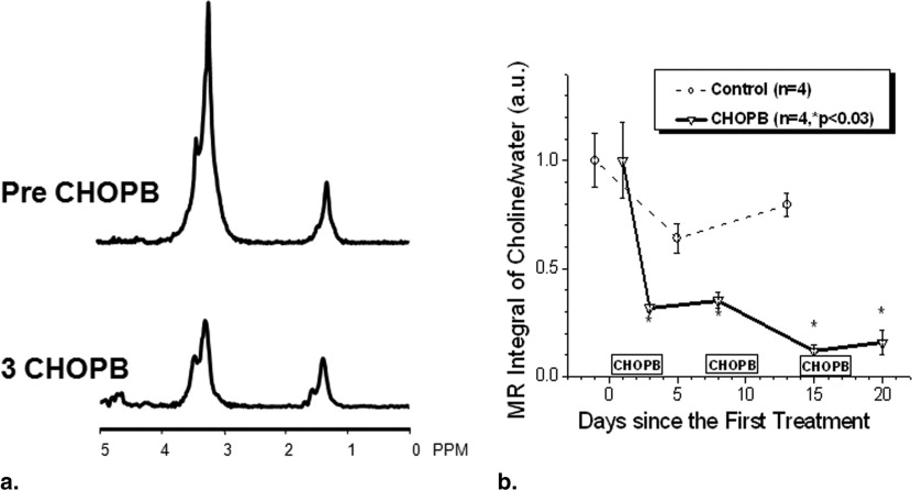
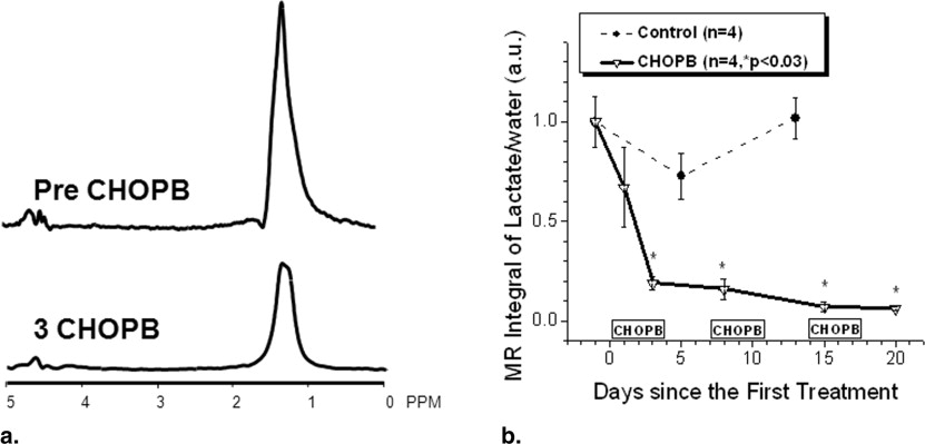
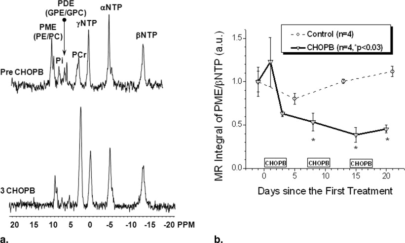
Combination chemotherapy with cyclophosphamide, hydroxy doxorubicin, Oncovin, prednisone, and bryostatin 1 (CHOPB) was administered to tumor-bearing SCID mice weekly for up to four cycles. Spectroscopic studies were performed before the initiation of treatment and after each cycle of the CHOPB. Proton MRS for detection of lactate and total choline was performed using a selective multiple-quantum-coherence-transfer (Sel-MQC) and a spin-echo–enhanced Sel-MQC (SEE-Sel-MQC) pulse sequence, respectively. Phosphorus-31 MRS using a nonlocalized, single-pulse sequence without proton decoupling was also performed on these animals.
Results
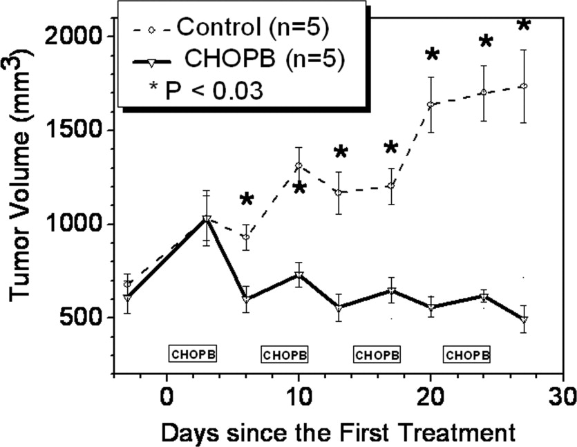
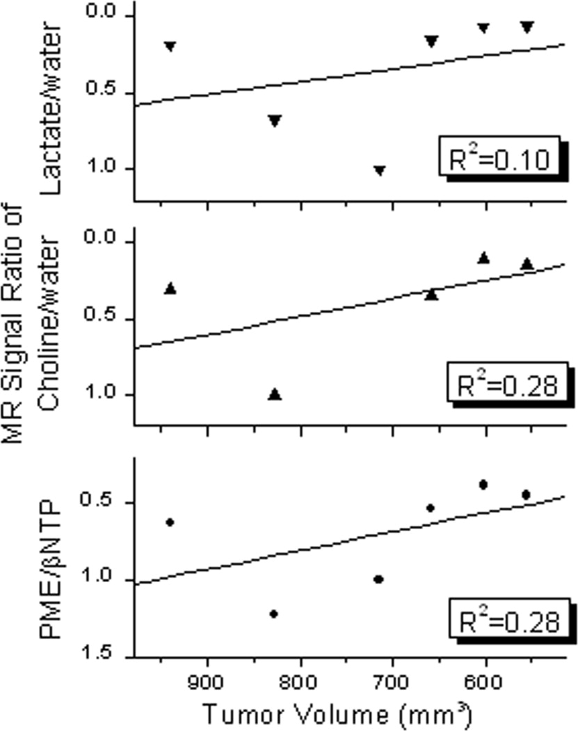
Significant decreases in lactate and total choline were detected in the DLCL2 tumors after one cycle of CHOPB chemotherapy. The ratio of phosphomonoesters to β-nucleoside triphosphate (PME/βNTP, measured by 31 P MRS) significantly decreased in the CHOPB-treated tumors after two cycles of CHOPB. The control tumors did not exhibit any significant changes in either of these metabolites.
Conclusions
This study demonstrates that 1 H and 31 P MRS can detect in vivo therapeutic response of NHL tumors and that lactate and choline offer a number of advantages over PMEs as markers of early therapeutic response.
Non-Hodgkin’s lymphoma (NHL) comprises a heterogeneous group of closely related malignancies of the lymphoid system in which the cells usually express either B-cell or T-cell markers, or both ( ). This disease results from disruption of normal development of the hematopoietic system at a precursor stage, probably because of immune deficiency, chronic inflammation, and chronic infection ( ). The incidence of NHL has been steeply rising (eg, by 3% per annum in the United States), increasing by 90% in the past 50 years ( ). An estimated 58,870 new cases of NHL are diagnosed in the United States annually. This disease ranks fifth and sixth in prevalence among cancers, and seventh and eighth as a cause of cancer death among females and males, respectively ( ); however, in terms of prevalence and numbers of cancer deaths, it affects males more than females and Caucasians more than other races. Furthermore, NHL affects the younger and middle-aged population and is the leading cause of cancer-related death among people between 20 and 40 years of age; it ranks fourth among all cancers in terms of total number of productive years lost ( ).
Only one-third of NHL cases are curable by standard chemotherapy ( ). The availability of noninvasive methods for prediction or early detection of therapeutic response of NHL tumors would be of considerable clinical value. Such methods would facilitate the rational design and individualization of therapy protocols. This would spare nonresponsive patients the unnecessary toxicity and expense of ineffective therapy and would offer them opportunities to explore more effective alternative treatment at an earlier time. However, an effective method of detecting early response of NHL tumors to the wide range of therapeutic agents available for treatment of this disease remains elusive because sensitive and specific markers of therapeutic response are still not available.
Get Radiology Tree app to read full this article<
Get Radiology Tree app to read full this article<
Get Radiology Tree app to read full this article<
Get Radiology Tree app to read full this article<
Materials and methods
Cell Line and Tumor Implantation
Get Radiology Tree app to read full this article<
Tumor Treatment
Get Radiology Tree app to read full this article<
Get Radiology Tree app to read full this article<
1 H and 31 P MRS Studies
Get Radiology Tree app to read full this article<
Get Radiology Tree app to read full this article<
Get Radiology Tree app to read full this article<
Get Radiology Tree app to read full this article<
Get Radiology Tree app to read full this article<
Statistical Analysis
Get Radiology Tree app to read full this article<
Results
Tumor Growth Delay
Get Radiology Tree app to read full this article<
Get Radiology Tree app to read full this article<
1 H MRS of Total Choline and Lactate
Get Radiology Tree app to read full this article<
Get Radiology Tree app to read full this article<
31 P MRS of DLCL2 Tumors
Get Radiology Tree app to read full this article<
Get Radiology Tree app to read full this article<
Discussions
Get Radiology Tree app to read full this article<
Get Radiology Tree app to read full this article<
Get Radiology Tree app to read full this article<
Get Radiology Tree app to read full this article<
Get Radiology Tree app to read full this article<
Get Radiology Tree app to read full this article<
Acknowledgments
Get Radiology Tree app to read full this article<
References
1. Ansell S.M., Armitage J.: Non-Hodgkin lymphoma: diagnosis and treatment. Mayo Clin Proc 2005; 80: pp. 1087-1097.
2. Rogers B.B.: Overview of non-Hodgkin’s lymphoma. Semin Oncol Nurs 2006; 22: pp. 67-72.
3. Zinzani P.L.: Lymphoma: diagnosis, staging, natural history, and treatment strategies. Semin Oncol 2005; 32: pp. S4-S10.
4. Jazirehi A.R., Bonavida B.: Cellular and molecular signal transduction pathways modulated by rituximab (rituxan, anti-CD20mAb) in non-Hodgkin’s lymphoma: implications in chemosensitization and therapeutic intervention. Oncogene 2005; 24: pp. 2121-2143.
5. American Cancer Society: Cancer facts & figures 2002.2002.American Cancer SocietyAtlanta
6. American Cancer Society: Cancer facts & figures 2006.2006.American Cancer SocietyAtlanta
7. Fisher R.: Cyclophosphamide, doxorubicin, vincristine, and prednisone versus intensive chemotherapy in non-Hodgkin’s lymphoma. Cancer Chemother Pharmacol 1997; 40: pp. S42-S46.
8. Juweid M.E., Stroobants S., Hoekstra O.S., et. al.: Use of positron emission tomography for response assessment of lymphoma: consensus of the Imaging Subcommittee of International Harmonization Project in Lymphoma. J Clin Oncol 2007; 25: pp. 571-578.
9. Glickson J.D.: Clinical NMR-spectroscopy of tumors—current status and future-directions. Invest Radiol 1989; 24: pp. 1011-1016.
10. Ng T.C., Evanochko W.T., Hiramoto R.N., et. al.: P-31 NMR-spectroscopy of in vivo tumors. J Magn Reson 1982; 49: pp. 271-286.
11. Li S.J., Wehrle J.P., Rajan S.S., et. al.: Response of radiation-induced fibrosarcoma-1 in mice to cyclophosphamide monitored by in vivo P-31 nuclear magnetic-resonance spectroscopy. Cancer Res 1988; 48: pp. 4736-4742.
12. Wehrle J.P., Li S.J., Rajan S.S., Steen R.G., et. al.: P-31 and H-1-NMR spectroscopy of tumors in vivo—untreated growth and response to chemotherapy. Ann NY Acad Sci 1987; 508: pp. 200-215.
13. Braunschweiger P.G., Kumar N., Constantinidis I., et. al.: Potentiation of interleukin 1-alpha mediated antitumor effects by ketoconazole. Cancer Res 1990; 50: pp. 4709-4717.
14. Shungu D.C., Bhujwalla Z.M., Wehrle J.P., et. al.: H-1-NMR spectroscopy of subcutaneous tumors in mice—preliminary studies of effects of growth, chemotherapy and blood-flow reduction. NMRBiomed 1992; 5: pp. 296-302.
15. Bhujwalla Z.M., Glickson J.D.: Detection of tumor response to radiation therapy by in vivo proton MR spectroscopy. Int J Radiat Oncol Biol Phys 1996; 36: pp. 635-639.
16. Griffiths J.R., Tate A.R., Howe F.A., et. al., The Multi-Institutional Group on MRS Application to Cancer: Magnetic resonance spectroscopy of cancer—practicalities of multi-centre trials and early results in non-Hodgkin’s lymphoma. Eur J Cancer 2002; 38: pp. 2085-2093.
17. Zakian K.L., Koutcher J.A.: Magnetic resonance spectroscopy and clinical cancer prognosis. Acad Radiol 2004; 11: pp. 365-367.
18. Sibtaina N.A., Howeb F.A., Saundersc D.E.: The clinical value of proton magnetic resonance spectroscopy in adult brain tumours. Clin Radiol 2007; 62: pp. 109-119.
19. He Q., Shungu D.C., van Zijl P.C., et. al.: Single-scan in vivo lactate editing with complete lipid and water suppression by selective multiple-quantum-coherence transfer (Sel-MQC)with application to tumors. J Magn Reson B 1995; 106: pp. 203-211.
20. Evanochko W.T., Sakai T.T., Ng T.C., et. al.: NMR-study of in vivo Rif-1 tumors—analysis of perchloric-acid extracts and identification of H-1-resonances, P-31-resonances and C-13-resonances. Biochim Biophys Acta 1984; 805: pp. 104-116.
21. Evelhoch J.L., Sapareto S.A., Nussbaum G.H., et. al.: Correlations between P-31 NMR-spectroscopy and O-15 perfusion measurements in the Rif-1 murine tumor in vivo. Radiat Res 1986; 106: pp. 122-131.
22. Negendank W.G., Padavicshaller K.A., Li C.W., et. al.: Metabolic characterization of human non-Hodgkins-lymphomas in-vivo with the use of proton-decoupled phosphorus magnetic-resonance spectroscopy. Cancer Res 1995; 55: pp. 3286-3294.
23. McPhail L.D., Chung Y.-L., Madhu B., et. al.: Tumor dose response to the vascular disrupting agent,5,6-dimethylxanthenone-4-acetic acid, using in vivo magnetic resonance spectroscopy. Clin Cancer Res 2005; 11: pp. 3705-3713.
24. Arias-Mendoza F., Smith M.R., Brown T.R.: Predicting treatment response in non-Hodgkin’s lymphoma from the pretreatment tumor content of phosphoethanolamine plus phosphocholine. Acad Radiol 2004; 11: pp. 368-376.
25. Bourne R.M., Stanwell P., Stretch J.R., et. al.: In vivo and ex vivo proton MR spectroscopy of primary and secondary melanoma. Eur J Radiol 2005; 53: pp. 506-513.
26. Chang L., Miller B.L., McBride D., et. al.: Brain lesions in patients with AIDS: H-1 MR spectroscopy. Radiology 1995; 197: pp. 525-531.
27. Panigrahy A., Krieger M.D., Gonzalez-Gomez I., et. al.: Untreated pediatric primitive neuroectodermal tumor in vivo: quantitation of taurine with MR Spectroscopy. Radiology 2005; 236: pp. 1020-1025.
28. Star-Lack J., Spielman D., Adalsteinsson E., et. al.: In vivo lactate editing with simultaneous detection of choline, creatine, NAA, and lipid singlets at 1.5 T using PRESS excitation with applications to the study of brain and head and neck tumors. J Magn Reson B 1998; 133: pp. 243-254.
29. Kurhanewicz J., Vigneron D.B., Hricak H., et. al.: Three-dimensional H-1 MR spectroscopic imaging of the in situ human prostate with high (0.24–0.7cm 3 ) spatial resolution. Radiology 1996; 198: pp. 795-805.
30. Stanwell P., Gluch L., Clark D., et. al.: Specificity of choline metabolites for in vivo diagnosis of breast cancer using 1H MRS at 1.5 T. Eur Radiol 2005; 15: pp. 1037-1043.
31. Mackinnon W.B., Barry P.A., Malycha P.L., et. al.: Fine-needle biopsy specimens of benign breast lesions distinguished from invasive cancer ex vivo with proton MR spectroscopy. Radiology 1997; 204: pp. 661-666.
32. Dixon R.M., Frahm J.: Localized proton MR spectroscopy of the human kidney in vivo by means of short echo time STEAM sequences. Magn Reson Med 1994; 31: pp. 482-487.
33. Tyszka J.M., Silverman J.M.: Navigated single-voxel proton spectroscopy of the human liver. Magn Reson Med 1998; 39: pp. 1-5.
34. Mohammad R.M., Wall N.R., Dutcher J.A., et. al.: The addition of Bryostatin 1 to cyclophosphamide, doxorubicin, vincristine, and prednisone (CHOP) chemotherapy improves response in a CHOP-resistant human diffuse large cell lymphoma xenograft model. Clin Cancer Res 2000; 6: pp. 4950-4956.
35. Al-Katib A.M., Smith M.R., Kamanda W.S., et. al.: Bryostatin 1 down-regulates mdr1 and potentiates vincristine cytotoxicity in diffuse large cell lymphoma xenografts. Clin Cancer Res 1998; 4: pp. 1305-1314.
36. He Q., Shungu D.C., van Zijl P.C., et. al.: Single-scan in vivo lactate editing with complete lipid and water suppression by selective multiple-quantum-coherence transfer (Sel-MQC) with application to tumors. J Magn Reson B 1995; 106: pp. 203-211.
37. He Q., Bhujwalla Z.M., Glickson J.D.: Proton detection of choline and lactate in EMT6 tumors by spin-echo-enhanced selective multiple-quantum-coherence transfer. J Magn Reson B 1996; 112: pp. 18-25.
38. Ackerstaff E., Glunde K., Bhujwalla Z.M.: Choline phospholipid metabolism: a target in cancer cells?. J Cell Biochem 2003; 90: pp. 525-533.
39. Dyke J.P., Zakian K.L., Spees W.M., et. al.: Metabolic response of the CWR22 prostate tumor xenograft after 20 Gy of radiation studied by 1H spectroscopic imaging. Clin Cancer Res 2003; 9: pp. 4529-4536.
40. Jagannathan N.R., Kumar M., Seenu V., et. al.: Evaluation of total choline from in-vivo volume localized proton MR spectroscopy and its response to neoadjuvant chemotherapy in locally advanced breast cancer. Br J Cancer 2001; 84: pp. 1016-1022.
41. Bhujwalla Z.M., Shungu D.C., Glickson J.D.: Effects of blood flow modifiers on tumor metabolism observed in vivo by proton magnetic resonance spectroscopic imaging. Magn Reson Med 1996; 36: pp. 204-211.
42. Madhu B., Waterton J.C., Griffiths J.R., et. al.: The response of RIF-1 fibrosarcomas to the vascular-disrupting agent ZD6126 assessed by in vivo and ex vivo 1H magnetic resonance spectroscopy. Neoplasia 2006; 8: pp. 560-567.
43. Warburg O.: On the origin of cancer cells. Science 1956; 123: pp. 309-314.
44. Bhujwalla Z.M., Glickson J.D.: Detection of tumor response to radiation therapy by in vivo proton MR spectroscopy. Int J Radiat Oncol Biol Phys 1996; 36: pp. 635-639.
45. Poptani H., Bansal N., Graham R.A., et. al.: Detecting early response to cyclophosphamide treatment of RIF-1 tumors using selective multiple quantum spectroscopy (SelMQC) and dynamic contrast enhanced imaging. NMR Biomed 2003; 16: pp. 102-111.
46. Aboagye E.O., Bhujwalla Z.M., Shungu D.C., et. al.: Detection of tumor response to chemotherapy by 1H nuclear magnetic resonance spectroscopy: effect of 5-fluorouracil on lactate levels in radiation-induced fibrosarcoma 1 tumors. Cancer Res 1998; 58: pp. 1063-1067.
47. Aboagye E.O., Bhujwalla Z.M., He Q., et. al.: Evaluation of lactate as a 1H nuclear magnetic resonance spectroscopy index for noninvasive prediction and early detection of tumor response to radiation therapy in EMT6 tumors. Radiat Res 1998; 150: pp. 38-42.
48. Artemov D., Bhujwalla Z.M., Pilatus U., et. al.: Two-compartment model for determination of glycolytic rates of solid tumors by in vivo 13C NMR spectroscopy. NMR Biomed 1998; 11: pp. 395-404.
49. Poptani H., Bansal N., Jenkins W.T., et. al.: Cyclophosphamide treatment modifies tumor oxygenation and glycolytic rates of RIF-1 tumors: 13C magnetic resonance spectroscopy, Eppendorf electrode, and redox scanning. Cancer Res 2003; 63: pp. 8813-8820.
50. Rivenzon-Segal D., Margalit R., Degani H.: Glycolysis as a metabolic marker in orthotopic breast cancer, monitored by in vivo (13)C MRS. Am J Physiol Endocrinol Metab 2002; 283: pp. E623-E630.
51. Negendank W.: Studies of human tumors by MRS: a review. NMR Biomed 1992; 5: pp. 303-324.
52. Kumar M., Jagannathan N.R., Seenu V., et. al.: Monitoring the therapeutic response of locally advanced breast cancer patients: sequential in vivo proton MR spectroscopy study. J Magn Reson Imaging 2006; 24: pp. 325-332.