
Rationale and Objectives
Hepatic ischemia/reperfusion injury (IRI) occurs during certain hepatobiliary surgeries, hemorrhagic shock, and veno-occlusive disease. Biochemical changes caused by hepatic IRI lead to hepatocellular remodeling, including cellular regeneration or irreversible apoptosis. This study aims to characterize and monitor the metabolic changes in hepatic IRI using proton magnetic resonance spectroscopy ( 1 H MRS).
Materials and Methods
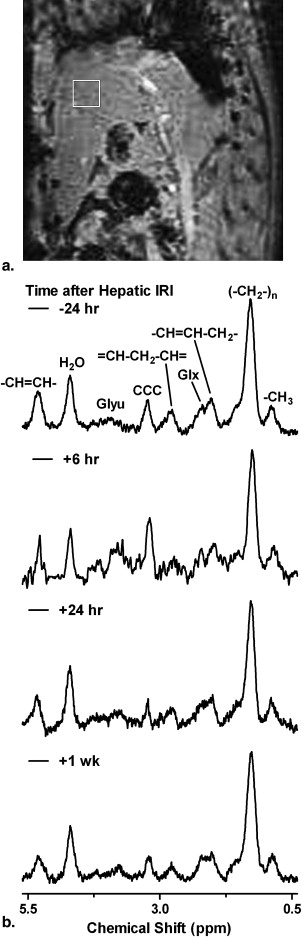
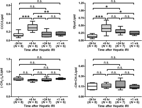
Sprague-Dawley rats ( n = 8) were scanned with 1 H MRS using 5.0 × 5.0 × 5.0 mm 3 voxel over a homogeneous liver parenchyma at 7 Tesla with a respiratory-gated point-resolved spectroscopy sequence at 1 day before, 6 hours, 1 day, and 1 week after 30 minutes total hepatic IRI. Signal integral ratios of choline-containing compounds (CCC), glycogen and glucose complex (Glyu), methylene proton ((-CH 2 -) n ), and methene proton (-CH=CH-) to lipid (integral sum of methyl proton (-CH 3 ), (-CH 2 -) n and -CH=CH-) were quantified by areas under peaks longitudinally.
Results
The CCC-to-lipid and Glyu-to-lipid ratios at 6 hours after IRI were significantly higher than those at 1 day before, 1 day, and 1 week after injury. The (-CH 2 -) n -to-lipid, and -CH=CH-to-lipid ratios showed no significant differences over different time points. Hepatocellular regeneration was observed at 6 hours after IRI in histology with immunohistochemical technique.
Conclusions
Changes in CCC-to-lipid and Glyu-to-lipid ratios likely reflect the hepatocellular remodeling and impaired glucose utilization upon hepatic IRI, respectively. The experimental findings in the current study demonstrated that 1 H MRS is a valuable tool for characterizing either global or regional metabolic changes in liver noninvasively and longitudinally. Such capability has the potential to lead to early diagnosis and detection of impaired liver function.
Hepatic ischemia/reperfusion injury (IRI) induced by vascular complications contributes to early organ failure and can lead to acute and chronic rejection after liver transplantation . Hepatic IRI also occurs during certain hepatobiliary resectional surgeries, hemorrhagic shock, and veno-occlusive disease. IRI is a major cause of acute liver failure, which is associated with high morbidity and mortality . Cellular damage can be induced not only during ischemia but also in reperfusion that follows, resulting in both local and systemic organ dysfunction . Biochemical changes caused by hepatic IRI lead to hepatocellular remodeling, including cellular regeneration or irreversible programmed cell death . Early diagnosis and detection of impaired liver function is vital for early and effective therapeutic interventions and thus prevents its progression to liver failure. Through assessing liver function, serology screenings of liver-specific transaminases (aspartate aminotransferase and alanine aminotransferase) have been widely used to monitor hepatic IRI .
Metabolic changes of liver can provide information and improve the characterization of liver abnormalities . Monitoring of liver metabolism through a microdialysis catheter placed in the liver tissue revealed profound metabolic changes during and after IRI, leading to potential evaluation of ischemic preconditioning and clinical application . However, microdialysis technique is invasive in nature because implantation of microdialysis probe needs to be accompanied by surgical procedures . Recently, proton magnetic resonance spectroscopy ( 1 H MRS) has been increasingly employed to investigate liver metabolism in vivo noninvasively in various diseases . Biochemical changes occurred during hepatic IRI were also investigated in liver extracts . However, in vivo and serial study of hepatic IRI model with such MRS technique has been limited.
Get Radiology Tree app to read full this article<
Materials and methods
Get Radiology Tree app to read full this article<
Animal preparation
Get Radiology Tree app to read full this article<
MR spectroscopy
Get Radiology Tree app to read full this article<
Data and statistical analysis
Get Radiology Tree app to read full this article<
Histology
Get Radiology Tree app to read full this article<
Results
Get Radiology Tree app to read full this article<
Get Radiology Tree app to read full this article<
Get Radiology Tree app to read full this article<
Get Radiology Tree app to read full this article<
Get Radiology Tree app to read full this article<

Get Radiology Tree app to read full this article<
Discussions
Get Radiology Tree app to read full this article<
Get Radiology Tree app to read full this article<
Get Radiology Tree app to read full this article<
Get Radiology Tree app to read full this article<
Get Radiology Tree app to read full this article<
Get Radiology Tree app to read full this article<
Get Radiology Tree app to read full this article<
Get Radiology Tree app to read full this article<
References
1. Kupiec-Weglinski J.W., Busuttil R.W.: Ischemia and reperfusion injury in liver transplantation. Transplant Proc 2005; 37: pp. 1653-1656.
2. Farmer D.G., Amersi F., Kupiec-Weglinski J., et. al.: Current status of ischemia and reperfusion injury in the liver. Transplant Rev 2000; 14: pp. 106-126.
3. Lentsch A.B., Yoshidome H., Cheadle W.G., et. al.: Chemokine involvement in hepatic ischemia/reperfusion injury in mice: roles for macrophage inflammatory protein-2 and KC. Hepatology 1998; 27: pp. 1172-1177.
4. Klaus S., Heringlake M., Gliemroth J., et. al.: Biochemical tissue monitoring during hypoxia and reoxygenation. Resuscitation 2003; 56: pp. 299-305.
5. Schlossberg H., Zhang Y., Dudus L., et. al.: Expression of c-fos and c-jun during hepatocellular remodeling following ischemia/reperfusion in mouse liver. Hepatology 1996; 23: pp. 1546-1555.
6. Lee S.H., Culberson C., Korneszczuk K., et. al.: Differential mechanisms of hepatic vascular dysregulation with mild vs. moderate ischemia-reperfusion. Am J Physiol Gastrointest Liver Physiol 2008; 294: pp. G1219-G1226.
7. Toledo-Pereyra L.H., Suzuki S.: Cellular and biomolecular mechanisms of liver ischemia and reperfusion injury. Transplant Proc 1994; 26: pp. 325-327.
8. Shaked A., Nunes F.A., Olthoff K.M., et. al.: Assessment of liver function: pre- and peritransplant evaluation. Clin Chem 1997; 43: pp. 1539-1545.
9. Kannerup A.S., Gronbaek H., Funch-Jensen P., et. al.: The influence of preconditioning on metabolic changes in the pig liver before, during, and after warm liver ischemia measured by microdialysis. Hepatol Int 2009; 3: pp. 310-315.
10. Kannerup A.S., Funch-Jensen P., Gronbaek H., et. al.: Metabolic changes in the pig liver during warm ischemia and reperfusion measured by microdialysis. J Gastrointest Surg 2008; 12: pp. 319-326.
11. Brunner M., Langer O.: Microdialysis versus other techniques for the clinical assessment of in vivo tissue drug distribution. AAPS J 2006; 8: pp. E263-E271.
12. Cho S.G., Kim M.Y., Kim H.J., et. al.: Chronic hepatitis: in vivo proton MR spectroscopic evaluation of the liver and correlation with histopathologic findings. Radiology 2001; 221: pp. 740-746.
13. Li C.W., Kuo Y.C., Chen C.Y., et. al.: Quantification of choline compounds in human hepatic tumors by proton MR spectroscopy at 3 T. Magn Reson Med 2005; 53: pp. 770-776.
14. Towner R.A., Foley L.M., Painter D.M.: Hepatocarcinogenesis tumor grading correlated with in vivo image-guided 1H-NMR spectroscopy in a rat model. Toxicol Appl Pharmacol 2005; 207: pp. 237-244.
15. Xu H., Li X., Yang Z.H., et. al.: In vivo 1H MR spectroscopy in the evaluation of the serial development of hepatocarcinogenesis in an experimental rat model. Acad Radiol 2006; 13: pp. 1532-1537.
16. Zhao W.D., Guan S., Zhou K.R., et. al.: In vivo detection of metabolic changes by 1H-MRS in the DEN-induced hepatocellular carcinoma in Wistar rat. J Cancer Res Clin Oncol 2005; 131: pp. 597-602.
17. Fischbach F., Bruhn H.: Assessment of in vivo 1H magnetic resonance spectroscopy in the liver: a review. Liver Int 2008; 28: pp. 297-307.
18. Hayakawa Y., Yoshioka Y., Yasuda N.: Effects of ligation and reperfusion of hepatic afferent vessels on the composition of liver cell membrane in the rat: 1H- and 31P-magnetic resonance spectroscopic analysis. NMR Biomed 1997; 10: pp. 257-262.
19. Nishida T., Ueshima S., Kazuo H., et. al.: Vagus nerve is involved in lack of blood reflow into sinusoids after rat hepatic ischemia. Am J Physiol Heart Circ Physiol 2000; 278: pp. H1565-H1570.
20. Cheung J.S., Fan S.J., Chow A.M., et. al.: In vivo DTI assessment of hepatic ischemia reperfusion injury in an experimental rat model. J Magn Reson Imaging 2009; 30: pp. 890-895.
21. Cheung J.S., Guo H., Leung J.C., et. al.: MRI visualization of rodent liver structure and peritoneal adhesion with dialyzate enhancement. Magn Reson Med 2008; 59: pp. 1170-1174.
22. Miyasaka N., Takahashi K., Hetherington H.P.: Fully automated shim mapping method for spectroscopic imaging of the mouse brain at 9.4 T. Magn Reson Med 2006; 55: pp. 198-202.
23. Chan K.C., So K.F., Wu E.X.: Proton magnetic resonance spectroscopy revealed choline reduction in the visual cortex in an experimental model of chronic glaucoma. Exp Eye Res 2009; 88: pp. 65-70.
24. Chan K.C., Khong P.L., Cheung M.M., et. al.: MRI of late microstructural and metabolic alterations in radiation-induced brain injuries. J Magn Reson Imaging 2009; 29: pp. 1013-1020.
25. Chan K.W., Chow A.M., Chan K.C., et. al.: Magnetic resonance spectroscopy of the brain under mild hypothermia indicates changes in neuroprotection-related metabolites. Neurosci Lett 2010; 475: pp. 150-155.
26. Bell J.D., Cox I.J., Sargentoni J., et. al.: A 31P and 1H-NMR investigation in vitro of normal and abnormal human liver. Biochim Biophys Acta 1993; 1225: pp. 71-77.
27. Tarasow E., Siergiejczyk L., Panasiuk A., et. al.: MR proton spectroscopy in liver examinations of healthy individuals in vivo. Med Sci Monit 2002; 8: pp. MT36-MT40.
28. Perman W.H., Balci N.C., Akduman I.: Review of magnetic resonance spectroscopy in the liver and the pancreas. Top Magn Reson Imaging 2009; 20: pp. 89-97.
29. Finkelstein S.D., Gilfor D., Farber J.L.: Alterations in the metabolism of lipids in ischemia of the liver and kidney. J Lipid Res 1985; 26: pp. 726-734.
30. Zhong Z., Schwabe R.F., Kai Y., et. al.: Liver regeneration is suppressed in small-for-size liver grafts after transplantation: involvement of c-Jun N-terminal kinase, cyclin D1, and defective energy supply. Transplantation 2006; 82: pp. 241-250.
31. Camargo C.A., Madden J.F., Gao W., et. al.: Interleukin-6 protects liver against warm ischemia/reperfusion injury and promotes hepatocyte proliferation in the rodent. Hepatology 1997; 26: pp. 1513-1520.
32. Corbin I.R., Buist R., Peeling J., et. al.: Hepatic 31P MRS in rat models of chronic liver disease: assessing the extent and progression of disease. Gut 2003; 52: pp. 1046-1053.
33. Lim A.K., Patel N., Hamilton G., et. al.: The relationship of in vivo 31P MR spectroscopy to histology in chronic hepatitis C. Hepatology 2003; 37: pp. 788-794.
34. Dezortova M., Taimr P., Skoch A., et. al.: Etiology and functional status of liver cirrhosis by 31P MR spectroscopy. World J Gastroenterol 2005; 11: pp. 6926-6931.
35. Wehmeyer N., Gunderson H., Nauman J., et. al.: Determination of the glycogen synthesis pathway by 13C nuclear magnetic resonance analysis. Metabolism 1994; 43: pp. 38-43.
36. Cohen S.M.: 13C NMR study of effects of fasting and diabetes on the metabolism of pyruvate in the tricarboxylic acid cycle and the utilization of pyruvate and ethanol in lipogenesis in perfused rat liver. Biochemistry 1987; 26: pp. 581-589.
37. Reo N.V., Siegfried B.A., Ackerman J.J.: Direct observation of glycogenesis and glucagon-stimulated glycogenolysis in the rat liver in vivo by high-field carbon-13 surface coil NMR. J Biol Chem 1984; 259: pp. 13664-13667.
38. Di Costanzo A., Trojsi F., Tosetti M., et. al.: High-field proton MRS of human brain. Eur J Radiol 2003; 48: pp. 146-153.
39. Dixon R.M.: NMR studies of phospholipid metabolism in hepatic lymphoma. NMR Biomed 1998; 11: pp. 370-379.
40. Cao Z., Wu L.P., Li Y.X., et. al.: Change of choline compounds in sodium selenite-induced apoptosis of rats used as quantitative analysis by in vitro 9.4T MR spectroscopy. World J Gastroenterol 2008; 14: pp. 3891-3896.
41. Ruiz-Cabello J., Cohen J.S.: Phospholipid metabolites as indicators of cancer cell function. NMR Biomed 1992; 5: pp. 226-233.
42. Yen C.L., Mar M.H., Meeker R.B., et. al.: Choline deficiency induces apoptosis in primary cultures of fetal neurons. FASEB J 2001; 15: pp. 1704-1710.
43. Martin K.: Concentrative accumulation of choline by human erythrocytes. J Gen Physiol 1968; 51: pp. 497-516.
44. Glinsmann W.H., Hern E.P., Lynch A.: Intrinsic regulation of glucose output by rat liver. Am J Physiol 1969; 216: pp. 698-703.
45. Moore M.C., Connolly C.C., Cherrington A.D.: Autoregulation of hepatic glucose production. Eur J Endocrinol 1998; 138: pp. 240-248.
46. Lampe E.W., Moberg A.W., Simmons R.L., et. al.: Impairment of glucose homeostasis after hepatic ischemia. J Surg Res 1971; 11: pp. 224-231.
47. Kurokawa T., Nonami T., Harada A., et. al.: Mechanism and prevention of ischemia-reperfusion injury of the liver. Semin Surg Oncol 1996; 12: pp. 179-182.
48. Hickman R., Rose-Innes C., Tyler M., et. al.: Energy charge as an indication of liver viability. A comparison of changes in livers that remained intact with those subjected to autografting. Transplantation 1992; 53: pp. 540-545.
49. Morikawa S., Inubushi T., Takahashi K., et. al.: Glucose and energy metabolism in rat liver after ischemic damage assessed by 13 C and 31 P NMR spectroscopy. J Surg Res 1996; 63: pp. 393-399.
50. Rej R.: Aminotransferases in disease. Clin Lab Med 1989; 9: pp. 667-687.
51. Bailey S.M., Reinke L.A.: Effect of low flow ischemia-reperfusion injury on liver function. Life Sci 2000; 66: pp. 1033-1044.
52. Nowak G., Ungerstedt J., Wernerman J., et. al.: Metabolic changes in the liver graft monitored continuously with microdialysis during liver transplantation in a pig model. Liver Transpl 2002; 8: pp. 424-432.
53. Silva M.A., Richards D.A., Bramhall S.R., et. al.: A study of the metabolites of ischemia-reperfusion injury and selected amino acids in the liver using microdialysis during transplantation. Transplantation 2005; 79: pp. 828-835.
54. Shimizu J., Dono K., Gotoh M., et. al.: Evaluation of regional liver function by gadolinium-EOB-DTPA-enhanced MR imaging. Dig Dis Sci 1999; 44: pp. 1330-1337.
55. Zhai Y., Shen X.D., O’connell R., et. al.: Cutting edge: TLR4 activation mediates liver ischemia/reperfusion inflammatory response via IFN regulatory factor 3-dependent MyD88-independent pathway. J Immunol 2004; 173: pp. 7115-7119.
56. Sun K., Liu Z.S., Sun Q.: Role of mitochondria in cell apoptosis during hepatic ischemia-reperfusion injury and protective effect of ischemic postconditioning. World J Gastroenterol 2004; 10: pp. 1934-1938.
57. Barone S., Okaya T., Rudich S., et. al.: Distinct and sequential upregulation of genes regulating cell growth and cell cycle progression during hepatic ischemia-reperfusion injury. Am J Physiol Cell Physiol 2005; 289: pp. C826-C835.
58. Kanoria S., Glantzounis G., Jalan R., et. al.: A model to study total hepatic ischemia-reperfusion injury. Transplant Proc 2004; 36: pp. 2586-2589.
59. Reimer P., Allkemper T., Bremer C., et. al.: Assessment of reperfusion injury by means of MR contrast agents in rat liver. J Magn Reson Imaging 1997; 7: pp. 490-494.
60. Chouker A., Lizak M., Schimel D., et. al.: Comparison of Fenestra VC Contrast-enhanced computed tomography imaging with gadopentetate dimeglumine and ferucarbotran magnetic resonance imaging for the in vivo evaluation of murine liver damage after ischemia and reperfusion. Invest Radiol 2008; 43: pp. 77-91.