
Rationale and Objectives
Computer-aided detection and diagnosis (CAD) systems have been developed in the past two decades to assist radiologists in the detection and diagnosis of lesions seen on breast imaging exams, thus providing a second opinion. Mammographic databases play an important role in the development of algorithms aiming at the detection and diagnosis of mammary lesions. However, available databases often do not take into consideration all the requirements needed for research and study purposes. This article aims to present and detail a new mammographic database.
Materials and Methods
Images were acquired at a breast center located in a university hospital (Centro Hospitalar de S. João [CHSJ], Breast Centre, Porto) with the permission of the Portuguese National Committee of Data Protection and Hospital’s Ethics Committee. MammoNovation Siemens full-field digital mammography, with a solid-state detector of amorphous selenium was used.
Results
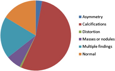
The new database—INbreast—has a total of 115 cases (410 images) from which 90 cases are from women with both breasts affected (four images per case) and 25 cases are from mastectomy patients (two images per case). Several types of lesions (masses, calcifications, asymmetries, and distortions) were included. Accurate contours made by specialists are also provided in XML format.
Conclusion
The strengths of the actually presented database—INbreast—relies on the fact that it was built with full-field digital mammograms (in opposition to digitized mammograms), it presents a wide variability of cases, and is made publicly available together with precise annotations. We believe that this database can be a reference for future works centered or related to breast cancer imaging.
According to the World Health Organization, breast cancer was responsible for approximately 519,000 deaths in 2004: 16% of all cancer incidence among women. In 2008, it was the most common form of cancer and cancer related death in women worldwide . In Portugal, 1500 women die every year from breast cancer, whereas in the European Union it is responsible for one in every six deaths from cancer in women . For this reason, early detection and diagnosis of breast cancer is essential to decrease its associated mortality rate. Therefore, mass screening is recommended by the medical community .
X-ray mammography is currently considered the best imaging method for breast cancer screening and the most effective tool for early detection of this disease . Screening mammographic examinations are performed annually on asymptomatic women to detect early, clinically unsuspected lesions. The age at which mass screening mammography is generally recommended in the United States is 40 . In Europe, screening at 40 to 50 years old is still not consensual . However, in women with genetic mutations or significant family history of breast cancer, screening should start earlier, usually 10 years earlier than the age of diagnosis of the youngest relative (never before 25) .
Get Radiology Tree app to read full this article<
 Open full size image
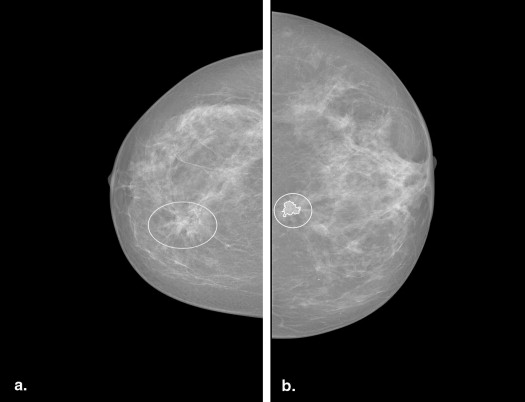
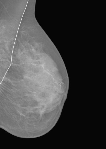
Figure 1
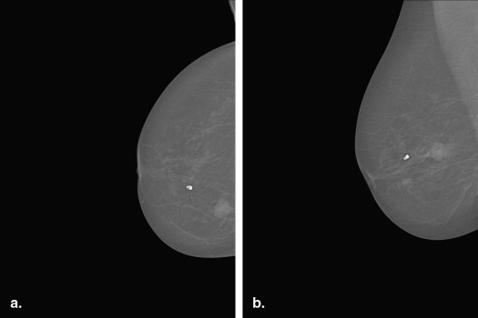
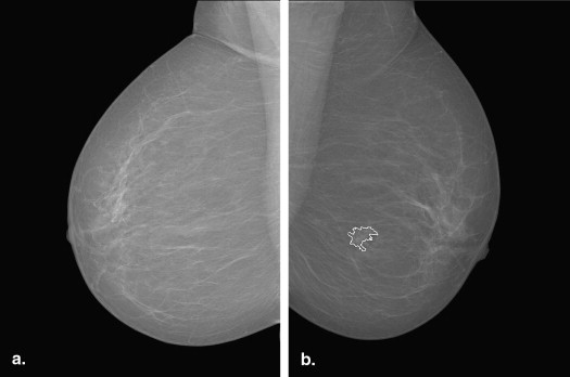
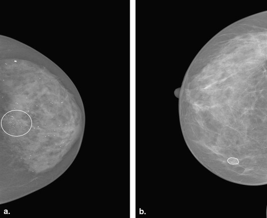
Mammogram examples: (a) craniocaudal (CC) view of the right breast; (b) CC view of the left breast; (c) mediolateral oblique (MLO) view of the right breast; (d) MLO view of the left breast.
Get Radiology Tree app to read full this article<
Get Radiology Tree app to read full this article<
Table 1
Breast Imaging Reporting and Data System Assessment Categories
Category Description 0 Needs additional imaging evaluation and/or prior mammograms for comparison 1 Negative 2 Benign finding(s) 3 Probably benign finding(s). Short-interval follow-up is suggested. 4 Suspicious anomaly. Biopsy should be considered. 5 Highly suggestive of malignancy. Appropriate action should be taken. 6 Biopsy proven malignancy
Get Radiology Tree app to read full this article<
Get Radiology Tree app to read full this article<
Get Radiology Tree app to read full this article<
Get Radiology Tree app to read full this article<
Requirements for a digital mammographic database
Get Radiology Tree app to read full this article<
Case Selection
Get Radiology Tree app to read full this article<
Ground Truth
Get Radiology Tree app to read full this article<
Associated Information
Get Radiology Tree app to read full this article<
Organization of Database
Get Radiology Tree app to read full this article<
Distribution of Database
Get Radiology Tree app to read full this article<
Available databases
Get Radiology Tree app to read full this article<
The Mammographic Image Analysis Society Digital Mammogram Database
Get Radiology Tree app to read full this article<
Get Radiology Tree app to read full this article<
Get Radiology Tree app to read full this article<
The Digital Database for Screening Mammography
Get Radiology Tree app to read full this article<
Get Radiology Tree app to read full this article<
The BancoWeb LAPIMO Database
Get Radiology Tree app to read full this article<
Get Radiology Tree app to read full this article<
Table 2
Most Used Databases in Literature
MIAS DDSM BancoWeb Origin UK USA Brazil Year 1994 1999 2010 Number of cases 161 2620 320 Views MLO MLO and CC MLO, CC, and other Number of images 322 10,480 1400 Mode of image acquisition Screen film Screen film Screen film Image type file PGM LJPEG TIFF Resolution 8 bits/pixel 8 or 16 bits/pixel 12 bits/pixel Lesion type All kinds (with special concentration of spiculated masses) All kinds All kinds Ground truth Center and radius of a circle around the interest area Pixel level boundary of the findings ROI is available in a few images only BI-RADS No Yes Yes Breast density Yes (not ACR) Yes (in ACR standard) Yes (not ACR) Clinical history No Age Yes Search system No Yes, but not functional Yes Access Yes Yes Yes Support No No Yes
ACR, American College of Radiology; CC, craniocaudal; DDSM, Digital Database for Screening Mammography; LJPEG, lossless JPEG (Joint Photographic Experts Group); MIAS, Mammographic Image Analysis Society Digital Mammogram Database; MLO, mediolateral oblique; PGM, portable gray map; ROI, region of interest; TIFF, tagged image file format; UK, United Kingdom; USA, United States of America.
Get Radiology Tree app to read full this article<
Get Radiology Tree app to read full this article<
Table 3
Other Databases Referred in Literature
Nijmegen Trueta IRMA MIRAcle LLNL Málaga NDMA Origin The Netherlands Spain Germany Greece USA Spain USA Year 1998 2008 2008 2009 Unknown Unknown Unknown Number of cases 21 89 Unknown 196 50 35 Unknown Views MLO and CC MLO and CC MLO and CC Unknown MLO and CC MLO and CC Unknown Number of images 40 320 10,509 204 198 Unknown 1,000,000 Mode of image acquisition Screen film FFDM Screen film Unknown Unknown Unknown Unknown Image type file Unknown DICOM Several Unknown ICS Raw Unknown Resolution 12 bits/pixel 12 bits/pixel Several Unknown 12 bits/pixel 12 bits/pixel Unknown Lesion type MCCs All kind All kind Unknown Calcifications Masses Unknown Ground truth Center and radius of a circle around the interest area Center and radius of a circle around the interest area Several Region of Interest Outline of calcifications Pixel level annotations Unknown BI-RADS Unknown Yes Yes Yes Unknown Unknown Unknown Breast density Unknown ACR ACR No Unknown Unknown Yes Clinical history Unknown Unknown Yes No Yes Unknown Unknown Search system No Unknown Unknown YES Unknown Unknown Unknown Access No No Yes Summer 2011 Paid Unknown No Support No Yes Yes Yes Unknown Unknown Unknown
ACR, American College of Radiology; BI-RADS, Breast Imaging Reporting and Data System; CC, craniocaudal; DICOM, Digital Imaging and Communications in Medicine; FFDM, full-field digital mammography; ICS, Image Cytometry Standard; IRMA, Image Retrieval in Medical Applications; LLNL, Lawrence Livermore National Laboratory; MCC, microcalcification; MLO, mediolateral oblique; NDMA, National Digital Medical Archive.
Get Radiology Tree app to read full this article<
Get Radiology Tree app to read full this article<
Inbreast database description
Get Radiology Tree app to read full this article<
Get Radiology Tree app to read full this article<
Get Radiology Tree app to read full this article<
Get Radiology Tree app to read full this article<
Get Radiology Tree app to read full this article<
Get Radiology Tree app to read full this article<
Get Radiology Tree app to read full this article<
Get Radiology Tree app to read full this article<
Get Radiology Tree app to read full this article<
Get Radiology Tree app to read full this article<
Get Radiology Tree app to read full this article<
Get Radiology Tree app to read full this article<
Get Radiology Tree app to read full this article<
Get Radiology Tree app to read full this article<
Get Radiology Tree app to read full this article<
Get Radiology Tree app to read full this article<
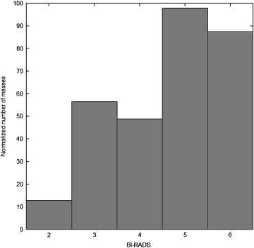
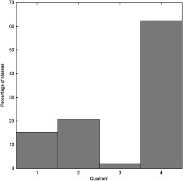
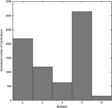
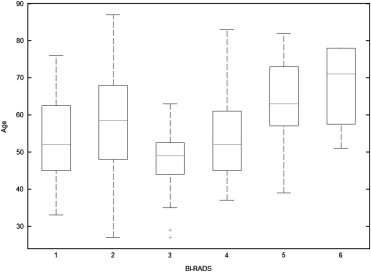
Findings characteristics
Get Radiology Tree app to read full this article<
Get Radiology Tree app to read full this article<
Get Radiology Tree app to read full this article<
Get Radiology Tree app to read full this article<
Get Radiology Tree app to read full this article<
Get Radiology Tree app to read full this article<
Get Radiology Tree app to read full this article<
Get Radiology Tree app to read full this article<
Get Radiology Tree app to read full this article<
Get Radiology Tree app to read full this article<
Performance evaluation
Get Radiology Tree app to read full this article<
Get Radiology Tree app to read full this article<
Get Radiology Tree app to read full this article<
Get Radiology Tree app to read full this article<
Get Radiology Tree app to read full this article<
d(referenceMCC,detectedMCC)=min(T2;d[referenceMCC,detectedMCC]) d
(
reference
MCC
,
detected
MCC
)
=
min
(
T
2
;
d
[
reference
MCC
,
detected
MCC
]
)
where T 2 is a saturation value (an alternative approach would be to use a sigmoid function to compress the Euclidean distance). The motivation for this saturation process is that erring by T 2 (eg, 100) is the same as erring by any value above T 2 (eg, 700).
Get Radiology Tree app to read full this article<
Discussion
Get Radiology Tree app to read full this article<
Get Radiology Tree app to read full this article<
Get Radiology Tree app to read full this article<
Get Radiology Tree app to read full this article<
Conclusions and future work
Get Radiology Tree app to read full this article<
Get Radiology Tree app to read full this article<
Get Radiology Tree app to read full this article<
Get Radiology Tree app to read full this article<
Get Radiology Tree app to read full this article<
References
1. World cancer report.2008.International Agency for Research and CancerLyon, France
2. Health statistics: atlas on mortality in the European Union.2009.EurostatLuxembourg
3. Fact sheet No. 297: Cancer.2009.World Health OrganizationFrance
4. Misra S., Solomon N.L., Moffat F.L., et. al.: Screening criteria for breast cancer. Adv Surg 2010; 44: pp. 87-100.
5. Lee C.H., Dershaw D.D., Kopans D., et. al.: Breast cancer screening with imaging: recommendations from the Society of Breast Imaging and the ACR on the use of mammography, breast MRI, breast ultrasound, and other technologies for the detection of clinically occult breast cancer. J Am Coll Radiol 2010; 7: pp. 18-27.
6. European Cancer Observatory. Breast cancer screening programmes in the EU Member States in 2006. Available at: http://eu-cancer.iarc.fr/cancer-13-display-text-562-564.html,en .
7. Karellas A, Vedantham S, Suryanarayan S. Digital mammography: an emerging technology. Business Briefing: Future Directions in Imaging 2006; 2:5. Available at: http://www.touchbriefings.com/pdf/1677/Future_imaging_06_Con.pdf .
8. Sampat M.P., Bovik A.C., Whitman G.J., et. al.: A model-based framework for the detection of speculated masses on mammography. Med Phys 2008; 35: pp. 2110-2123.
9. Orsi C.J.: The American College of Radiology mammography lexicon: an initial attempt to standardize terminology. AJR Am J Roentgenol 1996; 166: pp. 779-780.
10. Orel S.G., Kay N., Reynolds C., et. al.: BI-RADS categorization as a predictor of malignancy. Radiology 1999; 211: pp. 845-850.
11. Mammography Accreditation Program: Submitting Clinical Images. Preston White Drive, Reston: American College of Radiology, 2009; 20191–4397.
12. Bird R.E., Wallace T.W., Yankaskas B.E.: Analysis of cancers missed at screening mammography. Radiology 1992; 184: pp. 612-617.
13. Kolb T.M., Lichy J., Newhouse J.H.: Performance of screening mammography among women with and without a first-degree relative with breast cancer. Ann Intern Med 2000; 133: pp. 855-863.
14. Zheng B., Hardesty L.A., Polle W.R., et. al.: Mammography with computer-aided detection: reproducibility assessment - initial experience. Radiology 2003; 228: pp. 58-62.
15. Castellino R.A.: Computer aided detection (CAD): an overview. Cancer Imaging 2005; 5: pp. 17-19.
16. Nishikawa R.M.: Mammographic databases. Breast Dis 1998; 10: pp. 137-150.
17. Orellana CJ. Study of a mammographic CAD performance dependence on the considered mammogram set. Proceedings of the International Conference of IEEE Engineering in Medicine and Biology Society, Vancouver, British Columbia, Canada 2008; 4776–4779.
18. Nishikawa R.M.: Development of a common database for digital mammography research.1996.University of ChicagoChicago, IL
19. Antoniou AC. A web-accessible mammographic image database dedicated to combined training and evaluation of radiologists and machines. 9th International Conference on Information Technology and Applications in Biomedicine, Cyprus, 2009.
20. Oliver A., Freixenet J., Martí J., et. al.: A review of automatic mass detection and segmentation in mammographic images. Medical Image Anal 2010; 14: pp. 87-110.
21. Bernarding J., Thiel A., Decker I., et. al.: Implementation of a dynamic platform-independent DICOM-server. Comput Methods Programs Biomed 2001; 65: pp. 71-78.
22. Singh S., Bovis K.: An evaluation of contrast enhancement techniques for mammographic breast masses. IEEE Trans Inform Technol Biomed 2005; 9: pp. 109-119.
23. Song E., Xu S., Xu X., et. al.: Hybrid Segmentation of Mass in Mammograms Using Template Matching and Dynamic Programming. Academic Radiology 2010; vol. 17: pp. 1414-1424.
24. Rangayyan R.M., Mudigonda N., Desautels J.: Boundary modelling and shape analysis methods for classification of mammographic masses. Med Biol Eng Comp 2000; 38: pp. 487-496.
25. Suckling J. The Mammographic Image Analysis Society Digital Mammogram Database. In Exerpta Medica. International Congress Series 1069, York, England. 1994; 1069:375–378.
26. Oliver A., Lladó X., Pérez E., et. al.: A statistical approach for breast density segmentation. J Digital Imaging 2010; 23: pp. 527-537.
27. Domínguez AR, Nandi AK. Detection of masses in mammograms using enhanced multilevel-thresholding segmentation and region selection based on rank. In: IASTED International Conference on Biomedical Engineering. Austria: Innsbruck, 2007; 1:370-375.
28. Reljin B., Milosevic Z., Stojic T., et. al.: Computer aided system for segmentation and visualization of microcalcifications in digital mammograms. Folia Histochem Cytobiol 2009; 47: pp. 525-532.
29. Llobet R, Paredes R, Perez Cortes JC. Comparison of feature extraction methods for breast cancer detection. In Iberian Conference on Pattern Recognition and Image Analysis (IbPRIA). 2005; II:495.
30. Heath M, Bowyer K, Kopans D, et al. Current status of the Digital Database for Screening Mammography. In Digital Mammography 1998; 457–460.
31. Netsch T., Peitgen H.O.: Scale-space signatures for the detection of clustered microcalcifications in digital mammograms. IEEE Trans Med Imaging (MedImg) 1999; 18: pp. 774-786.
32. Joseph JYL, Gavrielides M, Markey M, et al. Computer-aided classification of breast microcalcification clusters: merging of features from image processing and radiologists. Medical Imaging Conference, San Diego, CA. Proc SPIE 2003; 5032:882–889. Available at: http://adsabs.harvard.edu//abs/2003SPIE.5032..882L .
33. Wang D.F., Shi L., Heng P.A.: Automatic detection of breast cancers in mammograms using structured support vector machines. Neurocomputing 2009; 72: pp. 3296-3302.
34. Sakellaropoulos P., Costaridou L.I., Panayiotakis G.: A wavelet-based spatially adaptive method for mammographic contrast enhancement. Phys Med Biol 2003; 48: pp. 787-803.
35. Cheng H.D., Cui M.: Mass lesion detection with a FUZZY neural network. Patt Recognit 2004; 37: pp. 1189-1200.
36. Matheus B.R., Schiabel H.: Online mammographic images database for development and comparison of CAD schemes. J Digital Imaging 2010; 24: pp. 500-506.
37. Lo CS, Chung PC, Chang CI, et al. A computerized system for detection and segmentation of clustered microcalcifications. Joint Conference of International Computer Symposium 1996; 247–253.
38. Oliver A.: Automatic mass segmentation in mammographic images. PhD thesis, Universitat de Girona (España) 2008;
39. Oliveira J.E.E., Gueld M.O., de Araújo A., et. al.: Towards a standard reference database for computer-aided mammography.2008.Med imaging Computer-aided diagnosisSan Diego, CA 6915. Available at: http://spie.org/x648.html?product_id=770325
40. Gardner WD. Breast cancer database provides faster access to patient records. InformationWeek, 2005. Available online at: http://www.informationweek.com/story/showArticle.jhtml;jsessionid=NBXENMVY3EP5AQSNDBGCKHSCJUMEKJVN?articleID=174400322 . Accessed Jan. 18, 2011.
41. Tangaro S., Bellotti R., De Carlo F., et. al.: MAGIC-5: an Italian mammographic database of digitised images for research. Radiol Med 2008; 113: pp. 477-485.
42. Warren R., Solomonides T., del Frate C., et. al.: MammoGrid: a prototype distributed mammographic database for Europe. Clin Radiol 2007; 62: pp. 1044-1051.
43. D’Orsi CJ, Mendelson EB, Ikeda DM, et al. Breast Imaging Reporting and Data System: ACR BI-RADS - Breast Imaging Atlas. Reston, VA: American College of Radiology; 2003. Available at: http://www.acr.org/secondarymainmenucategories/quality_safety/biradsatlas/biradsfaqs.aspx .
44. Danikas D., STheodorou S.J.V., Kokkalis G., et. al.: Mammographic findings following reduction mammoplasty. Aesthetic Plast Surg 2001; 25: pp. 283-285.
45. Sun L, Li L, Xu W, et al. A novel classification scheme for breast masses based on multi-view information fusion. In Bioinformatics and Biomedical Engineering (iCBBE), 2010 4th International Conference, 2010; 1–4.
46. Liu G., Haralick R.M.: Optimal matching problem in detection and recognition performance evaluation. Patt Recog 2002; 35: pp. 2125-2139.
47. Zheng B., Wang X., Lederman D., et. al.: Computer-aided detection: the effect of training databases on detection of subtle breast masses. Acad Radiol 2010; 17: pp. 1401-1408.