
Rationale and Objectives
To evaluate whether a computer-aided diagnosis (CADx) technique can accurately classify breast calcifications in full-field digital mammograms (FFDMs) as malignant or benign. The computer technique was developed previously on screen-film mammograms (SFMs) in which individual calcifications were identified manually. The present study evaluated the computer technique independently on a new database of FFDM images with automatic detection of the individual calcifications.
Materials and Methods
We analyzed 49 consecutive FFDM cases (19 cancers) that showed suspicious calcifications. Four mammography radiologists read soft-copy mammograms retrospectively and electronically indicated the region of calcifications in each image. The computer then automatically detected the individual calcifications within the indicated region and analyzed eight features of calcification morphology and distribution to arrive at an estimated likelihood of malignancy. The radiologists entered Breast Imaging Report and Data System assessments before and after seeing the computer results. Performance was analyzed using receiver operating characteristic analysis.
Results
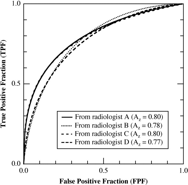
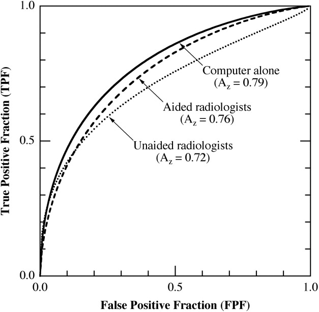
Despite variability in radiologist-indicated regions of calcifications, the computer achieved consistently high performance taking input from the four radiologists (receiver operating characteristic curve area, A z : 0.80, 0.80, 0.78, and 0.77; differences not statistically significant). Previous results showed that the computer technique achieved an A z value of 0.80 on SFMs, which improved radiologists’ performance significantly.
Conclusions
The computer technique appears to maintain consistently high performance in classifying calcifications in FFDMs as malignant or benign without requiring substantial modification from its initial development on SFMs. The computer performance appears to be robust with respect to variations in radiologists’ input.
Breast cancer is the most commonly diagnosed non–skin cancer among women in the United States, with approximately 274,900 new cases (including carcinoma in situ) expected in 2006. It is the second leading cause of female cancer deaths, killing more than 40,970 women every year ( ). Early detection is important for reducing mortality from breast cancer and, currently, screening mammography is the most effective method for detecting breast cancer early. Recently, full-field digital mammography (FFDM) and computer-aided detection (CADe) have been adopted clinically as a possible improvement to the conventional screen-film mammography (SFM). FFDM offers potentially better breast cancer detection ( ) and better facilitates the application of CADe by eliminating the film-digitization process necessary for SFMs to be analyzed by a computer ( ).
Whereas the goal of CADe is to help radiologists detect suspect lesions in a mammogram, the goal of computer-aided diagnosis (CADx) is to help radiologists determine whether a breast lesion is malignant when the results of computer analysis of the mammogram are provided to the radiologist as a second opinion ( ). Previous research indicates that CADx can potentially help radiologists increase the number of biopsies performed on mildly suspicious but histologically malignant lesions and reduce the number of biopsies performed on mildly suspicious but histologically benign lesions, thereby improving, simultaneously, both sensitivity and specificity of diagnostic mammography ( ). Research ( ) suggests that CADe and CADx, together, could serve as an alternative to double reading by radiologists for improving the diagnostic accuracy and consistency of radiologists’ mammogram interpretation ( ).
Get Radiology Tree app to read full this article<
Get Radiology Tree app to read full this article<
Materials and methods
Study Cases
Get Radiology Tree app to read full this article<
Get Radiology Tree app to read full this article<
Table 1
Image View Angles of the Study Cases
First Image Second Image Number of Cases Mediolateral/lateromedial Craniocaudal 18 Mediolateral oblique Craniocaudal 9 Mediolateral/lateromedial Lateromedial/mediolateral 5 Craniocaudal Craniocaudal/laterally exaggerated craniocaudal 2 Mediolateral oblique/mediolateral/lateromedial N/A 13 Craniocaudal N/A 2 Total 49
Note. —N/A = not available.
Get Radiology Tree app to read full this article<
Observer Study
Get Radiology Tree app to read full this article<
Get Radiology Tree app to read full this article<
Get Radiology Tree app to read full this article<
Get Radiology Tree app to read full this article<
Computer Detection of Individual Calcifications
Get Radiology Tree app to read full this article<
Table 2
Algorithm for Computer to Estimate the Number of Calcifications Within a Region of Interest in which the Computer Makes Four Presumptions of the Number of Calcifications and then Determines the Most Likely Number Range Based on Detection Results Under Each Presumption
Presumed Number of Calcifications Detection Results Under Each Presumption 3–5 Close to 5 Close to 5 Close to 5 Any 6–10 Close to 10 Close to 10 Any Close to 6 11–30 Close to 30 Any Close to 11 Close to 11 >31 Any Close to 31 Close to 31 Close to 31 Most likely number range >31 11–30 6–10 3–5
Note. —When the computer detection results does not fit into one of these four patterns, the computer asks the observer to select a number range for the calcifications.
Get Radiology Tree app to read full this article<
Computer Classification of Calcifications as Malignant or Benign
Get Radiology Tree app to read full this article<
Data Analysis
Get Radiology Tree app to read full this article<
Get Radiology Tree app to read full this article<
Results
Get Radiology Tree app to read full this article<
Get Radiology Tree app to read full this article<
Get Radiology Tree app to read full this article<
Get Radiology Tree app to read full this article<
Get Radiology Tree app to read full this article<
Discussion
Get Radiology Tree app to read full this article<
Get Radiology Tree app to read full this article<
Get Radiology Tree app to read full this article<
Get Radiology Tree app to read full this article<
Get Radiology Tree app to read full this article<
Acknowledgments
Get Radiology Tree app to read full this article<
Get Radiology Tree app to read full this article<
Get Radiology Tree app to read full this article<
Get Radiology Tree app to read full this article<
References
1. Jemal A., Siegel R., Ward E., et. al.: Cancer statistics, 2006. CA Cancer J Clin 2006; 56: pp. 106-130.
2. Lewin J.M., D’Orsi C.J., Hendrick R.E., et. al.: Clinical comparison of full-field digital mammography and screen-film mammography for detection of breast cancer. AJR Am J Roentgenol 2002; 179: pp. 671-677.
3. Skaane P., Skjennald A.: Screen-film mammography versus full-field digital mammography with soft-copy reading: randomized trial in a population-based screening program—the Oslo II study. Radiology 2004; 232: pp. 197-204.
4. Pisano E.D., Gatsonis C., Hendrick E., et. al.: Diagnostic performance of digital versus film mammography for breast-cancer screening. N Engl J Med 2005; 353: pp. 1773-1783.
5. Pisano E.D., Kuzmiak C., Koomen M.: Perspective on digital mammography. Semin Roentgenol 2001; 36: pp. 195-200.
6. Nishikawa R.M.: Computer-aided detection in digital mammography.Pisano E.D.Yaffe M.J.Kuzmiak C.M.Digital mammography.2003.Lippincott Williams and WilkinsPhiladelphia:pp. 43-48.
7. Doi K., MacMahon H., Katsuragawa S., et. al.: Computer-aided diagnosis in radiology: potential and pitfalls. Eur J Radiol 1999; 31: pp. 97-109.
8. Jiang Y., Nishikawa R.M., Schmidt R.A., et. al.: Improving breast cancer diagnosis with computer-aided diagnosis. Acad Radiol 1999; 6: pp. 22-33.
9. Jiang Y., Nishikawa R.M., Schmidt R.A., et. al.: Potential of computer-aided diagnosis to reduce variability in radiologists’ interpretations of mammograms depicting microcalcifications. Radiology 2001; 220: pp. 787-794.
10. Chan H.P., Doi K., Vyborny C.J., et. al.: Improvement in radiologists’ detection of clustered microcalcifications on mammograms. Invest Radiol 1990; 25: pp. 1102-1110.
11. Kegelmeyer W.P., Pruneda J.M., Bourland P.D., et. al.: Computer-aided mammographic screening for spiculated lesions. Radiology 1994; 191: pp. 331-337.
12. Freer T.W., Ulissey M.J.: Screening mammography with computer-aided detection: prospective study of 12,860 patients in a community breast center. Radiology 2001; 220: pp. 781-786.
13. Gur D., Sumkin J.H., Rockette H.E., et. al.: Changes in breast cancer detection and mammography recall rates after the introduction of a computer-aided detection system. J Natl Cancer Inst 2004; 96: pp. 185-190.
14. Getty D.J., Pickett R.M., D’Orsi C.J., et. al.: Enhanced interpretation of diagnostic images. Invest Radiol 1988; 23: pp. 240-252.
15. Jiang Y., Nishikawa R.M., Wolverton D.E., et. al.: Malignant and benign clustered microcalcifications: automated feature analysis and classification. Radiology 1996; 198: pp. 671-678.
16. Baker J.A., Kornguth P.J., Lo J.Y., et. al.: Artificial neural network: improving the quality of breast biopsy recommendations. Radiology 1996; 198: pp. 131-135.
17. Chan H.P., Sahiner B., Helvie M.A., et. al.: Improvement of radiologists’ characterization of mammographic masses by using computer-aided diagnosis: an ROC study. Radiology 1999; 212: pp. 817-827.
18. Veldkamp W.J., Karssemeijer N., Otten J.D., et. al.: Automated classification of clustered microcalcifications into malignant and benign types. Med Phys 2000; 27: pp. 2600-2608.
19. Huo Z., Giger M.L., Vyborny C.J., et. al.: Breast cancer: effectiveness of computer-aided diagnosis observer study with independent database of mammograms. Radiology 2002; 224: pp. 560-568.
20. Jiang Y., Nishikawa R.M., Schmidt R.A., et. al.: Comparison of independent double readings and computer-aided diagnosis (CAD) for the diagnosis of breast calcifications. Acad Radiol 2006; 13: pp. 84-94. (erratum in Acad Radiol 2006; 13:534–535)
21. Salfity M.F., Nishikawa R.M., Jiang Y., et. al.: The use of a priori information in the detection of mammographic microcalcifications to improve their classification. Med Phys 2003; 30: pp. 823-831.
22. Burgess A.: On the noise variance of a digital mammography system. Med Phys 2004; 31: pp. 1987-1995.
23. Metz C.E., Herman B.A., Shen J.H.: Maximum likelihood estimation of receiver operating characteristic (ROC) curves from continuously-distributed data. Stat Med 1998; 17: pp. 1033-1053.
24. Dorfman D.D., Berbaum K.S., Metz C.E.: Receiver operating characteristic rating analysis. Invest Radiol 1992; 27: pp. 723-731.
25. Metz C.E.: Some practical issues of experimental design and data analysis in radiological ROC studies. Invest Radiol 1989; 24: pp. 234-245.
26. Li H., Giger M.L., Yuan Y., et. al.: Comparison of computerized image analyses for digitized screen-film mammograms and full-field digital mammography images.Astley S.M.Brady M.Rose C. et. al.Digital mammography.2006.SpringerNew York:pp. 569-575.
27. Ge J., Sahiner B., Hadjiiski L.M., et. al.: Computer aided detection of clusters of microcalcifications on full field digital mammograms. Med Phys 2006; 33: pp. 2975-2988.
28. Jiang Y., Nishikawa R.M., Papaioannou J.: Dependence of computer classification of clustered microcalcifications on the correct detection of microcalcifications. Med Phys 2001; 28: pp. 1949-1957.