
Rational and Objectives
The liver is the most common organ for tumor metastasis. The development of new methods to depress hepatic metastasis is of great importance in improving survival. The aim of this study was to observe the effects of high–mechanical index ultrasound contrast on hepatic metastasis of colorectal cancer.
Materials and Methods
Hepatic metastasis models were established by injecting human colon carcinoma LoVo cells into the spleens of Sprague-Dawley rats. The rats were divided into a control group, a microbubble plus ultrasound group, a simple ultrasound group, and a simple microbubble group. The ultrasound contrast agent SonoVue (1 mL/kg) was injected via the tail vein, and high–mechanical index ultrasound contrast (frequency, 1.5 MHz; mechanical index, 1.7) was performed on the spleen intermittently for 2 minutes. The animals were sacrificed after 10 days, and the sizes and number of hepatic metastases were measured and compared. Histologic pathology and splenic ultrastructure were observed.
Results
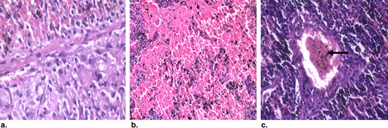
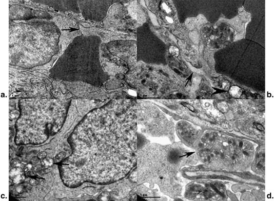
The number and sizes of hepatic metastases patently decreased in rats in the microbubble plus ultrasound group ( P < .01). There were no obvious differences among the control group, simple ultrasound group, and simple microbubble group in hepatic metastases ( P > .05). Histologic pathology showed that the number of tumor cells in the spleens decreased considerably, and massive necroses, hemorrhages, and thrombi were observed in the tumor and spleen tissues of rats in the microbubble plus ultrasound group. Transmission electron microscopy showed that the mitochondria of tumor cells and endothelial cells were clearly swelled, and there were gaps among endothelial cells and platelets aggregated in capillary vessels.
Conclusion
This research shows that intermittent high–mechanical index ultrasound contrast may inhibit the hepatic metastasis of cancer in a rat model.
Hepatic metastasis of malignant tumors is accomplished mainly by diffusion metastasis, lymphatic metastasis, blood-route metastasis, and implantation metastasis caused by malignant cell shedding. The liver is the largest parenchymatous organ in the human body and is considered the major location of many malignant tumor metastases because of its double blood supply. Hepatic metastasis is commonly seen in the blood-route metastasis of colon cancer. It was reported that in patients with colorectal cancer, about 10% to 25% were found to have hepatic metastases when they underwent primary surgery, another 25% were found to have hepatic metastases during follow-up, and >50% had hepatic metastasis . Therefore, the inhibition of hepatic metastasis is of great importance for the improvement of survival.
Compared to enhanced computed tomography and magnetic resonance imaging, ultrasound contrast is a new diagnostic technique. In recent years, with the development of the ultrasound contrast technique and the application of new ultrasound contrast agents, the effects of microbubble contrast agents on human tissues and organs have aroused great concern. Ultrasound contrast has been extensively used in the diagnosis of liver diseases and has made great progress . Yumita et al found that ultrasound cavitation caused by ultrasound contrast could damage tumor cells.
Get Radiology Tree app to read full this article<
Materials and methods
Materials and Equipment
Get Radiology Tree app to read full this article<
Cell Culture
Get Radiology Tree app to read full this article<
Establishment of the Animal Model
Get Radiology Tree app to read full this article<
Empirical Method
Get Radiology Tree app to read full this article<
Get Radiology Tree app to read full this article<
Statistical Analysis
Get Radiology Tree app to read full this article<
Results
Tumor Growth
Get Radiology Tree app to read full this article<
Get Radiology Tree app to read full this article<
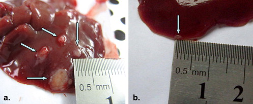
Table 1
Comparison of the Number and Sizes of Liver Metastases Among the Study Groups
Group Number Diameter (mm) Control 10.33 ± 4.62 3.75 ± 1.29 Microbubbles plus ultrasound 3.42 ± 1.38 ∗ 1.58 ± 0.67 ∗ Simple ultrasound 9.58 ± 3.42 † 3.58 ± 1.24 † Simple microbubbles 11.33 ± 3.73 † 3.75 ± 1.22 †
Data are expressed as mean ± standard deviation.
Get Radiology Tree app to read full this article<
Get Radiology Tree app to read full this article<
Get Radiology Tree app to read full this article<
Histopathologic Changes
Get Radiology Tree app to read full this article<
Get Radiology Tree app to read full this article<
Transmission Electron Microscopy
Get Radiology Tree app to read full this article<
Get Radiology Tree app to read full this article<
Discussion
Get Radiology Tree app to read full this article<
Get Radiology Tree app to read full this article<
Get Radiology Tree app to read full this article<
Get Radiology Tree app to read full this article<
Get Radiology Tree app to read full this article<
Get Radiology Tree app to read full this article<
Get Radiology Tree app to read full this article<
Conclusions
Get Radiology Tree app to read full this article<
Get Radiology Tree app to read full this article<
References
1. Cromheecke M., de Jong K.P., Hoekstra H.J.: Current treatment for colorectal cancer metastatic to liver. Eur J Surg Oncol 1999; 25: pp. 451-463.
2. Tellez C., Benson A.B., Lyster M.T., et. al.: Phase II trial of chemoembolization for the treatment of metastatic colorectal carcinoma to the liver and review of the literature. Cancer 1998; 82: pp. 1250-1259.
3. Cosgrove D.: Ultrasound contrast agents: an overview. Eur J Radiol 2006; 60: pp. 324-330.
4. Konopke R., Bunk A., Kersting S.: The role of contrast-enhanced ultrasound for focal liver lesion detection: an overview. Ultrasound Med Biol 2007; 33: pp. 1515-1526.
5. Yumita N., Okuyama N., Sasaki K., et. al.: Sonodynamic therapy on chemically induced mammary tumor: pharmacokinetics, tissue distribution and sonodynamically induced antitumor effect of porfimer sodium. Cancer Sci 2004; 95: pp. 765-769.
6. Hwang J.H., Brayman A.A., Reidy M.A., et. al.: Vascular effects induced by combined 1-MHz ultrasound and microbubble contrast agent treatments in vivo. Ultrasound Med Biol 2005; 31: pp. 553-564.
7. Miller D.L., Thomas R.M.: Contrast-agent gas bodies enhance hemolysis induced by lithotriptersho waves and high intensity focused ultrasound in whole blood. Ultrasound Med Biol 1996; 22: pp. 1089-1095.
8. Roberts W.W., Hall T.L., Ives K., et. al.: Pulsed cavitational ultrasound: a noninvasive technology for controlled tissue ablation (histotripsy) in the rabbit kidney. J Urol 2006; 175: pp. 447-453.
9. Frizzell W.L.A.: Biological effects of acoustic cavitation in ultrasound: its chemical, physical, and biological effects.Suslick K.S.Ultrasound: its chemical, physical, and biological effects.1988.VCHNew York:pp. 287-303.
10. Kozlowski J.M., Fidler I.J., Campbell D., et. al.: Metastatic behavior of human tumor cell lines grown in the nude mouse. Cancer Res 1984; 44: pp. 3522-3529.
11. Wang Q., Shi G.F., Zhang J.J., et. al.: Walker-256 small metastasis in rat liver: model establishment, pathologic and imaging study. Chin J Med Imaging Technol 2006; 22: pp. 541-544.
12. Wood A.K., Bunte R.M., Cohen J.D., et. al.: The antivascular action of physiotherapy ultrasound on a murine tumor: role of a microbubble contrast agent. Ultrasound Med Biol 2007; 33: pp. 1901-1910.
13. Gupta K., Zhang J.: Angiogenesis: a curse or cure?. Postgrad Med J 2005; 81: pp. 236-242.
14. Takeda A., Stoeltzing O., Ahmad S.A., et. al.: Role of angiogenesis in the development and growth of liver metastasis. Ann Surg Oncol 2002; 9: pp. 610-616.
15. Miller D.L., Pislaru S.V., Greenleaf J.E.: Sonoporation: mechanical DNA delivery by ultrasonic cavitation. Somat Cell Mol Genet 2002; 27: pp. 115-134.
16. Kobayashi N., Yasu T., Yamada S., et. al.: Endothelial cell injury in venule and capillary induced by contrast ultrasonography. Ultrasound Med Biol 2002; 28: pp. 949-956.
17. Folkman J., Browder T., Palmblad J.: Angiogenesis research: guidelines for translation to clinical application. Thromb Haemost 2001; 86: pp. 23-33.