
Rationale and Objectives
We have developed a microwave tomography system for experimental breast imaging.
Materials and Methods
In this article, we illustrate a strategy for optimizing the coupling liquid for the antenna array based on in vivo measurement data. We present representative phantom experiments to illustrate the imaging system’s ability to recover accurate property distributions over the range of dielectric properties expected to be encountered clinically. To demonstrate clinical feasibility and assess the microwave properties of the normal breast in vivo, we summarize our initial experience with microwave breast exams of 43 women with negative mammography according to the Breast Imaging Reporting and Data System (BI-RADS 1).
Results
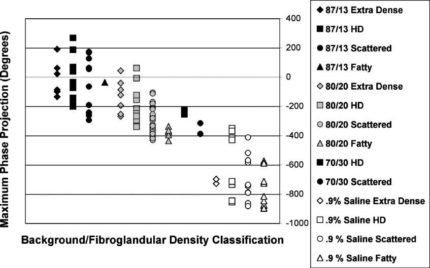
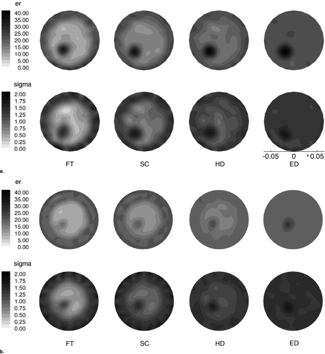
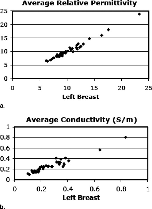
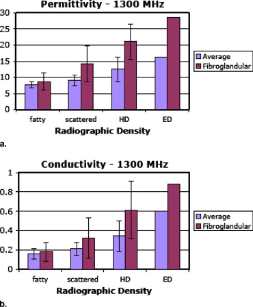
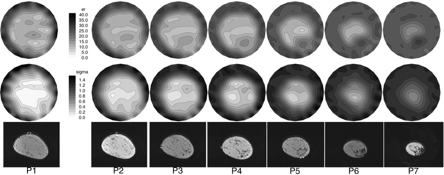
The clinical results show a high degree of bilateral symmetry in the whole breast average microwave properties. Focal assessments of microwave properties are associated with breast tissue composition evaluated through radiographic density categorization verified through magnetic resonance image correlation in selected cases. Specifically, both whole-breast average and local microwave properties increase with increasing radiographic density, in which the latter exhibits a more substantial rise.
Conclusion
These findings support our hypothesis that water content variations in the breast play an influential role in dictating the overall dielectric property distributions and indicate that the microwave properties in the breast are more heterogeneous than previously believed based on ex vivo property measurements reported in the literature.
In 2004 alone, more than 200,000 new cases of breast cancer were diagnosed in the United States, with roughly 40,000 women dying from the disease, making breast cancer the second largest cause of female cancer deaths in the United States ( ). There have been numerous reports demonstrating that early detection is the single most significant predictor of long-term survival ( ); therefore, improvements in detection may help to reduce the high mortality rates that exist. Mammography is the front-line screening modality, but its weaknesses in terms of sensitivity and specificity are well documented ( ). Although magnetic resonance (MR) and ultrasound are used primarily in a diagnostic setting, there is room for improvement in differential diagnosis as well.
Over the past decade, there has been a steady increase in the interest in using microwave imaging as a way of detecting breast cancers. Ex vivo studies have indicated that there is significant contrast in the dielectric properties of normal and malignant breast tissue ( ). However, it is well known that the breast is a heterogeneous organ whose composition varies significantly depending on a variety of factors ( ). For example, Woodard and White ( ) have shown that the mammary glands typically contain significantly more water than adipose tissue. Thus fibroglandular breast tissue would be expected to have higher dielectric properties than fat, but with properties that are still lower than high water-content tissues. Further, studies have shown that the amount of fibroglandular tissue can vary widely from fattier breasts (with very little) to dense breast (with substantially larger proportions), suggesting that the baseline electrical properties of the normal breast may be highly variable ( ).
Get Radiology Tree app to read full this article<
Get Radiology Tree app to read full this article<
Methods
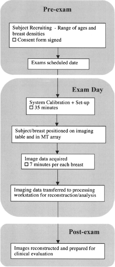
Imaging Procedure
Get Radiology Tree app to read full this article<
Table 1
Summary of the Age and Breast Density Distributions for the 43 Women Recruited for this Imaging Study
Age Density Total Fatty Scattered Heterogeneously Dense Extremely Dense 40–49 0 7 3 1 11 50–59 2 4 6 0 12 60–69 1 8 3 0 12 7079 2 4 2 0 8 Total 5 23 14 1 43
Table 2
Summary of Microwave Tomography System Performance Specifications
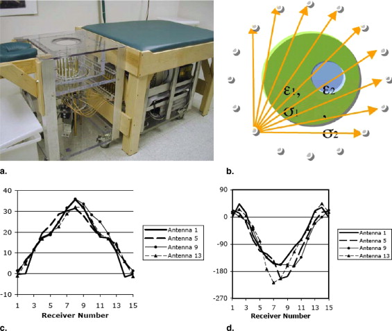
Performance Specifications Exam geometry Breast pendant in liquid bath Imaging planes 7 Coupling bath 80:20 and 86:14 glycerin:water mixtures Total number of images 49 (7 frequencies × 7 planes) Exam time 7 min Frequencies 500–1,700 MHz in 200-MHz increments Power transmitted <1 mW Measurement sensitivity −130 dBm Antenna array 16 monopole antennas on a 15.2-cm diameter circle, noncontacting Measurements/image set 240 (16 transmitters × 15 receivers per transmitter) Image properties Electrical permittivity (ε r ) and conductivity (σ) Reconstruction time <5 min/image
Get Radiology Tree app to read full this article<
Phantom Experiments
Get Radiology Tree app to read full this article<
Get Radiology Tree app to read full this article<
In Vivo Tissue Property Study
Get Radiology Tree app to read full this article<
Get Radiology Tree app to read full this article<
Get Radiology Tree app to read full this article<
Get Radiology Tree app to read full this article<
Get Radiology Tree app to read full this article<
Water Content and MR Comparison
Get Radiology Tree app to read full this article<
Results
Get Radiology Tree app to read full this article<
Background Liquid Selection
Get Radiology Tree app to read full this article<
Get Radiology Tree app to read full this article<
Get Radiology Tree app to read full this article<
Phantom Results
Get Radiology Tree app to read full this article<
Get Radiology Tree app to read full this article<
Get Radiology Tree app to read full this article<
In Vivo Normal Subject Study
Get Radiology Tree app to read full this article<
Get Radiology Tree app to read full this article<
Get Radiology Tree app to read full this article<

Get Radiology Tree app to read full this article<
Get Radiology Tree app to read full this article<
Get Radiology Tree app to read full this article<

Get Radiology Tree app to read full this article<
MR Comparison Results
Get Radiology Tree app to read full this article<
Get Radiology Tree app to read full this article<
Get Radiology Tree app to read full this article<
Discussion
Get Radiology Tree app to read full this article<
Get Radiology Tree app to read full this article<
Get Radiology Tree app to read full this article<
Get Radiology Tree app to read full this article<
Get Radiology Tree app to read full this article<
References
1. American Cancer Society: 2004.American Cancer SocietyAtlanta, GA
2. Joy J.E., Penhoet E.E., Petitti D.B.: 2005.The National Academy PressWashington DC
3. Tabar L., Yen M.F., Vitak B., et. al.: Mammography service screening and mortality in breast cancer patients: 20-year follow-up before and after introduction of screening. Lancet 2003; 361: pp. 1405-1410.
4. Chaudhary S.S., Mishra R.K., Swarup A., et. al.: Dielectric properties of normal and malignant human breast tissues at radiowave and microwave frequencies. Ind J Biochem Biophys 1984; pp. 2176-2179.
5. Joines W.T., Zhang Y., Li C., et. al.: The measured electrical properties of normal and malignant human tissues from 50 to 900 MHz. Med Phys 1994; 21: pp. 547-550.
6. Duck F.A.: 1990.Academic PressLondon, UK
7. Kumar V., Abbas A.K., Fausto N., et. al.: Pathologic basis of disease.2005.Elsevier SaundersPhiladelphia, PA:pp. 1119-1154.
8. Woodard H.Q., White D.R.: The composition of body tissues. Br J Radiol 1986; 59: pp. 1209-1219.
9. Wei J., Chan H.-P., Helvie M.A., et. al.: Correlation between mammographic density and volumetric fibroglandular tissue estimated on breast MR images. Med Phys 2004; 31: pp. 933-942.
10. Xu L., Bond E.J., Van Veen B.D., et. al.: An overview of ultra-wideband microwave imaging via space-time beamforming for early-stage breast-cancer detection. IEEE Antennas Propagation 2005; 47: pp. 19-34.
11. Davis S.K., Tandradinata H., Hagness S.C., et. al.: Ultrawideband microwave breast cancer detection: a detection-theoretic approach using the generalized likelihood ratio test. IEEE Trans Biomed Engineer 2005; 52: pp. 1237-1250.
12. Fear E.C., Li X., Hagness S.C., et. al.: Confocal microwave imaging for breast cancer detection: localization of tumors in three dimensions. IEEE Trans Biomed Engineer 2002; 49: pp. 812-822.
13. Yun X., Fear E.C., Johnston R.H.: Compact antenna for radar-based breast cancer detection. IEEE Trans Antennas Propagation 2005; 53: pp. 2374-2380.
14. Benjamin R., Craddock I.J., Hilton G.S., et. al.: Microwave detection of buried mines using non-contact, synthetic near-field focusing. IEEE Proc Radar Sonar Navig 2001; 148: pp. 233-240.
15. Bolomey J.-C., Izadnegahdar A., Jofre L., et. al.: Microwave diffraction tomography for biomedical applications. IEEE Trans Microwave Theory Tech 1982; 82: pp. 1998-2000.
16. Bolomey J.-C., Jofre L., Peronnet G.: On the possible use of microwave-active imaging for remote thermal sensing. IEEE Trans Microwave Theory Tech 1983; 83: pp. 777-781.
17. Bolomey J.-C., Michel Y.: 1985.
18. Liewei S., Nolte L.W., Zhong Q.Z., et. al.: Performance analysis for Bayesian microwave imaging in decision aided breast tumor diagnosis. Proc 2002 IEEE Int Symp Biomed Imaging 2002; pp. 1039-1042.
19. Souvorov A.E., Bulyshev A.E., Semenov S.Y., et. al.: Two-dimensional computer analysis of a microwave flat antenna array for breast cancer tomography. IEEE Trans Microwave Theory Tech 2000; 48: pp. 1413-1415.
20. Bulyshev A.E., Semenov S.Y., Souvorov A.E., et. al.: Computational modeling of three-dimensional microwave tomography of breast cancer. IEEE Trans Biomed Eng 2001; 48: pp. 1053-1056.
21. Meaney P.M., Fanning M.W., Li D., et. al.: A clinical prototype for active microwave imaging of the breast. IEEE Trans Microwave Theory Tech 2000; 48: pp. 1841-1853.
22. Meaney P.M., Pendergrass S.A., Fanning M.W., et. al.: Importance of using a reduced contrast coupling medium in 2D microwave breast imaging. J Electromagn Waves Appl 2003; 17: pp. 333-355.
23. Ciocan R., Jiang H.: Model-based microwave image reconstruction: simulations and experiments. Med Phys 2004; 31: pp. 3231-3241.
24. Poplack S.P., Paulsen K.D., Hartov A., et. al.: Electromagnetic breast imaging – normal tissue property values. Radiology 2004; 231: pp. 571-580.
25. Meaney P.M., Paulsen K.D., Ryan T.P.: Two-dimensional hybrid element image reconstruction for TM illumination. IEEE Trans Antenna Prop 1995; 43: pp. 239-247.
26. Meaney P.M., Paulsen K.D., Pogue B.W., et. al.: Microwave image reconstruction utilizing log-magnitude and unwrapped phase to improve high-contrast object recovery. IEEE Trans Med Imaging 2001; 20: pp. 104-116.
27. Paulsen K.D., Meaney P.M., Moskowitz M.J., et. al.: A dual mesh scheme for finite element based reconstruction algorithms. IEEE Trans Med Imaging 1995; 14: pp. 504-514.
28. Paulsen K.D., Meaney P.M.: Compensation for nonactive array element effects in a microwave imaging system: part I—forward solution vs. measured data comparison. IEEE Trans Med Imaging 1999; 18: 496–50
29. Li D., Meaney P.M., Raynolds T., et. al.: A parallel-detection microwave spectroscopy system for breast imaging. Rev Sci Instruments 2004; 75: pp. 2305-2313.
30. Meaney PM, Fang Q, Demidenko E, et al. Residual error analysis in applying a log transformation to the microwave parameter estimation problem. IEEE Trans Med Imaging. In press.
31. Fang Q., Meaney P.M., Paulsen K.D.: Multi-dimensional phase unwrapping: definition and properties. IEEE Trans Image Proc 2006; 15: pp. 3311-3324.
32. American College of Radiology (ACR): ACR BI-RADS—Mammography.ACR breast imaging reporting and data system, breast imaging atlas.2003.American College of RadiologyReston, VA:
33. Poplack S.P., Tosteson A.N., Grove M.R., et. al.: Mammography in 53,803 women from the New Hampshire Mammography Network. Radiology 2000; 217: pp. 832-840.
34. Foster K.R., Schepps J.L.: Dielectric properties of tumor and normal tissues at radio through microwave frequencies. J Microwave Power 1981; 16: pp. 107-119.
35. Kopans D.B.: 2nd ed.1997.Williams & WilkinsLippincott NY, NY
36. Pastorino M., Massa A., Caorsi S.: A microwave inverse scattering technique for image reconstruction based on a genetic algorithm. IEEE Trans Instrum Measure 2000; 49: pp. 573-578.
37. Caorsi S., Massa A., Pastorino M.: A computational technique based on a real-coded genetic algorithm for microwave imaging purposes. IEEE Trans Geosci Remote Sensing 2000; 38: pp. 1697-1708.
38. Semenov S.Y., Bulyshev A.E., Abubakar A., et. al.: Microwave-tomographic imaging of the high dielectric-contrast objects using different image-reconstruction approaches. IEEE Trans Microwave Theory Tech 2005; 53: pp. 2284-2294.
39. Fricke H.: A mathematical treatment of the electrical conductivity and capacity of disperse systems ii. Phys Rev 1925; 26: pp. 678-681.
40. Schepps J.L., Foster K.R.: The UHF and microwave dielectric properties of normal and tumour tissues: variation in dielectric properties with tissue water. Phys Med Biol 1980; 25: pp. 1149-1159.