
Rationale and Objectives
The effects of locally delivered angiogenic factors or stem cells on the coronary artery perfusion territory are not well defined. Therefore, the aim of this study was to determine the ability of the selective injection of magnetic resonance contrast media (MR-CM) to map and quantify the territories of the major coronary arteries.
Materials and Methods
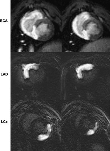
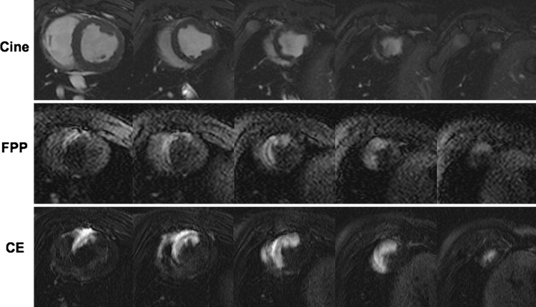
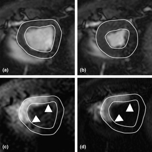
Selective coronary catheterization ( n = 16 pigs) was performed under x-ray and magnetic resonance imaging (MRI) fluoroscopy in an x-ray and magnetic resonance suite. Catheters were placed in the left anterior descending (LAD), circumflex, or right coronary artery. The coronary perfusion territories were mapped by the intracoronary injection of MR-CM using first-pass perfusion (FPP) and early contrast-enhanced (CE) MRI. Cine MRI was used to quantify left ventricular (LV) mass. In 12 animals, the LAD coronary artery was occluded by microspheres to create infarctions. Infarct size was measured on delayed enhanced (DE) MRI after the intravenous injection of MR-CM.
Results
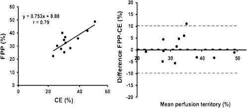
X-ray and magnetic resonance fluoroscopy were successfully used to catheterize the coronary arteries. The perfusion territories of the coronary arteries were defined as hyperenhanced regions on FPP and CE MRI. The LAD coronary artery territory was 33.7 ± 2.2% of LV mass on FPP MRI and 33.0 ± 2.3% on CE MRI ( P = .63). Bland-Altman analysis showed close agreement between the two methods (0.7 ± 5.0%). DE MRI demonstrated the infarcted myocardium as hyperenhanced subregions of the perfusion territory (7.5 ± 1.2% of LV mass).
Conclusions
Interventional cardiac x-ray and magnetic resonance fluoroscopy can be used to map and quantify the perfusion territory of each coronary artery. This experimental method can be used before and after the local delivery of angiogenic factors and stem cell therapy to determine their efficacy.
Coronary angioplasty or bypass surgery is routinely applied to restore blood flow to ischemic myocardium. Nevertheless, many patients with end-stage coronary artery disease continue to experience disabling angina. This problem has led to increasing interest in alternative revascularization strategies, such as therapeutic angiogenesis by the exogenous administration of angiogenic growth factors ( ) or stem cells ( ). Obstacles to effective treatment have been the difficulties in providing a clear delineation of the status and extent of the injury and the morbidity and mortality associated with surgical procedures that might be used for delivering the therapy.
Contrast-enhanced (CE) magnetic resonance imaging (MRI) has the ability to define the target for therapy, quantify the size of acute and chronic myocardial infarctions, and measure myocardial perfusion and left ventricular (LV) function ( ). Therefore, MRI has been used in experimental studies assessing the efficacy of angiogenic growth factors ( ), genes ( ), and stem cells ( ). MRI was able to demonstrate the beneficial effects of the intramyocardial delivery of genes on perfusion, infarct resorption, and LV function. In these experimental studies, the left anterior descending (LAD) coronary artery territory was not measured ( ). More recently, MRI has also been used in clinical studies to assess the effects of the above therapies. In the Bone Marrow Transfer to Enhance ST-Elevation Infarct Regeneration (BOOST) study ( ), 60 patients were enrolled to evaluate the effect of intracoronary autologous bone marrow cells after myocardial infarction. MRI showed a significant increase in ejection fraction from 50% to 57% in treated patients compared with 51% to 52% in untreated patients ( ) and that the beneficial effects were sustained at 18 months ( ).
Get Radiology Tree app to read full this article<
Materials and methods
Animal and Study Protocol
Get Radiology Tree app to read full this article<
Get Radiology Tree app to read full this article<
MRI Protocol
FPP Imaging
Get Radiology Tree app to read full this article<
Early CE Imaging
Get Radiology Tree app to read full this article<
Cine Imaging
Get Radiology Tree app to read full this article<
MRI Data Analysis
Get Radiology Tree app to read full this article<
Get Radiology Tree app to read full this article<
Postmortem
Get Radiology Tree app to read full this article<
Statistical Analysis
Get Radiology Tree app to read full this article<
Results
Get Radiology Tree app to read full this article<
Get Radiology Tree app to read full this article<
Get Radiology Tree app to read full this article<
Discussion
Get Radiology Tree app to read full this article<
Get Radiology Tree app to read full this article<
Get Radiology Tree app to read full this article<
Get Radiology Tree app to read full this article<
Get Radiology Tree app to read full this article<
Get Radiology Tree app to read full this article<
References
1. Yla-Herttuala S.: An update on angiogenic gene therapy: vascular endothelial growth factor and other directions. Curr Opin Mol Ther 2006; 8: pp. 295-300.
2. Taylor D.A., Atkins B.Z., Hungspreugs P., et. al.: Regenerating functional myocardium: improved performance after skeletal myoblast transplantation. Nat Med 1998; 4: pp. 929-933.
3. Zohlnhofer D., Ott I., Mehilli J., et. al.: Stem cell mobilization by granulocyte colony-stimulating factor in patients with acute myocardial infarction: a randomized controlled trial. JAMA 2006; 295: pp. 1003-1010.
4. Fernandez-Aviles F., San Roman J.A., Garcia-Frade J., et. al.: Experimental and clinical regenerative capability of human bone marrow cells after myocardial infarction. Circ Res 2004; 95: pp. 742-748.
5. Chen S.L., Fang W.W., Ye F., et. al.: Effect on left ventricular function of intracoronary transplantation of autologous bone marrow mesenchymal stem cell in patients with acute myocardial infarction. Am J Cardiol 2004; 94: pp. 92-95.
6. Wollert K.C., Meyer G.P., Lotz J., et. al.: Intracoronary autologous bone-marrow cell transfer after myocardial infarction: the BOOST randomised controlled clinical trial. Lancet 2004; 364: pp. 141-148.
7. Cleland J.G., Freemantle N., Coletta A.P., Clark A.L.: Clinical trials update from the American Heart Association: REPAIR-AMI, ASTAMI, JELIS, MEGA, REVIVE-II, SURVIVE, and PROACTIVE. Eur J Heart Fail 2006; 8: pp. 105-110.
8. Watzinger N., Saeed M., Wendland M.F., Akbari H., Lund G., Higgins C.B.: Myocardial viability: magnetic resonance assessment of functional reserve and tissue characterization. J Cardiovasc Magn Reson 2001; 3: pp. 195-208.
9. Pearlman J.D., Hibberd M.G., Chuang M.L., et. al.: Magnetic resonance mapping demonstrates benefits of VEGF-induced myocardial angiogenesis. Nat Med 1995; 1: pp. 1085-1089.
10. Carlsson M., Osman N.F., Ursell P.C., Martin A.J., Saeed M.: Quantitative MR measurements of regional and global left ventricular function and strain after intramyocardial transfer of VM202 into infarcted swine myocardium. Am J Physiol Heart Circ Physiol 2008; 295: pp. H522-H532.
11. Saeed M., Saloner D., Martin A., et. al.: Adeno-associated viral vector-encoding vascular endothelial growth factor gene: effect on cardiovascular MR perfusion and infarct resorption measurements in swine. Radiology 2007; 243: pp. 451-460.
12. Kraitchman D.L., Heldman A.W., Atalar E., et. al.: In vivo magnetic resonance imaging of mesenchymal stem cells in myocardial infarction. Circulation 2003; 107: pp. 2290-2293.
13. Serfaty J.M., Atalar E., Declerck J., et. al.: Real-time projection MR angiography: feasibility study. Radiology 2000; 217: pp. 290-295.
14. Omary R.A., Green J.D., Schirf B.E., Li Y., Finn J.P., Li D.: Real-time magnetic resonance imaging-guided coronary catheterization in swine. Circulation 2003; 107: pp. 2656-2659.
15. Saeed M., Martin A.J., Lee R.J., et. al.: MR guidance of targeted injections into border and core of scarred myocardium in pigs. Radiology 2006; 240: pp. 419-426.
16. Heiberg E., Engblom H., Engvall J., Hedstrom E., Ugander M., Arheden H.: Semi-automatic quantification of myocardial infarction from delayed contrast enhanced magnetic resonance imaging. Scand Cardiovasc J 2005; 39: pp. 267-275.
17. Tsekos N.V., Atalar E., Li D., Omary R.A., Serfaty J.M., Woodard P.K.: Magnetic resonance imaging-guided coronary interventions. J Magn Reson Imaging 2004; 19: pp. 734-749.
18. Saeed M., Henk C.B., Weber O., et. al.: Delivery and assessment of endovascular stents to repair aortic coarctation using MR and x-ray imaging. J Magn Reson Imaging 2006; 24: pp. 371-378.
19. Saeed M., Saloner D., Weber O., Martin A., Henk C., Higgins C.: MRI in guiding and assessing intramyocardial therapy. Eur Radiol 2005; 15: pp. 851-863.
20. Green J.D., Omary R.A., Schirf B.E., et. al.: Comparison of x-ray fluoroscopy and interventional magnetic resonance imaging for the assessment of coronary artery stenoses in swine. Magn Reson Med 2005; 54: pp. 1094-1099.