
Rationale and Objectives
To compare the diagnostic value of magnetic resonance (MR) and computed tomography (CT) for the detection of coronary artery disease (CAD) with special regard to calcifications.
Materials and Methods
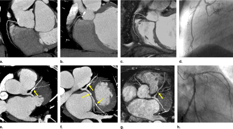
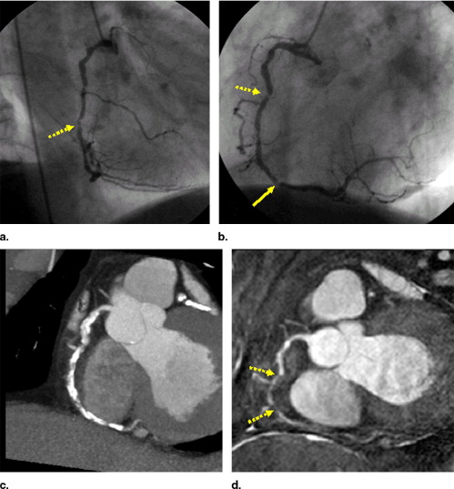
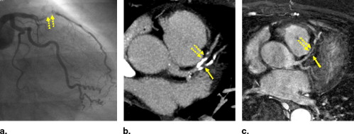
Twenty-seven patients with known CAD were examined with a targeted, navigator-gated, free-breathing, steady-state free precession MR angiography sequence (repetition time = 5.6 milliseconds, echo time = 2.8 milliseconds, flip angle 110°) and 16-slice coronary CT angiography. Segment-based sensitivity, specificity, and accuracy for the detection of stenoses larger than 50% were determined as defined by the gold standard catheter coronary angiography along with the subjective image quality (Grade 1–4). The degree of calcifications in each segment was quantified using a standard calcium scoring tool.
Results
Of 115 possible segments, 7% had to be excluded in MR imaging because of poor image quality. In CT, 3% were nondiagnostic because of image quality and 15% were not evaluable because of calcifications. Values for the detection of relevant coronary artery stenoses in the evaluated segments were: sensitivity: MR imaging 85% versus CT 96%; specificity: 88% versus 96%; accuracy: 87% versus. 96%. Average subjective image quality was 1.8 for MR imaging and 1.6 for CT. Of the 15% of segments that had to be excluded from CT evaluation because of calcifications, MR imaging provided the correct diagnosis segments in 67%.
Conclusions
CT provided a better image quality with superior accuracy for the detection of CAD. Despite its overall inferiority, MR imaging proved to be helpful method in interpreting coronary stenosis in severely calcified segments.
Computed tomography (CT) and magnetic resonance coronary angiography (MRA) are noninvasive imaging methods for the detection of coronary artery disease (CAD) in symptomatic and preclinical patients. Both methods have undergone clinical evaluations with comparisons to the gold standard, catheter coronary angiography (CCA). In these studies, good results with reliable diagnostic values were reported for both CT and magnetic resonance imaging (MRI) ( ). However, these studies differ in design (eg, number of evaluated segments, diameter of evaluated segments) and there are only two published studies on a direct comparison of CT and MRI ( ). One of the main disadvantages of computed tomography of the heart is the difficulty in assessing severely calcified segments ( ). Severe calcifications can impair the capability to correctly determinate stenoses due to beam hardening and partial volume artefacts. In MRI, calcifications may cause a signal loss and theoretically should not cause “blooming” artifacts. Therefore, MRI is supposed to be more consistent and not to be affected by calcifications, but this has not yet been investigated in a clinical study. The purpose of this study was to directly compare both modalities for the detection of coronary artery stenosis in an intraindividual patient study, paying special regard to calcified segments to investigate the value of alternative or additional MRI scans in the diagnosis of CAD.
Material and methods
Subjects
A total of 27 patients (21 male, mean age 59 ± 11) with suspected or known CAD were enrolled in this study. Exclusion criteria within this population were atrial fibrillation or other severe arrhythmias, claustrophobia, the presence of MRI-incompatible implants or other contraindication to MRI. Written informed consent was obtained from all participants.
Get Radiology Tree app to read full this article<
MRI
Get Radiology Tree app to read full this article<
Get Radiology Tree app to read full this article<
Get Radiology Tree app to read full this article<
Get Radiology Tree app to read full this article<
Get Radiology Tree app to read full this article<
CT
Get Radiology Tree app to read full this article<
Get Radiology Tree app to read full this article<
Get Radiology Tree app to read full this article<
CCA
Get Radiology Tree app to read full this article<
Data Evaluation
Get Radiology Tree app to read full this article<
Get Radiology Tree app to read full this article<
Get Radiology Tree app to read full this article<
Get Radiology Tree app to read full this article<
Get Radiology Tree app to read full this article<
Results
Get Radiology Tree app to read full this article<
Get Radiology Tree app to read full this article<
Get Radiology Tree app to read full this article<
Table 1
Image Quality, Diagnostic Accuracy of Coronary MRI and CT to Detect Stenoses of ≥50%
MRI CT Evaluated segments (of 115) (%) 103 (90%) 91 (79%) Not assessed 7% motion/misregistration 15% calcified 3% stent 3% motion 3% stent Image quality 1.8 ± 1.3 1.6 ± 0.6 ( P = NS) Sensitivity (%) 85 96 Specificity (%) 88 96 Accuracy (%) 87 96
MRI: magnetic resonance imaging; CT: computed tomography; NS: not significant.
Get Radiology Tree app to read full this article<
Get Radiology Tree app to read full this article<
Table 2
Diagnostic Accuracy for MRI and CT in Calcified Segments
Group A Group B Group C Number of segments (of 43) 21 14 8 Diagnostic accuracy CT 13 NA 4 NA 0 NA 7 of 8 right 8 of 10 right 6 of 8 right MRI 1 NA 1 NA 0 NA 16 of 20 right 9 of 13 right 6 of 8 right
MRI: magnetic resonance imaging; CT: computed tomography; NA: not assessable.
Get Radiology Tree app to read full this article<
Get Radiology Tree app to read full this article<
Get Radiology Tree app to read full this article<
Get Radiology Tree app to read full this article<
Discussion
Get Radiology Tree app to read full this article<
Get Radiology Tree app to read full this article<
Get Radiology Tree app to read full this article<
Get Radiology Tree app to read full this article<
Get Radiology Tree app to read full this article<
Get Radiology Tree app to read full this article<
Get Radiology Tree app to read full this article<
Get Radiology Tree app to read full this article<
References
1. Achenbach S., Ulzheimer S., Baum U., et. al.: Noninvasive coronary angiography by retrospectively ECG-gated multislice spiral CT. Circulation 2000; 102: pp. 2823-2828.
2. Kim W.Y., Danias P.G., Stuber M., et. al.: Coronary magnetic resonance angiography for the detection of coronary stenoses. N Engl J Med 2001; 345: pp. 1863-1869.
3. Manning W.J., Edelman R.R.: Magnetic resonance coronary angiography. Magn Reson Q 1993; 9: pp. 131-151.
4. Nieman K., Cademartiri F., Lemos P.A., et. al.: Reliable noninvasive coronary angiography with fast submillimeter multislice spiral computed tomography. Circulation 2002; 106: pp. 2051-2054.
5. Sommer T., Hofer U., Hackenbroch M., et. al.: [Submillimeter 3D coronary MR angiography with real-time navigator correction in 107 patients with suspected coronary artery disease]. Rofo Fortschr Geb Rontgenstr Neuen Bildgeb Verfahr 2002; 174: pp. 459-466.
6. Dewey M., Teige F., Schnapauff D., et. al.: Noninvasive detection of coronary artery stenoses with multislice computed tomography or magnetic resonance imaging. Ann Intern Med 2006; 145: pp. 407-415.
7. Kefer J., Coche E., Legros G., et. al.: Head-to-head comparison of three-dimensional navigator-gated magnetic resonance imaging and 16-slice computed tomography to detect coronary artery stenosis in patients. J Am Coll Cardiol 2005; 46: pp. 92-100.
8. Mitsutake R., Niimura H., Miura S., et. al.: Clinical significance of the coronary calcification score by multidetector row computed tomography for the evaluation of coronary stenosis in Japanese patients. Circ J 2006; 70: pp. 1122-1127.
9. Pundziute G., Schuijf J.D., Bax J.J., et. al.: Image assessment and post-processing with multislice CT angiography in highly calcified coronary arteries. Int J Cardiovasc Imaging 2006; 22: pp. 533-536.
10. Stuber M., Botnar R.M., Danias P.G., et. al.: Double-oblique free-breathing high resolution three-dimensional coronary magnetic resonance angiography. J Am Coll Cardiol 1999; 34: pp. 524-531.
11. Ozgun M., Hoffmeier A., Kouwenhoven M., et. al.: Comparison of 3D segmented gradient-echo and steady-state free precession coronary MRI sequences in patients with coronary artery disease. AJR Am J Roentgenol 2005; 185: pp. 103-109.
12. Spuentrup E., Buecker A., Stuber M., et. al.: Navigator-gated coronary magnetic resonance angiography using steady-state-free-precession: comparison to standard T2-prepared gradient-echo and spiral imaging. Invest Radiol 2003; 38: pp. 263-268.
13. Flohr T., Kuttner A., Bruder H., et. al.: Performance evaluation of a multi-slice CT system with 16-slice detector and increased gantry rotation speed for isotropic submillimeter imaging of the heart. Herz 2003; 28: pp. 7-19.
14. Baim D.S.: Grossman’s cardiac catheterization, angiography, and intervention.2005.Lippincott Williams and Wilkins
15. Ropers D., Baum U., Pohle K., et. al.: Detection of coronary artery stenoses with thin-slice multi-detector row spiral computed tomography and multiplanar reconstruction. Circulation 2003; 107: pp. 664-666.
16. Bohme E., Steinbigler P., Czernik A., et. al.: [Invasive versus noninvasive (MSCT) coronary angiography. Herz 2003; 28: pp. 36-43.
17. Dodge J.T., Brown B.G., Bolson E.L., et. al.: Intrathoracic spatial location of specified coronary segments on the normal human heart. Circulation 1988; 78: pp. 1167-1180.
18. Agatston A.S., Janowitz W.R., Hildner F.J., et. al.: Quantification of coronary artery calcium using ultrafast computed tomography. J Am Coll Cardiol 1990; 15: pp. 827-832.
19. Hong C., Becker C.R., Schoepf U.J., et. al.: Coronary artery calcium: absolute quantification in nonenhanced and contrast-enhanced multi-detector row CT studies. Radiology 2002; 223: pp. 474-480.
20. Budoff M.J., Achenbach S., Duerinckx A.: Clinical utility of computed tomography and magnetic resonance techniques for noninvasive coronary angiography. J Am Coll Cardiol 2003; 42: pp. 1867-1878.
21. Nieman K., van Geuns R.J., Wielopolski P., et. al.: Noninvasive coronary imaging in the new millennium: a comparison of computed tomography and magnetic resonance techniques. Rev Cardiovasc Med 2002; 3: pp. 77-84.
22. van Ooijen P.M., Dorgelo J., Oudkerk M.: Noninvasive coronary imaging: CT versus MR. Herz 2003; 28: pp. 143-149.
23. Cury R.C., Pomerantsev E.V., Ferencik M., et. al.: Comparison of the degree of coronary stenoses by multidetector computed tomography versus by quantitative coronary angiography. Am J Cardiol 2005; 96: pp. 784-787.
24. Mahnken A.H., Muhlenbruch G., Gunther R.W., et. al.: Cardiac CT: coronary arteries and beyond. Eur Radiol 2007; 17: pp. 994-1008.
25. Nikolaou K., Rist C., Wintersperger B.J., et. al.: Clinical value of MDCT in the diagnosis of coronary artery disease in patients with a low pretest likelihood of significant disease. AJR Am J Roentgenol 2006; 186: pp. 1659-1668.
26. Ropers D.: Multislice computer tomography for detection of coronary artery disease. J Interv Cardiol 2006; 19: pp. 574-582.
27. Stein P.D., Beemath A., Kayali F., et. al.: Multidetector computed tomography for the diagnosis of coronary artery disease: a systematic review. Am J Med 2006; 119: pp. 203-216.
28. Schuijf J.D., Bax J.J., Shaw L.J., et. al.: Meta-analysis of comparative diagnostic performance of magnetic resonance imaging and multislice computed tomography for noninvasive coronary angiography. Am Heart J 2006; 151: pp. 404-411.
29. Cordeiro M.A., Lardo A.C., et. al.: CT angiography in highly calcified arteries: 2D manual vs. modified automated 3D approach to identify coronary stenoses. Brito milliseconds Int J Cardiovasc Imaging 2006; 22: pp. 507-516.
30. Cademartiri F., Mollet N.R., Lemos P.A., et. al.: Impact of coronary calcium score on diagnostic accuracy for the detection of significant coronary stenosis with multislice computed tomography angiography. Am J Cardiol 2005; 95: pp. 1225-1227.
31. Taylor A.M., Jhooti P., Wiesmann F., et. al.: MR navigator-echo monitoring of temporal changes in diaphragm position: implications for MR coronary angiography. J Magn Reson Imaging 1997; 7: pp. 629-636.
32. Taylor A.M., Keegan J., Jhooti P., et. al.: Differences between normal subjects and patients with coronary artery disease for three different MR coronary angiography respiratory suppression techniques. J Magn Reson Imaging 1999; 9: pp. 786-793.
33. Nieman K., Rensing B.J., van Geuns R.J., et. al.: Non-invasive coronary angiography with multislice spiral computed tomography: impact of heart rate. Heart 2002; 88: pp. 470-474.
34. Jahnke C., Paetsch I., Achenbach S., et. al.: Coronary MR imaging: breath-hold capability and patterns, coronary artery rest periods, and beta-blocker use. Radiology 2006; 239: pp. 71-78.