
Rationale and Objectives
To evaluate the correlations of tracheal volume and collapsibility on inspiratory and end-expiratory computed tomography (CT) with lung volume and with lung function in smokers.
Materials and Methods
The institutional review board approved this study at each institution. 85 smokers (mean age 68, range 45–87 years; 40 females and 45 males) underwent pulmonary function tests and chest CT at full inspiration and end-expiration. On both scans, intrathoracic tracheal volume and lung volume were measured. Collapsibility of the trachea and the lung was expressed as expiratory/inspiratory (E/I) ratios of these volumes. Correlations of the tracheal measurements with the lung measurements and with lung function were evaluated by the linear regression analysis.
Results
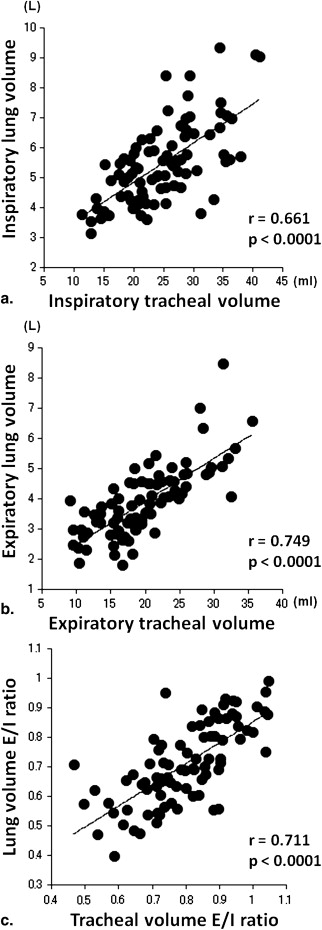
Tracheal volume showed moderate or strong, positive correlations with lung volume on both inspiratory ( r = 0.661, P < .0001) and end-expiratory ( r = 0.749, P < .0001) scans. The E/I ratio of tracheal volume showed a strong, positive correlation with the E/I ratio of lung volume ( r = 0.711, P < .0001). A weak, negative correlation was found between the E/I ratio of tracheal volume and the ratio of forced expiratory volume in the first second to forced vital capacity ( r = −0.436, P < .0001). Also, a weak, positive correlation was observed between the E/I ratio of tracheal volume and the ratio of residual volume to total lung capacity ( r = 0.253, P = .02).
Conclusions
Tracheal volume and collapsibility, measured by inspiratory and end-expiratory CT scans, is related to lung volume and collapsibility. The highly collapsed trachea on end-expiratory CT does not indicate more severe airflow limitation or air-trapping in smokers.
The meaning behind the highly collapsed trachea, observed on expiratory scans of computed tomography (CT), is still controversial. Although it has been acknowledged that the central airways markedly decrease in size on dynamic or end-expiratory CT scans in patients with tracheomalacia (TM) or tracheobronchomalacia (TBM) , some studies have demonstrated that the highly collapsed trachea can also be observed on these expiratory scans in subjects with normal lung function . In patients with chronic obstructive pulmonary disease (COPD), published information on the size of the trachea is more limited. Although some investigations have demonstrated the presence of the highly collapsed central airways in COPD using dynamic expiratory or end-expiratory scans , it still remains unclear whether or not the highly collapsed trachea indicates reduced lung function.
In 2003, Ederle and colleagues demonstrated that, in both normal subjects and subjects with obstructive pulmonary disease, cross-sectional area of the trachea correlated with mean lung density (MLD) and cross-sectional area of the lung on both inspiratory and end-expiratory CT scans . They also showed that the changes in tracheal cross-sectional area between inspiratory and expiratory scans significantly correlated to the changes in MLD. Further, more recent studies have demonstrated significant correlations between MLD and lung volume (LV) . Based on these observations, it can be predicted that the observations of Ederle and colleagues would be reproduced using volumetric measurements, such as LV and tracheal volume.
Get Radiology Tree app to read full this article<
Get Radiology Tree app to read full this article<
Materials and methods
Get Radiology Tree app to read full this article<
Subjects
Get Radiology Tree app to read full this article<
Get Radiology Tree app to read full this article<
Table 1
Clinical Characteristics of the 85 Subjects
Mean ± SD (Range) Age (y) 67.8 ± 8.0 (45−87) Smoking index (pack-years) 48.4 ± 32.9 (2−180) FEV 1 /FVC 0.56 ± 0.14 (0.17−0.83) FEV 1 (%predicted) 60.8 ± 23.6 (14−126) RV/TLC 0.50 ± 0.10 (0.32−0.73) TLC (L) 6.45 ± 1.34 (4.10−10.20) RV (L) 3.24 ± 0.97 (1.50−7.00)
FEV 1, forced expiratory volume in 1 second; FVC, forced vital capacity; RV, residual volume; SD, standard deviation; TLC, total lung capacity.
Get Radiology Tree app to read full this article<
Pulmonary Function Tests
Get Radiology Tree app to read full this article<
Get Radiology Tree app to read full this article<
Thin-section CT
Get Radiology Tree app to read full this article<
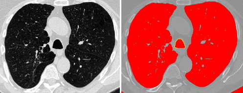
Measurements of the Trachea
Get Radiology Tree app to read full this article<
Get Radiology Tree app to read full this article<
Get Radiology Tree app to read full this article<
Get Radiology Tree app to read full this article<
Measurements of the Lung Volume
Get Radiology Tree app to read full this article<
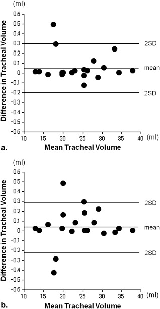
Reproducibility of Tracheal Volume Measurements
Get Radiology Tree app to read full this article<
Statistical Analysis
Get Radiology Tree app to read full this article<
Results
Reproducibility of Tracheal Volume Measurements
Get Radiology Tree app to read full this article<
Table 2
Reproducibility of Tracheal Volume Measurements
Mean Difference ± SD Limit of Agreement Tracheal volume (mL) Intraobserver error 0.05 ± 0.13 −0.20 to 0.30 Interobserver error 0.04 ± 0.15 −0.22 to 0.29
SD, standard deviation.
Get Radiology Tree app to read full this article<
Tracheal Measurements
Get Radiology Tree app to read full this article<
Table 3
Measurements of the Trachea and Lung Volume on Chest CT
Inspiration Expiration E/I ratio Cross-sectional area (mm 2 ) Upper trachea 273.3 ± 63.7 236.4 ± 60.2 0.87 ± 0.12 Lower trachea 273.1 ± 66.7 234.4 ± 62.1 0.86 ± 0.11 Tracheal volume (mL) 24.6 ± 6.8 19.5 ± 6.1 0.79 ± 0.13 Lung volume (l) 5.44 ± 1.35 3.83 ± 1.17 0.71 ± 0.14
CT, computed tomography; E/I, expiratory/inspiratory.
Get Radiology Tree app to read full this article<
CT-based Lung Volume
Get Radiology Tree app to read full this article<
Correlations between Tracheal Volume and CT-based Lung Volume
Get Radiology Tree app to read full this article<
Table 4
Correlations of Tracheal Volume with CT-based Lung Volume and with Lung Function
CT-based Lung Volume Lung Function Inspiration Expiration E/I Ratio FEV 1 /FVC FEV 1
(% predicted) RV/TLC Tracheal volume Inspiration 0.661 ∗ 0.472 ∗ −0.126 −0.221 ¶ −0.110 −0.182 Expiration 0.677 ∗ 0.749 ∗ 0.293 ‡ −0.445 ∗ −0.179 −0.020 E/I ratio 0.135 0.551 ∗ 0.711 ∗ −0.436 ∗ −0.333 † 0.253 ¶
CT, computed tomography; E/I, expiratory/inspiratory; FEV 1 , forced expiratory volume in 1 second; FVC, forced vital capacity; RV/TLC, ratio of residual volume to total lung capacity.
Get Radiology Tree app to read full this article<
Get Radiology Tree app to read full this article<
Get Radiology Tree app to read full this article<
Get Radiology Tree app to read full this article<
Get Radiology Tree app to read full this article<
Correlations between Tracheal Volume and Lung Function
Get Radiology Tree app to read full this article<
Discussion
Get Radiology Tree app to read full this article<
Get Radiology Tree app to read full this article<
Get Radiology Tree app to read full this article<
Get Radiology Tree app to read full this article<
Get Radiology Tree app to read full this article<
Get Radiology Tree app to read full this article<
Get Radiology Tree app to read full this article<
Get Radiology Tree app to read full this article<
Get Radiology Tree app to read full this article<
Get Radiology Tree app to read full this article<
References
1. Gilkeson R.C., Ciancibello L.M., Hejal R.B., et. al.: Tracheobronchomalacia: dynamic airway evaluation with multidetector CT. Am J Roentgenol 2001; 176: pp. 205-210.
2. Aquino S.L., Shepard J.A., Ginns L.C., et. al.: Acquired tracheomalacia: detection by expiratory CT scan. J Comput Assist Tomogr 2001; 25: pp. 394-399.
3. Zhang J., Hasegawa I., Feller-Kopman D., et. al.: Dynamic expiratory volumetric CT imaging of the central airways: comparison of standard-dose and low-dose techniques. Acad Radiol 2003; 10: pp. 719-724.
4. Carden K.A., Boiselle P.M., Waltz D.A., et. al.: Tracheomalacia and tracheobronchomalacia in children and adults: an in-depth review. Chest 2005; 127: pp. 984-1005.
5. Baroni R.H., Feller-Kopman D., Nishino M., et. al.: Tracheobronchomalacia: comparison between end-expiratory and dynamic expiratory CT for evaluation of central airway collapse. Radiology 2005; 235: pp. 635-641.
6. Lee K.S., Sun M.E., Ernst A., et. al.: Comparison of dynamic expiratory CT with bronchoscopy in diagnosing airway malacia. Chest 2007; 131: pp. 758-764.
7. Ferretti G.R., Jankowski A., Perrin M.A., et. al.: Multi-detector CT evaluation in patients suspected of tracheobronchomalacia: comparison of end-expiratory with dynamic expiratory volumetric acquisitions. Eur J Radiol 2008; 68: pp. 340-346.
8. Boiselle P.M., Ernst A.: Tracheal morphology in patients with tracheomalacia: prevalence of inspiratory “lunate” and expiratory “frown” shapes. J Thorac Imaging 2006; 21: pp. 190-196.
9. Boiselle P.M., O’Donnell C.R., Bankier A.A., et. al.: Tracheal collapsibility in healthy volunteers during forced expiration: assessment with multidetector CT. Radiology 2009; 252: pp. 255-262.
10. Thiriet M., Maarek J.M., Chartrand D.A., et. al.: Transverse images of the human thoracic trachea during forced expiration. J Appl Phsiol 1989; 67: pp. 1032-1040.
11. Ederle J.R., Heussel C.P., Hast J., et. al.: Evaluation of changes in central airway dimensions, lung area and mean lung density at paired inspiratory/expiratory high-resolution computed tomography. Eur Radiol 2003; 13: pp. 2454-2461.
12. Ochs R.A., Petkovska I., Kim H.J., et. al.: Prevalence of tracheal collapse in an emphysema cohort as measured with end-expiration CT. Acad Radiol 2009; 16: pp. 46-53.
13. Sverzellati N., Rastelli A., Chetta A., et. al.: Airway malacia in chronic obstructive pulmonary disease: prevalence, morphology and relationship with emphysema, bronchiectasis and bronchial wall thickening. Eur Radiol 2009; 19: pp. 1669-1678.
14. Lee C.J., Lee J.H., Song J.W., et. al.: Correlation of tracheal cross-sectional area with parameters of pulmonary function test in COPD. Tuberc Respir Dis 1999; 46: pp. 628-635.
15. Yamashiro T., Matsuoka S., Bartholmai B.J., et. al.: Collapsibility of lung volume by paired inspiratory and expiratory CT scans: correlations with lung function and mean lung density. Acad Radiol 2010; 17: pp. 489-495.
16. Camiciottoli G., Cavigli E., Grassi L., et. al.: Prevalence and correlates of pulmonary emphysema in smokers and former smokers. A densitometric study of participants in the ITALUNG trial. Eur Radiol 2009; 19: pp. 58-66.
17. Kauczor H.U., Heussel C.P., Fischer B., et. al.: Assessment of lung volumes using helical CT at inspiration and expiration: comparison with pulmonary function tests. Am J Roentgenol 1998; 171: pp. 1091-1095.
18. Zaporozhan J., Ley S., Eberhardt R., et. al.: Paired inspiratory/expiratory volumetric thin-section CT scan for emphysema analysis: comparison of different quantitative evaluations and pulmonary function test. Chest 2005; 128: pp. 3212-3220.
19. Iwano S., Okada T., Satake H., et. al.: 3D-CT volumetry of the lung using multidetector row CT: comparison with pulmonary function tests. Acad Radiol 2009; 16: pp. 250-256.
20. Griscom N.T., Wohl M.E.: Tracheal size and shape: effects of change in intraluminal pressure. Radiology 1983; 149: pp. 27-30.
21. Miller M.R., Hankinson J., Brusasco V., et. al.: ATS/ERS task force: standardisation of spirometry. Eur Respir J 2005; 26: pp. 319-338.
22. Stocks J., Quanjer P.H.: Reference values for residual volume, functional residual capacity and total lung capacity: ATS workshop on lung volume measurements/official statement of the European Respiratory Society. Eur Respir J 1995; 8: pp. 492-506.
23. Rabe K.F., Hurd S., Anzueto A., et. al.: Global strategy for the diagnosis, management, and prevention of chronic obstructive pulmonary disease: GOLD executive summary. Am J Respir Crit Care Med 2007; 176: pp. 532-555.
24. Bland J.M., Altman D.G.: Statistical methods for assessing agreement between two methods of clinical measurement. Lancet 1986; 1: pp. 307-310.
25. Hoffstein V.: Relationship between lung volume, maximal expiratory flow, forced expiratory volume in one second, and tracheal area in normal men and women. Am Rev Respir Dis 1986; 134: pp. 956-961.
26. Hoffstein V., Castile R.G., O’Donnell C.R., et. al.: In vivo estimation of tracheal distensibility and hysteresis in normal adults. J Appl Physiol 1987; 63: pp. 2482-2489.
27. Martin T.R., Castile R.G., Fredberg J.J., et. al.: Airway size is related to sex but not lung size in normal adults. J Appl Physiol 1987; 63: pp. 2042-2047.
28. Dolyniuk M.V., Fahey P.J.: Relationship of tracheal size to maximal expiratory airflow and density dependence. J Appl Physiol 1986; 60: pp. 501-515.
29. Heussel C.P., Ley S., Biedermann A., et. al.: Respiratory lumenal change of the pharynx and trachea in normal subjects and COPD patients: assessment by cine-MRI. Eur Radiol 2004; 14: pp. 2188-2197.
30. Akira M., Toyokawa K., Inoue Y., et. al.: Quantitative CT in chronic obstructive pulmonary disease: inspiratory and expiratory assessment. Am J Roentgenol 2009; 192: pp. 267-272.
31. Matsuoka S., Kurihara Y., Yagihashi K., et. al.: Airway dimensions at inspiratory and expiratory multisection CT in chronic obstructive pulmonary disease: correlation with airflow limitation. Radiology 2008; 248: pp. 1042-1049.
32. O’Donnell C.R., Loring S.H.: Comparison of plethysmographic, helium dilution and CT-derive total lung capacity. Am J Respir Crit Care Med 2005; 171: A293 (abstract)
33. Garfield J.L., Marchetti N., Gaughan J.P., et. al.: Lung volume by plethsmography and CT in advanced COPD. Am J Respir Crit Care Med 2009; 179: A2902 (abstract)