
Rationale and Objectives
Emergency department (ED) patients with acute low back pain (LBP) may present with ambiguous clinical findings that pose diagnostic challenges to exclude cauda equina syndrome (CES). As a proof of concept, we aimed to determine the efficacy of a rapid lumbar spine (LS) magnetic resonance imaging (MRI) screening protocol consisting of a single 3D-T2 SPACE FS (3D-T2 Sampling Perfection with Application optimized Contrasts using different flip angle Evolution fat saturated) sequence relative to conventional LS MRI to exclude emergently treatable pathologies in this complex patient population.
Materials and Methods
LS MRI protocol including a sagittal 3D-T2 SPACE FS pulse sequence was added to the routine for ED patients presenting with acute atypical LBP over a 12-month period. Imaging findings were categorically scored on the 3D-T2 SPACE FS sequence and separately on the reference standard conventional LS MRI sequences. Patients’ symptoms were obtained from review of the electronic medical record. Descriptive test statistics were performed.
Results
Of the 206 ED patients who obtained MRI for acute atypical LBP, 118 (43.3 ± 13.5 years of age; 61 female) were included. Specific pathologies detected on reference standard conventional MRI included disc herniation ( n = 30), acute fracture ( n = 3), synovial cyst ( n = 3), epidural hematoma ( n = 2), cerebrospinal fluid leak ( n = 1), and leptomeningeal metastases ( n = 1), and on multiple occasions these pathologies resulted in nerve root impingement ( n = 36), severe spinal canal stenosis ( n = 13), cord/conus compression ( n = 2), and cord signal abnormality ( n = 2). The 3D-T2 SPACE FS sequence was an effective screen for fracture (sensitivity [sens] = 100%, specificity [spec] = 100%), cord signal abnormality (sens = 100%, spec = 99%), and severe spinal canal stenosis (sens = 100%, spec = 96%), and identified cord compression not seen on reference standard. Motion artifact was not seen on the 3D-T2 SPACE FS but noted on 8.5% of conventional LS MRI.
Conclusions
The 3D-T2 SPACE FS sequence MRI is a rapid, effective screen for emergently actionable pathologies that might be a cause of CES in ED patients presenting with acute atypical LBP. As this abbreviated, highly sensitive sequence requires a fraction of the acquisition time of conventional LS MRI, it has the potential of contributing to increased efficiencies in the radiology department and improved ED throughput.
Introduction
In the emergency department (ED) setting, the main role of magnetic resonance imaging (MRI) of the lumbar spine (LS) is not to confirm chronic problems, but to detect emergently actionable pathologies that, if not treated expeditiously, could result in permanent neurological deficits. Cauda equina syndrome (CES) is the most important example of such a critical clinical and imaging diagnosis that warrants emergent exclusion to prevent permanent neurological damage. Clinical guidelines set forth in the radiology and emergency medicine literature , based upon the presence of specific clinical “red flags,” are useful for stratifying the risk of an underlying serious low back pain (LBP) etiology (e.g., malignancy, infection, and/or trauma). While the CES diagnosis is frequently suspected in ED patients complaining of LBP, multiple prior studies have demonstrated that true CES is quite uncommon, found in only 1%–2% of suspected ED patients . The surgical literature estimates that true CES accounts for only 1%–3% of patients requiring lumbar spinal surgery . The varied clinical presentation, sundry etiologies, potential gravity of findings, and medicolegal liability of a missed diagnosis all serve as driving forces for the increasing utilization of emergent LS MRI . In our institution, more than 200 emergent LS MRI examinations per year on average are performed to “rule out” CES in ED patients with acute LBP , and although this is disproportionate to the true incidence of this rare entity, imaging is crucial to exclude critical pathology.
We hypothesized that a single-sequence fat-saturated (FS) 3D-T2 turbo spin echo with variable flip angles (3D-T2 SPACE [3D-T2 Sampling Perfection with Application optimized Contrasts using different flip angle Evolution]; Siemens AG, Munich, Germany) can serve as a rapid and accurate screening examination for excluding potentially emergently actionable etiologies (i.e., fracture, cord compression, cord signal abnormality, and/or severe spinal canal stenosis) in these acute atypical LBP ED patients. As a proof of concept study, we present the sensitivity and specificity for a single-sequence 3D-T2 SPACE FS MRI screen compared to the reference standard conventional LS MRI to validate this screening study in ruling out important etiologies in this complicated patient population. Furthermore, given that this rapid, abbreviated MRI can be completed in a fraction of the time of conventional LS MRI, there is the real potential of improving efficiency in the radiology department and patient throughput in the ED. With limited charge for the abbreviated LS MRI protocol, there is also the potential for overall decreased healthcare costs.
Materials and Methods
Participants
Get Radiology Tree app to read full this article<
Magnetic Resonance Imaging
Get Radiology Tree app to read full this article<
Image Analysis
Get Radiology Tree app to read full this article<
Statistical Analysis
Get Radiology Tree app to read full this article<
Results
Demographic and Pathology Characteristics
Get Radiology Tree app to read full this article<
TABLE 1
Patient Demographics
Subjects Included_n_ = 118 Excluded_n_ = 88 Sex Female_n_ = 61 Male_n_ = 57 Age Mean 43.3 y 95% CI 29.8–56.8 y Range 18–69 y BMI Mean 30.1 95% CI 29.3–31.9 Range 18–52 Prior lumbar spine surgery Orthopedic hardware_n_ = 8 No orthopedic hardware_n_ = 6 Presenting symptom(s) Lower extremity weakness_n_ = 68 Incontinence_n_ = 56 Perineal anesthesia_n_ = 23 Focal neurological deficit_n_ = 9
CI, confidence interval.
TABLE 2
Pathologies Observed on Reference Standard LS MRI
Intraspinal mass_n_ = 37 Disc_n_ = 30 Synovial cyst_n_ = 3 Hematoma_n_ = 2 CSF leak (perineural and epidural T2 hyperintensity)n = 1 Metastases_n_ = 1 Nerve root impingement_n_ = 36 Severe spinal canal stenosis_n_ = 13 Marrow signal abnormality_n_ = 4 Acute fracture_n_ = 3 Cord signal abnormality_n_ = 2 Cord/conus medullaris compression_n_ = 2
CSF, cerebrospinal fluid; LS, lumbar spine; MRI, magnetic resonance imaging.
Get Radiology Tree app to read full this article<
Qualitative Analysis
Get Radiology Tree app to read full this article<
TABLE 3
Qualitative Results
Variable 3D-T2 SPACE FS Conventional LS MRI Obscured conus medullaris 2.5% ( n = 3) 0% ( n = 0) Obscured cauda equina 6.8% ( n = 8) 0.8% ( n = 1) Orthopedic hardware 6.8% ( n = 8) 6.8% ( n = 8) Hardware limited examination 5.9% ( n = 7) 4.2% ( n = 5) Motion artifact 0% ( n = 0) 8.5% ( n = 10)
3D-T2 SPACE FS, 3D-T2 Sampling Perfection with Application optimized Contrasts using different flip angle Evolution fat saturated; LS, lumbar spine; MRI, magnetic resonance imaging.
Get Radiology Tree app to read full this article<
Quantitative Analysis
Get Radiology Tree app to read full this article<
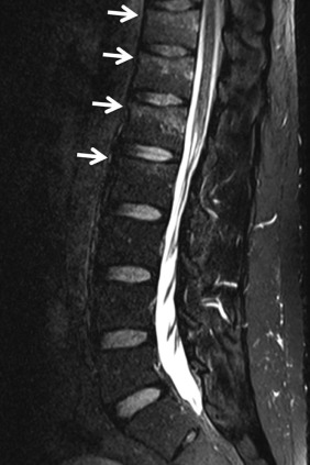
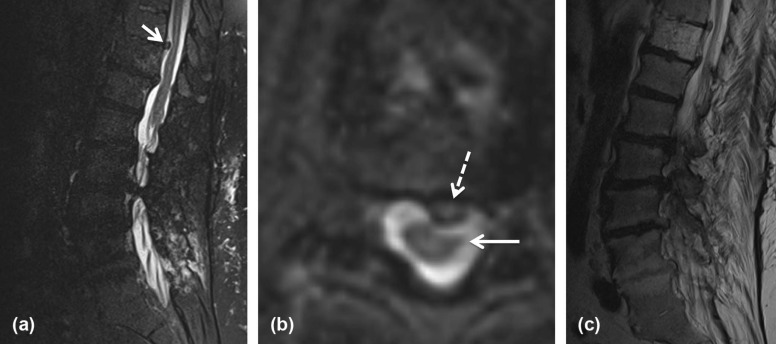
TABLE 4
Performance of 3D-T2 SPACE FS for Evaluation of CES in All Included Patients
Variable Sensitivity % (95% CI), n Specificity % (95% CI), n Acute fracture 100 (29–100), 3 \* 100 (97–100), 115 Cord signal abnormality 100 (2.5–100), 1 \* 99 (95–100), 117 Cord/conus medullaris compression † † 100 (2.5–100), 2 \* 99 (95–100), 117 Synovial cyst 100 (29–100), 3 \* 100 (97–100), 116 CSF leak 100 (2.5–100), 1 \* 100 (97–100), 117 Marrow signal abnormality 100 (40–100), 4 \* 98 (94–100), 114 Severe spinal canal stenosis 100 (75–100), 13 96 (91–99), 105 Nerve root impingement 69 (52–84), 36 90 (82–96), 82 Disc 67 (47–83), 30 89 (90–94), 88 Hematoma 50 (1.3–99), 2 \* 100 (97–100), 116 Metastasis 0 (0–98), 1 \* 100 (97–100), 117
3D-T2 SPACE FS, 3D-T2 Sampling Perfection with Application optimized Contrasts using different flip angle Evolution fat saturated; CES, cauda equina syndrome; CI, confidence interval; CSF, cerebrospinal fluid.
Get Radiology Tree app to read full this article<
Get Radiology Tree app to read full this article<
Get Radiology Tree app to read full this article<
Get Radiology Tree app to read full this article<
Inter-rater Agreement on 3D-T2 SPACE FS
Get Radiology Tree app to read full this article<
Get Radiology Tree app to read full this article<
Discussion
Get Radiology Tree app to read full this article<
Get Radiology Tree app to read full this article<
Get Radiology Tree app to read full this article<
Get Radiology Tree app to read full this article<
Get Radiology Tree app to read full this article<
Supplementary Data
Get Radiology Tree app to read full this article<
Appendix S1
Get Radiology Tree app to read full this article<
Get Radiology Tree app to read full this article<
Get Radiology Tree app to read full this article<
References
1. Patel N.D., Broderick D.F., Burns J., et. al.: ACR appropriateness criteria low back pain. J Am Coll Radiol 2016; 13: pp. 1069-1078.
2. Davis P.C., Wippold F.J., Brunberg J.A., et. al.: ACR appropriateness criteria on low back pain. J Am Coll Radiol 2009; 6: pp. 401-407.
3. Edlow J.A.: Managing nontraumatic acute back pain. Ann Emerg Med 2015; 66: pp. 148-153.
4. Shah L.M., Long D., Sanone D., et. al.: Application of ACR appropriateness guidelines for spine MRI in the emergency department. J Am Coll Radiol 2014; 11: pp. 1002-1004.
5. Shah L.M., Evans C., Osborn A.: Emergent lumbar spine imaging for cauda equina syndrome: what is appropriate?. American Society of Neuroradiology Annual Meeting; New York2011.
6. Fraser S., Roberts L., Murphy E.: Cauda equina syndrome: a literature review of its definition and clinical presentation. Arch Phys Med Rehabil 2009; 90: pp. 1964-1968.
7. Lavy C., James A., Wilson-MacDonald J., et. al.: Cauda equina syndrome. BMJ 2009; 338: pp. b936.
8. Spangfort E.V.: The lumbar disc herniation. A computer-aided analysis of 2,504 operations. Acta Orthop Scand Suppl 1972; 142: pp. 1-95.
9. Deyo R.A., Rainville J., Kent D.L.: What can the history and physical examination tell us about low back pain?. JAMA 1992; 268: pp. 760-765.
10. Gleave J.R., Macfarlane R.: Cauda equina syndrome: what is the relationship between timing of surgery and outcome?. Br J Neurosurg 2002; 16: pp. 325-328.
11. Roudsari B., Jarvik J.G.: Lumbar spine MRI for low back pain: indications and yield. AJR Am J Roentgenol 2010; 195: pp. 550-559.
12. Verbiest H.: Pathomorphologic aspects of developmental lumbar stenosis. Orthop Clin North Am 1975; 6: pp. 177-196.
13. Wilson E.B.: Probable interference, the law of succession, and statistical interference. J Am Stat Assoc 1927; 22: pp. 209-212.
14. Brown L.D., Cai T.T., DasGupta A.: Interval estimation for a binomial proportion. Stat Sci 2001; 16: pp. 101-133.
15. Byrt T., Bishop J., Carlin J.B.: Bias, prevalence and kappa. J Clin Epidemiol 1993; 46: pp. 423-429.
16. CDC : Behavioral risk factor surveillance system: prevalence and trend data–overweight and obesity. U.S. Obesity Trends, Trends by State; Available at: http://nccd.cdc.gov/NPAO_DTM/
17. Pepe M.S.: The statistical evaluation of medical tests for classification and prediction.2003.Oxford University PressNew York
18. FDA : Statistical guidance on reporting results from studies evaluating diagnostic tests.Health CfDaR 2007.
19. Tins B., Cassar-Pullicino V., Haddaway M., et. al.: Three-dimensional sampling perfection with application-optimised contrasts using a different flip angle evolutions sequence for routine imaging of the spine: preliminary experience. Br J Radiol 2012; 85: pp. e480-e489.
20. Lee S., Jee W.H., Jung J.Y., et. al.: MRI of the lumbar spine: comparison of 3D isotropic turbo spin-echo SPACE sequence versus conventional 2D sequences at 3.0 T. Acta Radiol 2015; 56: pp. 174-181.
21. Mahadevappa K., Persi A., Nesathurai S.: Acute cauda equina syndrome caused by a disk lesion: is emergent surgery the correct option?. Spine 2015; 40: pp. 636-638.
22. Macki M., Hernandez-Hermann M., Bydon M., et. al.: Spontaneous regression of sequestrated lumbar disc herniations: literature review. Clin Neurol Neurosurg 2014; 120: pp. 136-141.
23. Casey E.: Natural history of radiculopathy. Phys Med Rehabil Clin N Am 2011; 22: pp. 1-5.