
Rationale and Objectives
The aims of this study were to evaluate the effectiveness of low-dose, contrast-enhanced (CE), time-resolved, three-dimensional magnetic resonance angiography (MRA) in the assessment of the abdominal aorta and its major branches at 3 T and to compare the results with those of high–spatial resolution CE MRA.
Materials and Methods
Twenty-two consecutive patients (eight men, 14 women; mean age, 43.9 ± 17.9 years) underwent CE time-resolved three-dimensional MRA and high–spatial resolution three-dimensional MRA. Studies were performed using a 3-T magnetic resonance system; gadolinium-based contrast medium was administered at a dose of 3 to 5 mL for time-resolved MRA, followed by 0.1 mmol/kg gadopentetate dimeglumine for single-phase CE MRA. For analysis purposes, the abdominal arterial system was divided into 11 arterial segments, and image quality as well as the presence and degree of vascular pathology were evaluated by two independent magnetic resonance radiologists.
Results
A total of 242 arterial segments were visualized with good image quality. Time-resolved MRA was able to visualize the majority of arterial segments with good definition in the diagnostic range. Vascular pathologies (stenosis, occlusion) or abnormal vascular anatomy was detected in 19 arterial segments, with good interobserver agreement (κ = 0.78). All image findings were detected with time-resolved CE MRA by both observers and were confirmed by correlative imaging.
Conclusion
Low-dose, time-resolved MRA at 3 T yields rapid and important anatomic and functional information in the evaluation of the abdominal vasculature. Because of its limited spatial resolution, time-resolved MRA is inferior to CE MRA in demonstrating fine vascular details.
Three-dimensional (3D) contrast-enhanced (CE) magnetic resonance (MR) angiography (MRA) has made substantial technical advances in the past decade and is now widely used in many applications . Furthermore, CE MRA has been accepted in clinical routine as a substitute for the standard of reference, digital subtraction angiography (DSA), for evaluating the abdominal vasculature and its major branches .
Today most conventional CE MR angiographic methods aim to capture a single imaging volume at peak arterial contrast enhancement with minimal venous overlay. Therefore, these methods require that the acquisition and breath-hold coincide with the presence of the contrast bolus in the vessels of interest for optimal visualization of the arterial tree. But the timing of contrast material arrival may be crucial, because sufficient T1 shortening of blood is achieved only for few seconds during peak vascular concentration. As a result, delayed acquisition will lead to a substantial decrease in diagnostic accuracy. To guarantee accurate bolus timing, different strategies have been developed, including the test-bolus technique , automated detection of the contrast material , and real-time depiction of bolus arrival .
Get Radiology Tree app to read full this article<
Get Radiology Tree app to read full this article<
Get Radiology Tree app to read full this article<
Get Radiology Tree app to read full this article<
Materials and methods
Patient Population
Get Radiology Tree app to read full this article<
Get Radiology Tree app to read full this article<
MR Imaging
Get Radiology Tree app to read full this article<
Get Radiology Tree app to read full this article<
Time-resolved CE MRA
Get Radiology Tree app to read full this article<
Get Radiology Tree app to read full this article<
Get Radiology Tree app to read full this article<
Get Radiology Tree app to read full this article<
Get Radiology Tree app to read full this article<
Get Radiology Tree app to read full this article<
Single-phase CE MRA
Get Radiology Tree app to read full this article<
Table 1
Sequence Parameters of Time-resolved CE MRA and High–spatial resolution Single-phase CE MRA
Parameter Time-resolved 4D CE MRA Single-phase 3D CE MRA Orientation Coronal Coronal Repetition time (ms) 2.54 3.0 Echo time (ms) 1.05 1.14 Flip angle (°) 12–19 ∗ 19–23 ∗ Slice thickness (mm) 3.0 1.0 FOV read (mm) 420 420 FOV phase (%) 75 75 Base resolution 512 512 Phase resolution (%) 75 80 Slices per slab 32–36 ∗ 88–96 ∗ Matrix size 512 × 288 512 × 308 True voxel size (mm) 1.1 × 0.8 × 3.8 1.0 × 0.8 × 1.6 Interpolated voxel size (mm) 0.8 × 0.8 × 3.0 0.8 × 0.8 × 1.0 PAT mode GRAPPA GRAPPA Acceleration factor 3 3 Reference lines 24 24 Bandwidth (Hz/pixel) 750 650 Number of measurements 14–16 ∗ 1 Total scan time (s) 26 19
CE, contrast-enhanced; 4D, four-dimensional; FOV, field of view; GRAPPA, generalized autocalibrating partially parallel acquisitions; MRA, magnetic resonance angiography; PAT, parallel acquisition technique; 3D, three-dimensional.
Get Radiology Tree app to read full this article<
Get Radiology Tree app to read full this article<
Get Radiology Tree app to read full this article<
Get Radiology Tree app to read full this article<
Table 2
Demographic Data of Patient Population
Patient Age (y) Height (m) Weight (kg) BMI (kg/m 2 ) Reason for MRA Referral 1 56 1.52 73 31.60 RAS 2 46 1.75 66 21.55 Abdominal angina 3 59 1.78 90 28.41 RAS 4 40 1.71 83 28.38 RAS 5 19 1.65 60 22.04 RAS 6 63 1.64 61 22.68 RAS 7 37 1.90 77 21.33 RAS 8 51 1.72 83 28.06 KD 9 61 1.67 84 30.12 RAS 10 59 1.69 53 18.56 AAA 11 32 1.86 112 32.37 RAS 12 50 1.63 70 26.35 Abdominal angina 13 66 1.67 79 28.33 KD 14 65 1.80 75 23.15 Abdominal angina 15 33 1.73 70 23.39 RAS 16 58 1.65 63 23.14 RAS 17 63 1.64 62 23.05 RAS 18 52 1.66 86 31.21 RAS 19 26 1.78 83 26.20 KD 20 45 1.77 78 24.90 KD 21 54 1.70 69 23.88 KD 22 27 1.73 55 18.38 FMD
AAA, abdominal aortic aneurysm; BMI, body mass index; FMD, fibromuscular dysplasia; KD, kidney donation; MRA, magnetic resonance angiography; RAS, renal artery stenosis.
Get Radiology Tree app to read full this article<
Get Radiology Tree app to read full this article<
Image Analysis
Get Radiology Tree app to read full this article<
Get Radiology Tree app to read full this article<
Semiquantitative Analysis
Get Radiology Tree app to read full this article<
Get Radiology Tree app to read full this article<
Get Radiology Tree app to read full this article<
Get Radiology Tree app to read full this article<
Correlation with DSA
Get Radiology Tree app to read full this article<
Statistical Analysis
Get Radiology Tree app to read full this article<
Results
Get Radiology Tree app to read full this article<
Get Radiology Tree app to read full this article<
Get Radiology Tree app to read full this article<
Vascular Pathology
Get Radiology Tree app to read full this article<
Get Radiology Tree app to read full this article<
Vessel Depiction and Visibility Score
Get Radiology Tree app to read full this article<
Table 3
Results for Total Vessel Visibility Score on a per Patient Basis Calculated for Both Sequence Types
Time-resolved 4D CE MRA (TWIST) Single-phase 3D CE MRA Patient Reader 1 Reader 2 Mean Reader 1 Reader 2 Mean 1 34 35 34.5 40 39 39.5 2 30 30 30 34 35 34.5 3 26 28 27 40 40 40 4 24 24 24 39 37 38 5 24 23 23.5 33 35 34 6 28 29 28.5 36 36 36 7 21 22 21.5 40 39 39.5 8 25 24 24.5 40 38 39 9 26 26 26 40 40 40 10 25 25 25 44 42 43 11 20 21 20.5 40 40 40 12 35 36 35.5 42 40 41 13 37 36 36.5 41 41 41 14 35 37 36 44 44 44 15 24 25 24.5 31 32 31.5 16 31 32 31.5 42 43 42.5 17 32 31 31.5 39 39 39 18 22 23 22.5 36 38 37 19 19 20 19.5 35 34 34.5 20 28 31 29.5 40 39 39.5 21 29 31 30 40 38 39 22 28 32 30 40 40 40 Maximum 37 37 37 44 44 44 Minimum 19 20 19.5 31 32 31.5 Mean 27.4 28.2 27.8 38.9 38.6 38.8 SD 4.96 5.11 5.03 3.30 2.84 3.07
CE, contrast-enhanced; 4D, four-dimensional; MRA, magnetic resonance angiography; 3D, three-dimensional; SD, standard deviation; TWIST, time-resolved angiography with stochastic trajectories.
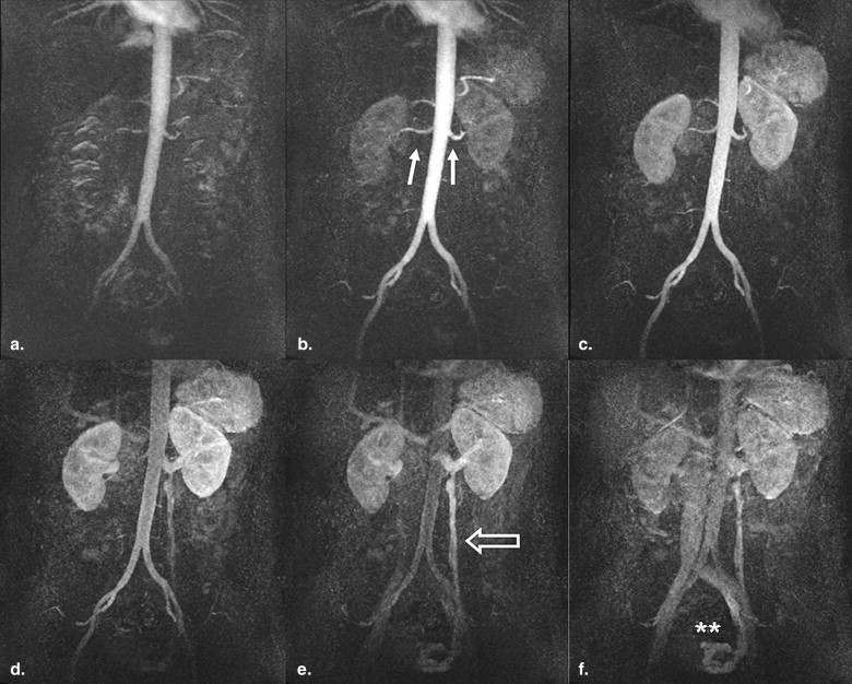
The mean visibility score for conventional single-phase 3D CE MRA was significantly higher than for time-resolved multiphase 4D CE MRA.
Table 4
Results for Total Vessel Visibility Score on a per Segment Basis Calculated for Both Sequence Types
Segment 1 2 3 4 5 6 7 8 9 10 11 4D 3D 4D 3D 4D 3D 4D 3D 4D 3D 4D 3D 4D 3D 4D 3D 4D 3D 4D 3D 4D 3D Sum 176 176 175 176 61 167 59 128 125 167 168 174 34 46 63 166 13 153 176 176 174 176 Visibility score 0 0 0 0 0 16 0 14 5 3 0 0 0 34 32 18 0 35 4 0 0 0 0 1 0 0 0 0 10 0 13 3 5 0 0 0 0 0 8 0 5 0 0 0 0 0 2 0 0 0 0 7 2 9 5 8 1 1 0 2 0 4 2 4 2 0 0 0 0 3 0 0 1 0 7 5 4 9 8 7 6 2 2 2 9 6 0 3 0 0 2 0 4 44 44 43 44 4 37 4 22 20 36 37 42 6 10 5 36 0 35 44 44 42 44
4D, four-dimensional; 3D, three-dimensional.
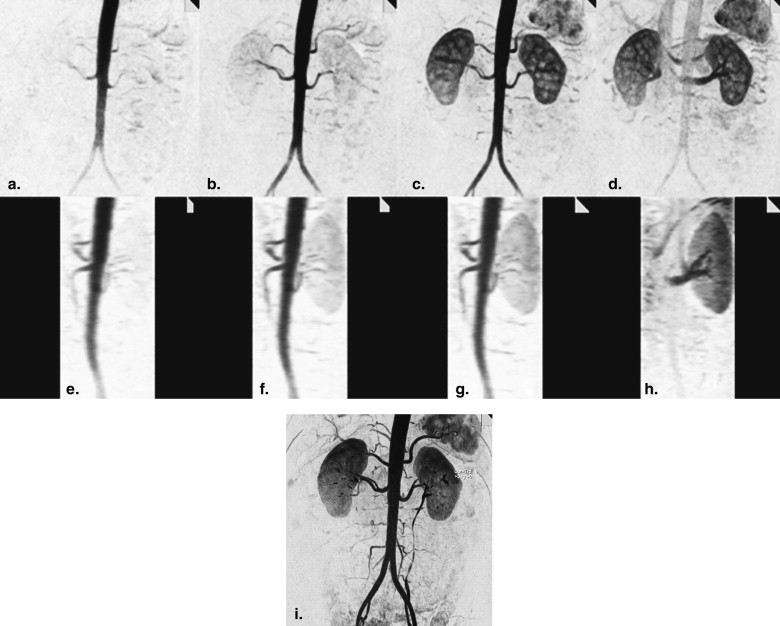
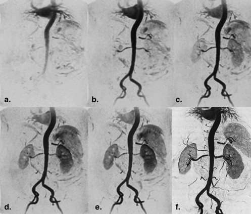
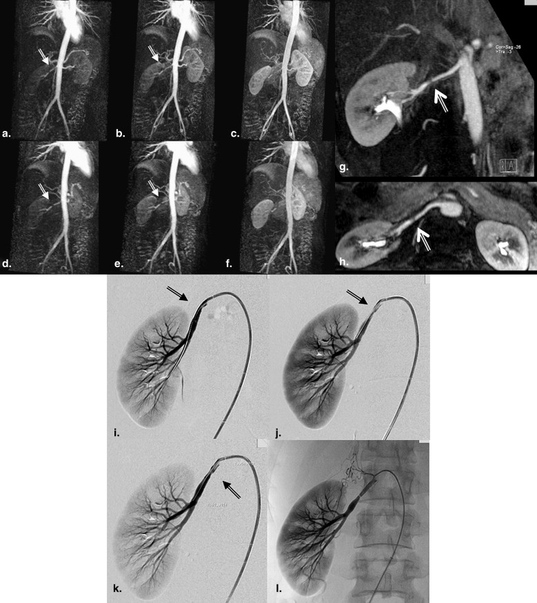
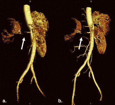
Vessel segments with smaller diameters, such as the hepatic artery (segment 5), splenic artery (segment 6), superior mesenteric artery (segment 8), and the inferior mesenteric artery (segment 9), were significantly better shown in detail on single-phase 3D contrast-enhanced magnetic resonance angiography.
Get Radiology Tree app to read full this article<
Get Radiology Tree app to read full this article<
Get Radiology Tree app to read full this article<
Get Radiology Tree app to read full this article<
Get Radiology Tree app to read full this article<
Image Artifacts
Get Radiology Tree app to read full this article<
Get Radiology Tree app to read full this article<
Discussion
Get Radiology Tree app to read full this article<
Technical Considerations
Get Radiology Tree app to read full this article<
Get Radiology Tree app to read full this article<
Get Radiology Tree app to read full this article<
Advantages of Higher Field Strength
Get Radiology Tree app to read full this article<
Get Radiology Tree app to read full this article<
Potential Clinical Applications
Get Radiology Tree app to read full this article<
Get Radiology Tree app to read full this article<
Get Radiology Tree app to read full this article<
Image Postprocessing and Multiplanar Reformatting
Get Radiology Tree app to read full this article<
Get Radiology Tree app to read full this article<
Limitations
Get Radiology Tree app to read full this article<
Get Radiology Tree app to read full this article<
Get Radiology Tree app to read full this article<
Get Radiology Tree app to read full this article<
Get Radiology Tree app to read full this article<
Get Radiology Tree app to read full this article<
Conclusion
Get Radiology Tree app to read full this article<
References
1. De Cobelli F., Venturini M., Vanzulli A., et. al.: Renal arterial stenosis: prospective comparison of color Doppler US and breath-hold, three-dimensional, dynamic, gadolinium-enhanced MR angiography. Radiology 2000; 214: pp. 373-380.
2. Fain S.B., King B.F., Breen J.F., et. al.: High-spatial-resolution contrast-enhanced MR angiography of the renal arteries: a prospective comparison with digital subtraction angiography. Radiology 2001; 218: pp. 481-490.
3. Leung D.A., Hagspiel K.D., Angle J.F., et. al.: MR angiography of the renal arteries. Radiol Clin North Am 2002; 40: pp. 847-865.
4. Fenchel M., Nael K., Deshpande V.S., et. al.: Renal magnetic resonance angiography at 3.0 Tesla using a 32-element phased-array coil system and parallel imaging in 2 directions. Invest Radiol 2006; 41: pp. 697-703.
5. Nael K., Saleh R., Lee M., et. al.: High-spatial-resolution contrast-enhanced MR angiography of abdominal arteries with parallel acquisition at 3.0 T: initial experience in 32 patients. AJR Am J Roentgenol 2006; 187: pp. W77-W85.
6. Schoenberg S.O., Rieger J., Weber C.H., et. al.: High-spatial-resolution MR angiography of renal arteries with integrated parallel acquisitions: comparison with digital subtraction angiography and US. Radiology 2005; 235: pp. 687-698.
7. Zhang H., Prince M.R.: Renal MR angiography. Magn Reson Imaging Clin N Am 2004; 12: pp. 487-503.
8. Kramer U., Wiskirchen J., Fenchel M.C., et. al.: Isotropic high-spatial-resolution contrast-enhanced 3.0-T MR angiography in patients suspected of having renal artery stenosis. Radiology 2008; 247: pp. 228-240.
9. Leung D.A., Hany T.F., Debatin J.F.: Three-dimensional contrast-enhanced magnetic resonance angiography of the abdominal arterial system. Cardiovasc Intervent Radiol 1998; 21: pp. 1-10.
10. Michaely H.J., Nael K., Schoenberg S.O., et. al.: The feasibility of spatial high-resolution magnetic resonance angiography (MRA) of the renal arteries at 3.0 T. Rofo 2005; 177: pp. 800-804.
11. Wilson G.J., Hoogeveen R.M., Willinek W.A., Muthupillai R., Maki J.H.: Parallel imaging in MR angiography. Top Magn Reson Imaging 2004; 15: pp. 169-185.
12. Schoenberg S.O., Bock M., Knopp M.V., et. al.: Renal arteries: optimization of three-dimensional gadolinium-enhanced MR angiography with bolus-timing-independent fast multiphase acquisition in a single breath hold. Radiology 1999; 211: pp. 667-679.
13. Shetty A.N., Bis K.G., Kirsch M., et. al.: Contrast-enhanced breath-hold three-dimensional magnetic resonance angiography in the evaluation of renal arteries: optimization of technique and pitfalls. J Magn Reson Imaging 2000; 12: pp. 912-923.
14. Prince M.R., Chenevert T.L., Foo T.K., et. al.: Contrast-enhanced abdominal MR angiography: optimization of imaging delay time by automating the detection of contrast material arrival in the aorta. Radiology 1997; 203: pp. 109-114.
15. Wilman A.H., Riederer S.J., King B.F., et. al.: Fluoroscopically triggered contrast-enhanced three-dimensional MR angiography with elliptical centric view order: application to the renal arteries. Radiology 1997; 205: pp. 137-146.
16. Fenchel M., Saleh R., Dinh H., et. al.: Juvenile and adult congenital heart disease: time-resolved 3D contrast-enhanced MR angiography. Radiology 2007; 244: pp. 399-410.
17. Korosec F.R., Frayne R., Grist T.M., Mistretta C.A.: Time-resolved contrast-enhanced 3D MR angiography. Magn Reson Med 1996; 36: pp. 345-351.
18. Pinto C., Hickey R., Carroll T.J., et. al.: Time-resolved MR angiography with generalized autocalibrating partially parallel acquisition and time-resolved echo-sharing angiographic technique for hemodialysis arteriovenous fistulas and grafts. J Vasc Interv Radiol 2006; 17: pp. 1003-1009.
19. Mistretta C.A., Grist T.M., Korosec F.R., et. al.: 3D time-resolved contrast-enhanced MR DSA: advantages and tradeoffs. Magn Reson Med 1998; 40: pp. 571-581.
20. Michaely H.J., Herrmann K.A., Kramer H., et. al.: High-resolution renal MRA: comparison of image quality and vessel depiction with different parallel imaging acceleration factors. J Magn Reson Imaging 2006; 24: pp. 95-100.
21. Kramer U., Nael K., Laub G., et. al.: High-resolution magnetic resonance angiography of the renal arteries using parallel imaging acquisition techniques at 3.0 T: initial experience. Invest Radiol 2006; 41: pp. 125-132.
22. Griswold M.A., Jakob P.M., Heidemann R.M., et. al.: Generalized autocalibrating partially parallel acquisitions (GRAPPA). Magn Reson Med 2002; 47: pp. 1202-1210.
23. Pruessmann K.P., Weiger M., Scheidegger M.B., Boesiger P.: SENSE: sensitivity encoding for fast MRI. Magn Reson Med 1999; 42: pp. 952-962.
24. Werder R., Nanz D., Lutz A.M., et. al.: Assessment of the abdominal aorta and its visceral branches by contrast-enhanced dynamic volumetric hepatic parallel magnetic resonance imaging: feasibility, reliability and accuracy. Eur Radiol 2007; 17: pp. 541-551.
25. Nael K., Michaely H.J., Lee M., et. al.: Dynamic pulmonary perfusion and flow quantification with MR imaging, 3.0T vs. 1.5T: initial results. J Magn Reson Imaging 2006; 24: pp. 333-339.
26. Swan J.S., Carroll T.J., Kennell T.W., et. al.: Time-resolved three-dimensional contrast-enhanced MR angiography of the peripheral vessels. Radiology 2002; 225: pp. 43-52.
27. Kramer H., Michaely H.J., Requardt M., et. al.: Effects of injection rate and dose on image quality in time-resolved magnetic resonance angiography (MRA) by using 1.0M contrast agents. Eur Radiol 2007; 17: pp. 1394-1402.
28. Du J., Fain S.B., Korosec F.R., et. al.: Time-resolved contrast-enhanced carotid imaging using undersampled projection reconstruction acquisition. J Magn Reson Imaging 2007; 25: pp. 1093-1099.
29. Wieben O., Grist T.M., Hany T.F., et. al.: Time-resolved 3D MR angiography of the abdomen with a real-time system. Magn Reson Med 2004; 52: pp. 921-926.
30. Nael K., Ruehm S.G., Michaely H.J., et. al.: High spatial-resolution CE-MRA of the carotid circulation with parallel imaging: comparison of image quality between 2 different acceleration factors at 3.0 Tesla. Invest Radiol 2006; 41: pp. 391-399.
31. Kramer U., Thiel C., Seeger A., et. al.: Preoperative evaluation of potential living related kidney donors with high-spatial-resolution magnetic resonance (MR) angiography at 3 Tesla: comparison with intraoperative findings. Invest Radiol 2007; 42: pp. 747-755.
32. Campeau N.G., Huston J., Bernstein M.A., et. al.: Magnetic resonance angiography at 3.0 Tesla: initial clinical experience. Top Magn Reson Imaging 2001; 12: pp. 183-204.
33. Grobner T.: Gadolinium—a specific trigger for the development of nephrogenic fibrosing dermopathy and nephrogenic systemic fibrosis?. Nephrol Dial Transplant 2006; 21: pp. 1104-1108.
34. Perez-Rodriguez J., Lai S., Ehst B.D., et. al.: Nephrogenic systemic fibrosis: incidence, associations, and effect of risk factor assessment—report of 33 cases. Radiology 2009; 250: pp. 371-377.
35. Mihai G., Chung Y.C., Kariisa M., et. al.: Initial feasibility of a multi-station high resolution three-dimensional dark blood angiography protocol for the assessment of peripheral arterial disease. J Magn Reson Imaging 2009; 30: pp. 785-793.
36. Michaely H.J., Kramer H., Dietrich O., et. al.: Intraindividual comparison of high-spatial-resolution abdominal MR angiography at 1.5 T and 3.0 T: initial experience. Radiology 2007; 244: pp. 907-913.
37. Cohen E.I., Weinreb D.B., Siegelbaum R.H., et. al.: Time-resolved MR angiography for the classification of endoleaks after endovascular aneurysm repair. J Magn Reson Imaging 2008; 27: pp. 500-503.
38. Volk M., Strotzer M., Lenhart M., et. al.: Time-resolved contrast-enhanced MR angiography of renal artery stenosis: diagnostic accuracy and interobserver variability. AJR Am J Roentgenol 2000; 174: pp. 1583-1588.
39. Baskaran V., Pereles F.S., Nemcek A.A., et. al.: Gadolinium-enhanced 3D MR angiography of renal artery stenosis: a pilot comparison of maximum intensity projection, multiplanar reformatting, and 3D volume-rendering postprocessing algorithms. Acad Radiol 2002; 9: pp. 50-59.
40. Vasbinder G.B., Nelemans P.J., Kessels A.G., et. al.: Accuracy of computed tomographic angiography and magnetic resonance angiography for diagnosing renal artery stenosis. Ann Intern Med 2004; 141: pp. 674-682.