
Rationale and Objectives
The purpose of this study was to assess the frequency and spectrum of abnormalities on routine screening chest radiographs among inpatients and outpatients with “positive purified protein derivative (PPD)” in a large tertiary care academic medical center in a country with low prevalence of tuberculosis (TB).
Materials and Methods
The reports of all chest radiographs of general inpatients and outpatients referred for positive PPD (2010–2014) were evaluated for the frequency of evidence of active or latent TB and the spectrum of imaging findings. The results of additional chest radiographs and computed tomography scans were recorded, as were additional relevant clinical histories and symptoms.
Results
Of the 2518 patients who underwent chest radiography for positive PPD, the radiographs were normal in 91.3%. The vast majority of the abnormal radiographs demonstrated findings consistent with old tuberculous disease. There were three cases (0.1%) of active TB, all of which were either recent immigrants from an endemic area or had other relevant histories or clinical symptoms suggestive of the disease.
Conclusions
Universal chest radiography in general inpatient and outpatient populations referred for positive PPD is of low yield for detecting active disease in a country with low prevalence of TB.
Introduction
According to the Centers for Disease Control and Prevention, there were 9557 cases of active tuberculosis (TB) (a rate of three cases per 100,000 persons) reported in the United States during 2015, representing a 1.6% increase compared to the prior year. However, the number of TB deaths reported annually continues to decrease every year (11% from 2013 to 2014), with a 71% decrease in the annual death rate since 1992. The case rate among foreign-born persons is approximately 13 times higher than among U.S.-born persons (15.1 vs 1.2 cases per 100,000 persons) .
In countries with low TB prevalence such as the United States, about 90% of new cases of TB arise from reactivation of dormant foci of infection . Consequently, treatment of latent tuberculosis infection (LTBI) is a major component of the national strategy for eliminating the disease in the United States . Purified protein derivative (PPD), also known as tuberculin skin test, is the most common means of detecting prior TB infection. For those who test positively, the American Thoracic Society and the Centers for Disease Control and Prevention recommend routine screening chest radiography to exclude clinically active TB or to detect evidence of old healed disease to assess reactivation risk .
Get Radiology Tree app to read full this article<
Get Radiology Tree app to read full this article<
Materials and Methods
Get Radiology Tree app to read full this article<
Get Radiology Tree app to read full this article<
Get Radiology Tree app to read full this article<
Get Radiology Tree app to read full this article<
Results
Get Radiology Tree app to read full this article<
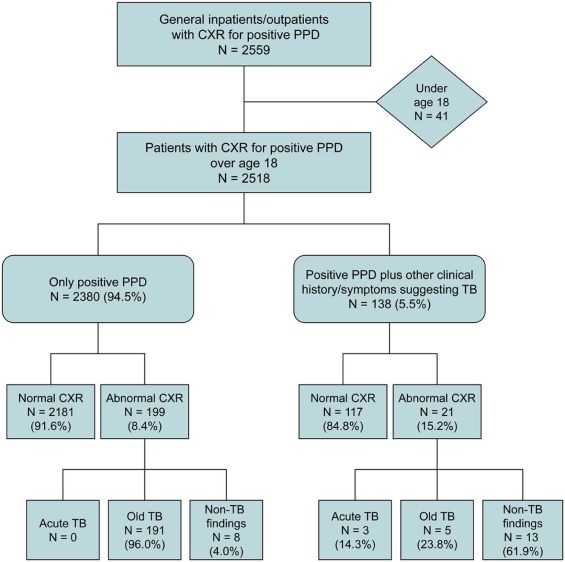
Table 1
Demographic Information and Clinical Symptoms
Characteristics Mean ± Standard Deviation (Range) Age (y) 42 ± 15(18–93) Gender Female 1539/2518(61.1) Male 979//2518(38.9) Any clinical symptoms 138/251(5.5) Cough 54/138(39.1) Fever 19/138(13.8) Night sweats 12/138(8.7) Hemoptysis 9/138(6.5) Unintended weight loss 9/138(6.5)
Continuous variables are displayed as mean ± standard deviation (range); categorical variables are displayed as numerator over denominator (%).
Get Radiology Tree app to read full this article<
Get Radiology Tree app to read full this article<
Get Radiology Tree app to read full this article<
Get Radiology Tree app to read full this article<
Get Radiology Tree app to read full this article<
Get Radiology Tree app to read full this article<
Get Radiology Tree app to read full this article<
Discussion
Get Radiology Tree app to read full this article<
Get Radiology Tree app to read full this article<
Get Radiology Tree app to read full this article<
Get Radiology Tree app to read full this article<
Get Radiology Tree app to read full this article<
Get Radiology Tree app to read full this article<
Conclusion
Get Radiology Tree app to read full this article<
References
1. Center for Disease Control and Prevention : Fact sheet—trends in tuberculosis. Available at: https://www.cdc.gov/tb/publications/factsheets/statistics/tbtrends.htm Accessed December 15, 2016
2. Sia I.G., Wieland M.L.: Current concepts in the management of tuberculosis. Mayo Clin Proc 2011; 86: pp. 348-361.
3. Horsburgh C.R.: Priorities for the treatment of latent tuberculous infection in the United States. N Engl J Med 2004; 350: pp. 2060-2067.
4. Targeted tuberculin testing and treatment of latent tuberculosis infection: a joint statement of the American Thoracic Society and the Centers for Disease Control and Prevention. Am J Respir Crit Care Med 2000; 161: pp. S221-S247.
5. Eisenberg R.L., Pollock N.R.: Low yield of chest radiography in a large tuberculosis screening program. Radiology 2010; 250: pp. 998-1004.
6. Jeong Y.J., Lee K.S.: Pulmonary tuberculosis: up-to-date imaging and management. AJR 2008; 191: pp. 834-844.
7. Jasmer R.M., Nahid P., Hopewell P.C.: Latent tuberculosis infection. N Engl J Med 2002; 347: pp. 1860-1866.
8. Gottridge J.A., Meyer R., Schwartz N.S., et. al.: The nonutility of chest roentgenographic examination in asymptomatic patients with positive tuberculin test results. Arch Intern Med 1989; 149: pp. 1660-1662.
9. Schaefer G.: Female genital tuberculosis. Clin Obstet Gynec 1976; 19: pp. 223-239.