
Rationale and Objectives
We aimed to determine the capability of magnetic resonance (MR) lymphography using gadofluorine M and to demonstrate normal lymph drainage from the mammary glands in mice.
Materials and Methods
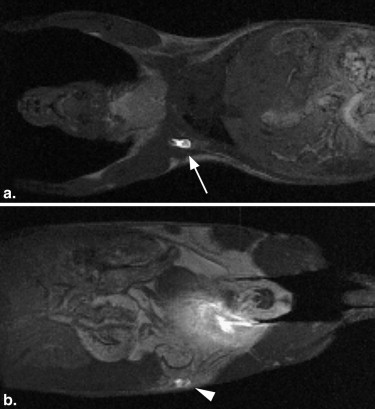
Three mice were intradermally injected with gadofluorine M near the papilla of the right fifth mammary gland, and subsequently underwent serial MR imaging to determine the appropriate method for assessment of the lymphatic pathway. MR lymphography was performed in 10 mice for the five right mammary glands to assess lymph drainage from each gland.
Results
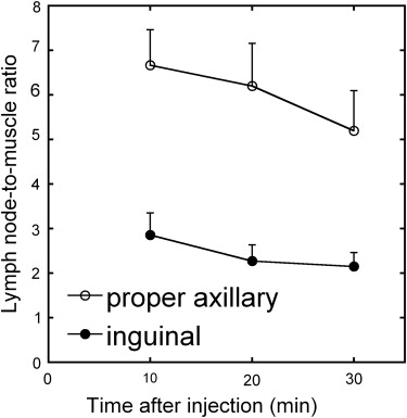
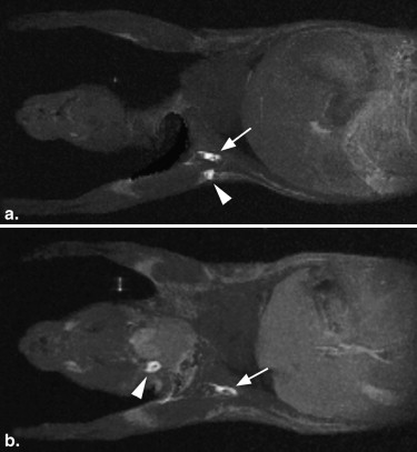
After intradermal injection near the right fifth papilla, high signal intensities representing lymph drainage were clearly demonstrated in the right inguinal and right proper axillary lymph nodes in all mice. The contrast between the lymph node and adjacent muscles was highest 10 minutes after injection and was still evident at 30 minutes. The lymph pathways from the five right mammary glands were successfully revealed, and no contralateral lymph nodes received lymph flow. Although variations in lymph drainage patterns from the first and second mammary glands existed among mice, injection in the third, fourth, and fifth glands gave consistent results. Lymphatics from the third gland drained exclusively into the proper axillary lymph node, and those from the fourth and fifth glands drained into the inguinal and proper axillary nodes.
Conclusion
MR lymphography with gadofluorine M allows noninvasive visualization of lymph drainage from the mammary glands in healthy mice.
Regional lymph node metastasis has an essential prognostic implication and is an important determinant in the treatment of patients with breast cancer . Animal experiments play a significant role in investigating the pathophysiology of diseases and predicting the efficacy of therapeutic strategies. Mice are the most popular platforms for small animal experiments because of their ease of handling, short lifespan, and fast reproductive turnover, and mouse models of breast cancer with lymph node metastasis are indispensable to breast cancer research . The normal lymph pathway from the mammary glands has been extensively studied in large animals such as dogs and cats . Such basic knowledge of lymph drainage pattern is important for understanding lymph node metastasis in animal models; however, this information is limited in mice. Harrell et al studied lymph drainage in mice using Evans blue dye, but failed to demonstrate drainage from the mammary glands . The small size of the lymph nodes in healthy mice may prevent investigation of the normal lymph pathway.
Imaging technologies are increasingly being used in small animal experiments. Among them, magnetic resonance (MR) imaging offers high-resolution three-dimensional (3D) images and is accepted as a potent tool for small animal research. Lymph drainage patterns may be assessed by interstitial MR lymphography . Contrast agents administered locally enter the lymphatic capillaries and are transported with the lymph flow to the lymph nodes, allowing the identification of lymph drainage from the injection site. Various contrast agents, including extracellular agents, ultrasmall superparamagnetic iron oxide particles, liposomes, dendrimers, and micelle-forming agents, have been used in MR lymphography. MR lymphography has been reported to allow the assessment of lymph drainage from the mammary gland using dendrimers as contrast agents ; however, this technique has not been used for the assessment of the lymph pathway from various levels of the mammary glands.
Get Radiology Tree app to read full this article<
Materials and methods
Animals
Get Radiology Tree app to read full this article<
Imaging Procedures
Get Radiology Tree app to read full this article<
Get Radiology Tree app to read full this article<
Technical Assessment of MR Lymphography for the Mammary Glands
Get Radiology Tree app to read full this article<
Get Radiology Tree app to read full this article<
Lymph Drainage from Mammary Glands
Get Radiology Tree app to read full this article<
Get Radiology Tree app to read full this article<
Results
Technical Assessment of MR Lymphography for the Mammary Glands
Get Radiology Tree app to read full this article<
Get Radiology Tree app to read full this article<
Lymph Drainage from Mammary Glands
Get Radiology Tree app to read full this article<
Table 1
Lymph Nodes Enhanced after Contrast Injection
Lymph Node
Region Number of Positive Results First gland Second gland Third gland Fourth gland Fifth gland Superficial cervical 3 0 0 0 0 Proper axillary 10 10 10 10 10 Accessory axillary 8 2 0 0 0 Inguinal 0 0 0 10 10
Number of positive results, the number of mice (among 10 mice studied) showing contrast enhancement in the given lymph node region after injection of gadofluorine M in the indicated mammary gland.
Get Radiology Tree app to read full this article<
Discussion
Get Radiology Tree app to read full this article<
Get Radiology Tree app to read full this article<
Get Radiology Tree app to read full this article<
Get Radiology Tree app to read full this article<
Acknowledgment
Get Radiology Tree app to read full this article<
References
1. Goldhirsch A., Glick J.H., Gelber R.D., et. al.: Meeting highlights: International Consensus Panel on the Treatment of Primary Breast Cancer. Seventh International Conference on Adjuvant Therapy of Primary Breast Cancer. J Clin Oncol 2001; 19: pp. 3817-3827.
2. Vantyghem S.A., Allan A.L., Postenka C.O., et. al.: A new model for lymphatic metastasis: development of a variant of the MDA-MB-468 human breast cancer cell line that aggressively metastasizes to lymph nodes. Clin Exp Metastasis 2005; 22: pp. 351-361.
3. Dadiani M., Kalchenko V., Yosepovich A., et. al.: Real-time imaging of lymphogenic metastasis in orthotopic human breast cancer. Cancer Res 2006; 66: pp. 8037-8041.
4. Patsikas M.N., Dessiris A.: The lymph drainage of the mammary glands in the bitch: a lymphographic study. Part I: The 1st, 2nd, 4th and 5th mammary glands. Anat Histol Embryol 1996; 25: pp. 131-138.
5. Raharison F., Sautet J.: Lymph drainage of the mammary glands in female cats. J Morphol 2006; 267: pp. 292-299.
6. Harrell M.I., Iritani B.M., Ruddell A.: Lymph node mapping in the mouse. J Immunol Methods 2008; 332: pp. 170-174.
7. Misselwitz B.: MR contrast agents in lymph node imaging. Eur J Radiol 2006; 58: pp. 375-382.
8. Kobayashi H., Kawamoto S., Sakai Y., et. al.: Lymphatic drainage imaging of breast cancer in mice by micro-magnetic resonance lymphangiography using a nano-size paramagnetic contrast agent. J Natl Cancer Inst 2004; 96: pp. 703-708.
9. Misselwitz B., Platzek J., Weinmann H.J.: Early MR lymphography with gadofluorine M in rabbits. Radiology 2004; 231: pp. 682-688.
10. Raatschen H.J., Swain R., Shames D.M., et. al.: MRI tumor characterization using Gd-GlyMe-DOTA-perfluorooctyl-mannose-conjugate (Gadofluorine M), a protein-avid contrast agent. Contrast Media Mol Imaging 2006; 1: pp. 113-120.
11. Meding J., Urich M., Licha K., et. al.: Magnetic resonance imaging of atherosclerosis by targeting extracellular matrix deposition with Gadofluorine M. Contrast Media Mol Imaging 2007; 2: pp. 120-129.
12. Choi S.H., Han M.H., Moon W.K., et. al.: Cervical lymph node metastases: MR imaging of gadofluorine M and monocrystalline iron oxide nanoparticle-47 in a rabbit model of head and neck cancer. Radiology 2006; 241: pp. 753-762.
13. Haishi T., Uematsu T., Matsuda Y., et. al.: Development of a 1.0 T MR microscope using a Nd-Fe-B permanent magnet. Magn Reson Imaging 2001; 19: pp. 875-880.
14. Inoue Y., Nomura Y., Haishi T., et. al.: Imaging living mice using a 1-T compact MRI system. J Magn Reson Imaging 2006; 24: pp. 901-907.
15. Kiryu S., Inoue Y., Yoshikawa K., et. al.: Diet and gastrointestinal signal on T1-weighted magnetic resonance imaging of mice. Magn Reson Imaging 2010; 28: pp. 273-280.
16. Ruehm S.G., Schroeder T., Debatin J.F.: Interstitial MR lymphography with gadoterate meglumine: initial experience in humans. Radiology 2001; 220: pp. 816-821.
17. Shimada M., Yoshikawa K., Suganuma T., et. al.: Interstitial magnetic resonance lymphography: comparative animal study of gadofluorine 8 and gadolinium diethylenetriamine-pentaacetic acid. J Comput Assist Tomogr 2003; 27: pp. 641-646.
18. McMasters K.M., Wong S.L., Martin R.C., et. al.: Dermal injection of radioactive colloid is superior to peritumoral injection for breast cancer sentinel lymph node biopsy: results of a multiinstitutional study. Ann Surg 2001; 233: pp. 676-687.
19. Suga K., Ogasawara N., Yuan Y., et. al.: Visualization of breast lymphatic pathways with an indirect computed tomography lymphography using a nonionic monometric contrast medium iopamidol: preliminary results. Invest Radiol 2003; 38: pp. 73-84.
20. Lin X., Yue P., Chen Z., et. al.: Hepatic triglyceride contents are genetically determined in mice: results of a strain survey. Am J Physiol Gastrointest Liver Physiol 2005; 288: pp. G1179-G1189.