
Rationale and Objectives
To evaluate neurochemical alterations in Heschl’s gyri and determine the most affected side in case of unilateral acoustic neuroma using magnetic resonance spectroscopy (MRS).
Materials and Methods
Fifteen patients with unilateral acoustic neuroma were studied. Following routine cranial MRI sequences, MRS of Heschl’s gyri on tumor and nontumor sides was obtained. MRS metabolite values of both Heschl’s gyri were statistically compared.
Results
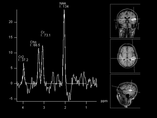
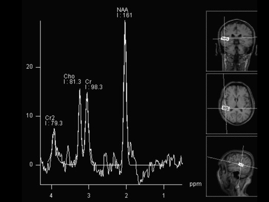
The values of N-acetylaspartate (NAA) and Cr on nontumor side Heschl’s gyrus (HG) were significantly lower than that on tumor side.
Conclusions
We found nontumor side HG more affected with lower NAA and Cr values, suggesting neuronal damage and decreased energy metabolism compared to the tumoral side.
Acoustic neuromas arise from the vestibular nerve and are caused by an overproliferation of Schwann cells. The incidence of acoustic neuroma is estimated to be about 0.7–1 per 100,000 per year. The mean age of those affected is usually about 45–50 years . The most common symptom observed in patients with acoustic neuroma is a progressive asymmetric or unilateral sensorineural hearing loss. About 90% of patients present with gradual progressive hearing loss in one ear. However, about 5% will present with a sudden hearing loss and many of them experience tinnitus in one ear . About 3% of patients with acoustic neuroma will have normal hearing at presentation . Tumor growth within the internal auditory canal (IAC) and resultant compression of cranial nerves VII and VIII cause associated symptoms . There is a significant correlation between the site of origin of the tumor and the incidence of subjective hearing loss .
The mechanisms of hearing loss and affected regions by the disease are still unclear. Hearing impairment in patients with acoustic neuroma has been associated with a retrolabyrinthine disturbance because the tumor originates in the vestibular nerve and impairs cochlear nerve function. Tumor-related hearing loss may be due to direct cochlear nerve damage, sensory organ impairment resulting from disruption of the circulation by vascular compression in the IAC, or a combination of these two factors . Because of the bilateral projections from the ears, it is believed that unilateral lesions have no effect on hearing, although minor and transient hearing loss in the contralateral ear resulted from acoustic neuroma. The effect of acoustic neuroma on the primary auditory cortex is critical. Because lesions in the Heschl’s gyrus (HG) may cause symptoms similar to those caused by lesions in other parts of the auditory pathway, radiologists may need to localize the lesions in relation to the HG .
Get Radiology Tree app to read full this article<
Get Radiology Tree app to read full this article<
Material and methods
Get Radiology Tree app to read full this article<
Get Radiology Tree app to read full this article<
Get Radiology Tree app to read full this article<
Get Radiology Tree app to read full this article<
Get Radiology Tree app to read full this article<
Results
Get Radiology Tree app to read full this article<
Get Radiology Tree app to read full this article<
Table 1
Mean Values of Metabolites of Heschl’s Gyrus From Tumor and Nontumor Sides in Patients with Acoustic Neuroma ∗
Groups N-acetylaspartate Choline Creatine Mean ± Standard Deviation (SD) Mean ± SD Mean ± SD Tumor side 137.87 ± 29 69.91 ± 10 79.99 ± 11 Nontumor side 120.67 ± 32 62.29 ± 14 69.68 ± 13P <.05 † >.05 <.05 †
Get Radiology Tree app to read full this article<
Get Radiology Tree app to read full this article<
Get Radiology Tree app to read full this article<
Get Radiology Tree app to read full this article<
Discussion
Get Radiology Tree app to read full this article<
Get Radiology Tree app to read full this article<
Get Radiology Tree app to read full this article<
Get Radiology Tree app to read full this article<
Get Radiology Tree app to read full this article<
Get Radiology Tree app to read full this article<
Get Radiology Tree app to read full this article<
Get Radiology Tree app to read full this article<
Get Radiology Tree app to read full this article<
References
1. Berrettini S., Ravecca F., Sellari-Franceschini S., et. al.: Acoustic neuroma: correlations between morphology and otoneurological manifestations. J Neurol Sci 1996; 144: pp. 24-33.
2. Anderson T.D., Loevner L.A., Bigelow D.C., et. al.: Prevalence of unsuspected acoustic neuroma found by magnetic resonance imaging. Otolaryngol. Head and Neck Surgery 2000; 122: pp. 643-646.
3. Heier L.A., Comunale J.P., Lavyne M.H.: Sensorineural hearing loss and cerebellopontine angle lesions not always an acoustic neuroma—a pictorial essay. Clin Imaging 1997; 21: pp. 213-223.
4. Raj-Koziak D., Bartnik G., Fabijanska A., et. al.: Tinnitus as a symptom of acoustic neuroma. International Congress Series 2003; 1240: pp. 313-315.
5. Ho S.Y., Kveton J.F.: Acoustic neuroma assessment and management. Otolaryngol Clin North Am 2002; 35: pp. 393-404.
6. Yokoyama K., Nishida H., Noguchi Y., et. al.: Hearing impairment in patients with acoustic neuroma—analysis by electrocochleography. Auris Nasus Larynx 1999; 26: pp. 401-409.
7. Yoshiura T., Higano S., Rubio A., et. al.: Heschl and superior temporal gyri: low signal intensity of the cortex on T2-weighted MR images of the normal brain. Radiology 2000; 214: pp. 217-221.
8. Held P., Fellner C., Fellner F., et. al.: MRI of inner ear anatomy using 3D MP-RAGE and 3D CISS sequences. Br J Radiol 1997; 70: pp. 465-472.
9. Held P., Fellner C., Seitz J., et. al.: The value of T2 (*)-weighted MR images for the diagnosis of acoustic neuromas. Eur J Radiol 1999; 30: pp. 237-244.
10. Bitsch A., Bruhn H., Vougioukas V., et. al.: Inflammatory CNS demyelination: histopathologic with in vivo quantitative proton MR spectroscopy. AJNR Am J Neuroradiol 1999; 20: pp. 1619-1627.
11. Helmchen C., Klinkenstein J.C., Krüger A., et. al.: Structural brain changes following peripheral vestibulo-cochlear lesion may indicate multisensory compensation. J Neurol Neurosurg Psychiatry 2011; 82: pp. 309-316.
12. Bense S., Bartenstein P., Lochmann M., et. al.: Metabolic changes in vestibular and visual cortices in acute vestibular neuritis. Ann Neurol 2004; 56: pp. 624-630.
13. Barta P.E., Petty R.G., McGilchrist I., et. al.: Asymmetry of the planum temporale: methodological considerations and clinical associations. Psychiatr Res Neuroimaging 1995; 61: pp. 137-150.
14. Sörös P., Dzewas R., Manemann E., et. al.: No indication of brain reorganization after unilateral ischemic lesions of the auditory cortex. Neurology 2006; 67: pp. 1059-1061.
15. Lessard N., Lepore F., Poirier P., et. al.: Sound localization in hemispherectomized subjects: the contribution of crossed and uncrossed cortical afferents. Experimental Brain Research 2000; 134: pp. 344-352.
16. de Bode S., Sininger Y., Healy E.W., et. al.: Dichotic listening after cerebral hemispherectomy: methodological and theoretical observations. Neuropsychologia 2007; 45: pp. 2461-2466.
17. Khosla D., Ponton C.W., Eggermont J.J., et. al.: Differential ear effects of profound unilateral deafness on the adult human central auditory system. J Assoc Res Otolaryngol 2003; 4: pp. 235-249.
18. Langers D.R., van Dijk P., Backes W.H.: Lateralization, connectivity and plasticity in the human central auditory system. Neuroimage 2005; 28: pp. 490-499.
19. Paiement P., Champoux F., Bacon B.A., et. al.: Functional reorganization of the human auditory pathways following hemispherectomy: an fMRI demonstration. Neuropsychologia 2008; 46: pp. 2936-2942.
20. Popelar J.E., Aran J.M., Cazals Y.: Plastic changes in the ipsi-contralateral differences of auditory cortex and inferior colliculus evoked potentials after injury to one ear in the adult guinea pig. Hearing Research 1994; 72: pp. 125-134.
21. Irvine D.R., Rajan R.: Injury- and use-related plasticity in the primary sensory cortex of adult mammals: possible relationship of perceptual learning. Clin Exp Pharmacol Physiol 1996; 23: pp. 939-947.
22. Rajan R., Irvine D.R., Wise L.Z., et. al.: Effects of unilateral partial cochlear lesions in adult cats on the representation of lesioned and unlesioned cochleas in the primary auditory cortex. J Comp Neurol 1993; 338: pp. 17-49.
23. Jäncke L., Wüstenberg T., Schulze K., et. al.: Asymmetric hemodynamic responses of the human auditory cortex to monaural and binaural stimulation. Hearing Res 2002; 170: pp. 166-178.
24. Loveless N., Vasama J.P., Mäkelä J., et. al.: Human auditory cortical mechanisms of sound lateralization; III. Monaural and binaural shift responses. Hear Res 1994; 81: pp. 91-99.
25. Schlindwein P., Mueller M., Bauermann T., et. al.: Cortical representation of saccular vestibular stimulation. VEMPs in fMRI. Neuroimage 2008; 39: pp. 19-31.
26. Dieterich M., Brandt T.: Functional brain imaging of peripheral and central vestibular disorders. Brain 2008; 131: pp. 2538-2552.
27. Cecil K.M., Jones B.V.: Magnetic resonance spectroscopy of the pediatric brain. Top Magn Reson Imaging 2001; 12: pp. 435-452.
28. Krouwer H.G., Kim T.A., Rand S.D., et. al.: Single-voxel proton MR spectroscopy of nonneoplastic brain lesions suggestive of a neoplasm. AJNR Am J Neuroradiol 1998; 19: pp. 1695-1703.
29. Miller B.L.: A review of chemical issues in 1H NMR spectroscopy: N -acetyl- L-aspartate, creatine and choline. NMR Biomed 1991; 2: pp. 47-52.
30. Demougeot C., Garnier P., Mossiat C., et. al.: N -acetylaspartate, a marker of both cellular dysfunction and neuronal loss: its relevance to studies of acute brain injury. J Neurochem 2001; 77: pp. 408-415.