
Rationale and Objectives
The aim of this study is to determine the role of magnetization transfer ratio (MTR) in the early period of Parkinson disease (PD).
Materials and Methods
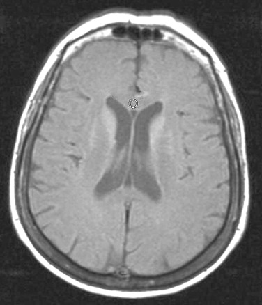
Clinically diagnosed 33 patients with PD in the first year of diagnosis, and 30 healthy volunteers were assessed. Magnetic resonance imaging (MRI) was performed without and with magnetization transfer (MT) imaging. Signal intensity measurements were obtained from 15 anatomic regions (substantia nigra pars compacta [SNPC], substantia nigra pars reticulate [SNPR], red nucleus, dentate nucleus, cerebellum, pons, globus pallidus, putamen, caudate nucleus, thalamus, internal capsule posterior horn, forceps major, forceps minor, and genu and splenium of corpus callosum) and MTR was calculated. Comparisons of the findings between each anatomic location of the patients with PD and normal subjects were performed.
Results
Most prominent decrease of MTR was found in SNPC ( p < 0.001). A significant decrease of MTR was also found in the SNPR ( p = 0.006), red nucleus ( p = 0.037), and pons ( p = 0.046). The other regions lack significance.
Conclusion
MTR analysis is a useful technique for initial PD assessment. Even in the first year of diagnosis, significant reduction of MTR is found in substantia nigra, red nucleus, and pons compared with that of the control group.
Parkinson disease (PD) is a slow neurodegenerative process with progressive neuronal loss of pigmented nuclei, prominently in substantia nigra (SN) ( ). Conventional magnetic resonance imaging (MRI) may demonstrate signal changes or thickness reduction of SN ( ), which is insufficient on the diagnosis of presymptomatic patients and the progress of the disease. Thus new studies are needed to diagnose the presymptomatic patients for admission of neuroprotective agents in early treatment ( ). Magnetization transfer ratio (MTR) imaging, a technique based on interactions between protons that reside in a relatively free environment and those where motion is restricted, is reported to be sensitive on myelin destruction and axonal loss ( ). The purpose of our study is to asses the MTR MRI in the early period of PD disease and to compare the findings with those in normal healthy volunteers.
Materials and methods
Clinically diagnosed 33 patients with PD (age range 46–82 years; mean 66.9 ± 8.49; 9 females, 24 males) and 30 healthy volunteers (age range 42–78 years; mean 66.8 ± 8.575; 15 females, 15 males) were included prospectively and consecutively in the study.
Get Radiology Tree app to read full this article<
Get Radiology Tree app to read full this article<
Get Radiology Tree app to read full this article<
Get Radiology Tree app to read full this article<
Get Radiology Tree app to read full this article<
Get Radiology Tree app to read full this article<
Results
Get Radiology Tree app to read full this article<
Get Radiology Tree app to read full this article<
Table 1
Mean MTR Values and P Values of Both Groups
Anatomic Location Mean MTR_P_ value PD Group Control Group SNPC 0.342 0.364 <0.001 SNPR 0.354 0.374 0.006 Red nucleus 0.350 0.362 0.037 Dentate nucleus 0.391 0.395 0.170 Cerebellum 0.339 0.341 0.847 Pons 0.368 0.380 0.046 Globus pallidus 0.393 0.394 0.880 Putamen 0.370 0.379 0.278 Caudat nucleus 0.341 0.351 0.165 Thalamus 0.362 0.365 0.643 Internal capsule posterior horn 0.379 0.380 0.821 Forceps major 0.368 0.375 0.304 Forceps minor 0.374 0.383 0.143 Corpus callosum, genu 0.376 0.391 0.146 Corpus callosum, splenium 0.385 0.394 0.328
MTR: magnetization transfer ratio; PD: Parkinson disease; SNPC: substantia nigra pars compacta; SNPR: substantia nigra pars reticulate.
Get Radiology Tree app to read full this article<
Discussion
Get Radiology Tree app to read full this article<
Get Radiology Tree app to read full this article<
Get Radiology Tree app to read full this article<
Get Radiology Tree app to read full this article<
Get Radiology Tree app to read full this article<
Conclusion
Get Radiology Tree app to read full this article<
References
1. Tambasco N., Pelliccioli G.P., Chiarini P., et. al.: Magnetization transfer changes of grey and white matter in Parkinson’s disease. Neuroradiology 2003; 45: pp. 224-230.
2. Braffman B.H., Grossman R.I., Goldberg H.I., et. al.: MR imaging of Parkinson disease with spin-echo and gradient-echo sequences. Am J Roentgenol 1989; 152: pp. 159-165.
3. Brooks D.J.: The early diagnosis of Parkinson’s disease. Ann Neurol 1998; 44: pp. 10-18.
4. Hutchinson M., Raff U.: Parkinson’s disease: a novel MRI method for determining structural changes in the substantia nigra. J Neurol Neurosurg Psychiatry 1999; 67: pp. 815-818.
5. Anik Y., Kural Z., Demirci A., et. al.: Magnetization transfer ratio in neuro-Behcet disease. Neuroradiology 2005; 47: pp. 108-113.
6. Mehta R.C., Pike G.B., Enzmann D.R.: Magnetization transfer MR of the normal adult brain. Am J Neuroradiol 1995; 16: pp. 2085-2091.
7. Hutchinson M., Raff U., Lebedev S.: MRI correlates of pathology in parkinsonism: segmented inversion recovery ratio imaging (SIRRIM). Neuroimage 2003; 20: pp. 1899-1902.
8. Adachi M., Hosoya T., Haku T., et. al.: Evaluation of the substantia nigra in patients with Parkinsonian syndrome accomplished using multishot diffusion-weighted MR imaging. Am J Neuroradiol 1999; 20: pp. 1500-1506.
9. Oikawa H., Sasaki M., Tamakawa Y., et. al.: The substantia nigra in Parkinson disease: proton density-weighted spin-echo and fast short inversion time inversion-recovery MR findings. Am J Neuroradiol 2002; 23: pp. 1747-1756.
10. Savoiardo M.: Differential diagnosis of Parkinson’s disease and atypical parkinsonian disorders by magnetic resonance imaging. Neurol Sci 2003; 24: pp. 35-37.
11. Antonini A., Leenders K.L., Meier D., et. al.: T2 relaxation time in patients with Parkinson’s disease. Neurology 1993; 43: pp. 697-700.
12. Gorell J.M., Ordidge R.J., Brown G.G., et. al.: Increased iron-related MRI contrast in the substantia nigra in Parkinson’s disease. Neurology 1995; 45: pp. 1138-1143. Erratum in: Neurology 1995; 45:1420.
13. Duguid J.R., De La Paz R., DeGroot J.: Magnetic resonance imaging of the midbrain in Parkinson’s disease. Ann Neurol 1986; 20: pp. 744-747.
14. Huber S.J., Chakeres D.W., Paulson G.W., et. al.: Magnetic resonance imaging in Parkinson’s disease. Arch Neurol 1990; 47: pp. 735-737.
15. Nakahara M., Hayashi H., Kumazaki T., et. al.: Value of magnetization transfer contrast as a sensitive technique to reflect histopathological changes in the white matter adjacent to the frontal horns of lateral ventricles. Nippon Ika Daigaku Zasshi 1999; 66: pp. 245-252.
16. Rutledge J.N., Hilal S.K., Silver A.J., et. al.: Study of movement disorders and brain iron by MR. Am J Neuroradiol 1987; 149: pp. 365-379.