
Rationale and Objectives
A hands-on stations-based approach to teaching anatomy to third-year medical students is used at Boston University. The goal of our study was to demonstrate that such an interactive, team-based approach to teaching anatomy would be well received and be helpful in recall, comprehension, and reinforcement of anatomy learned in the first year of medical school.
Materials and Methods
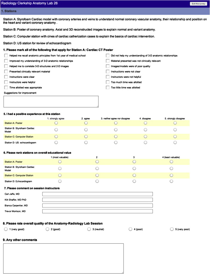
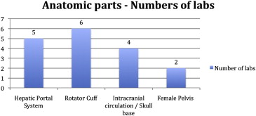
Each radiology-anatomy correlation lab was focused on one particular anatomic part, such as skull base, pelvis, coronary anatomy, etc. Four stations, including a three-dimensional model, computer, ultrasound, and posters, were created for each lab. Informed consent was obtained before online survey dissemination to assess the effectiveness and quality of radiology-anatomy correlation lab. This study was approved by our institutional institutional review board, and data were analyzed using a χ 2 test.
Results
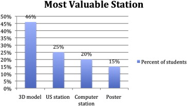
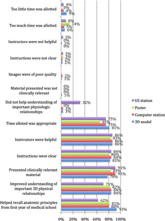
Survey data were collected from February 2010 through March 2012. The response rate was 33.5%. Overall, the highest percentage of students (46%) found the three-dimensional model station to be the most valuable. The computer station was most helpful in recall of the anatomic principles from the first year of medical school. Regarding the quality of the anatomy lab, less than 2% of the students thought that the images were of poor quality or the material presented was not clinically relevant.
Discussion
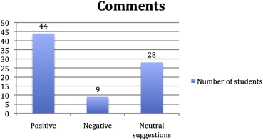
Our results indicate that an interactive, team-based approach to teaching anatomy was well received by the medical students. It was engaging and students were able to benefit from it in multiple ways.
A strong working knowledge of anatomy is essential to learn radiology, for radiologists as well as clinicians reviewing cases with their patients or interpreting imaging reports. In a traditional medical school curriculum, human anatomy is taught in the first year, whereas radiology clerkships, if offered, are generally in the clinical third or fourth years . Studies have shown that retention of anatomic knowledge from the first year to clinical third or fourth years is poor . In studies examining retention of anatomic knowledge in Dutch medical students, it was shown that learning anatomy in a clinically oriented manner and revisiting topics later in the clinical curriculum led to improved test performance . From these studies, it can be inferred that medical students would benefit from revisiting key anatomic concepts during the radiology clerkship to enhance their level of understanding and retention. Based on this belief, radiology-anatomy correlation labs (RACL) were developed in collaboration between the Anatomy Department and the Radiology Department, to allow third-year medical students to revisit key concepts learned in first-year anatomy course and place them in the context of a clinical setting. Our group has previously published the description of these active learning exercises, which are integrated into RACL stations .
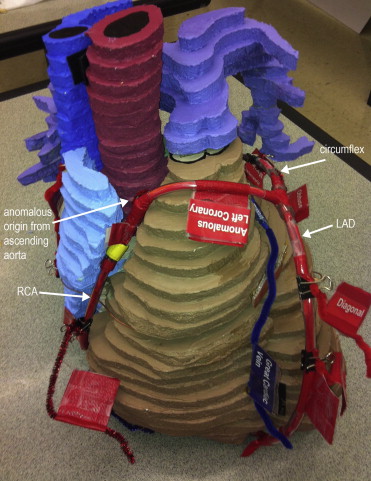
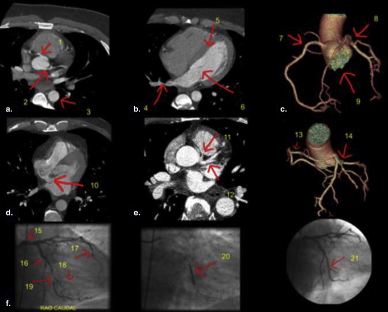
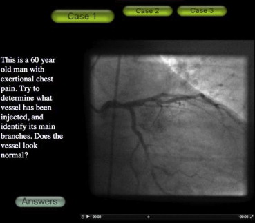
At our institution, radiology is a required month-long clinical clerkship for third-year medical students. RACL takes place once during the radiology clerkship, giving students a chance to rotate through the lab once during their rotation. Each RACL session, which lasts 1.5–2 hours, focuses on one specific anatomic region. Labs designed to date include brachial plexus, pelvic anatomy, coronary circulation, skull base anatomy, liver/portal system, and upper/lower extremity vascular access sites. One month, the group rotating through the radiology clerkship may participate in the pelvic anatomy RACL, while during another month, another group may participate in a skull base anatomy RACL, and so on. For the RACL, students are divided into four groups that rotate among four task-oriented stations, which are focused on a single anatomic part: (a) a station of 3D (three-dimensional) model construction, where students assemble a complex part of the body from component parts; (b) a poster station with typical imaging of the body area being studied for identification; (c) a computer station with video clips that allow scrolling through cross-sectional exams or angiography/fluoroscopy in real-time; and (d) a hands-on ultrasound (US) station for students to practice scanning techniques pertinent to the specific anatomic part. Each RACL is lead by three or four instructors, which may include radiology residents, radiology attendings, and anatomy department faculty (The instructors were recruited on a voluntary basis via an e-mail, which included information on the anatomy module being covered.) The goal of our study was to demonstrate that such an interactive, team-based approach to teaching anatomy would be well received and would be helpful in recall, comprehension, and reinforcement of anatomy learned in the first year of medical school.
Methods
Get Radiology Tree app to read full this article<
Get Radiology Tree app to read full this article<
Get Radiology Tree app to read full this article<
Get Radiology Tree app to read full this article<
Get Radiology Tree app to read full this article<
Get Radiology Tree app to read full this article<
Get Radiology Tree app to read full this article<
Get Radiology Tree app to read full this article<
Get Radiology Tree app to read full this article<
Get Radiology Tree app to read full this article<
Results
Get Radiology Tree app to read full this article<
Get Radiology Tree app to read full this article<
Get Radiology Tree app to read full this article<
Get Radiology Tree app to read full this article<
Get Radiology Tree app to read full this article<
Get Radiology Tree app to read full this article<
Discussion
Get Radiology Tree app to read full this article<
Get Radiology Tree app to read full this article<
Get Radiology Tree app to read full this article<
Get Radiology Tree app to read full this article<
Get Radiology Tree app to read full this article<
Get Radiology Tree app to read full this article<
Get Radiology Tree app to read full this article<
Get Radiology Tree app to read full this article<
Get Radiology Tree app to read full this article<
References
1. Ganske I., Su T., Loukas M., et. al.: Teaching methods in anatomy courses in North American medical schools the role of radiology. Acad Radiol 2006; 13: pp. 1038.
2. Feigin D.S., Magid D., Smirniotopoulos J.G., et. al.: Learning and retaining normal radiographic chest anatomy: does preclinical exposure improve student performance?. Acad Radiol 2007; 14: pp. 1137.
3. Prince K.J., Scherpbier A.J., Van Mameren H., et. al.: Do students have sufficient knowledge of clinical anatomy?. Med Educ 2005; 39: pp. 326-332.
4. Bergman E.M., Prince K.J., Drukker J., et. al.: How much anatomy is enough?. Anat Sci Educ 2008; 1: pp. 184-188.
5. Zumwalt A.C., Lufler R.S., Monteiro J., et. al.: Building the body: active learning laboratories that emphasize practical aspects of anatomy and integration with radiology. Anat Sci Educ 2010; 3: pp. 134-140.
6. Faraone K.L., Garrett P.H., Romberg E.: A blended learning approach to teaching pre-clinical complete denture prosthodontics. Eur J Dent Educ 2013; 17: pp. e22-e27.
7. Garrison D.R., Kanuka H.: Blended learning: uncovering its transformative potential in higher education. Internet Higher Educ 2004; 7: pp. 95-105.
8. Lujan H.L., DiCarlo S.E.: First-year medical students prefer multiple learning styles. Adv Physiol Educ 2006; 30: pp. 13-16.
9. Shaffer K., Small J.E.: Blended learning in medical education: use of an integrated approach with web-based small group modules and didactic instruction for teaching radiologic anatomy. Acad Radiol 2004; 11: pp. 1059.
10. Rosenbaum P.E., Mikalsen Ø., Lygre H.: A blended learning course design in clinical pharmacology for post-graduate dental students. Open Dent J 2012;
11. Terrell M.: Anatomy of learning: instructional design principles for the anatomical sciences. Anat Rec Part B New Anatomist 2006; 289: pp. 252-260.
12. Lufler R.S., Zumwalt A.C., Romney C.A., et. al.: Effect of visual-spatial ability on medical students’ performance in a gross anatomy course. Anat Sci Educ 2012; 5: pp. 3-9.
13. Sugand K., Abrahams P., Khurana A.: The anatomy of anatomy: a review for its modernization. Anat Sci Educ 2010; 3: pp. 83-93.
14. Rodt T., Ratiu P., Becker H., et. al.: 3D visualisation of the middle ear and adjacent structures using reconstructed multi-slice CT datasets, correlating 3D images and virtual endoscopy to the 2D cross-sectional images. Neuroradiology 2002; 44: pp. 783-790.
15. Gabard D.L., Lowe D.L., Chang J.W.: Current and future instructional methods and influencing factors in anatomy instruction in physical therapy and medical schools in the US. J Allied Health 2012; 41: pp. 53-62.
16. Azer S.A.: Cost consciousness and medical education. N Engl J Med 2010; 363: pp. 890. author reply 890-891
17. Vozenilek J., Huff J.S., Reznek M., et. al.: See one, do one, teach one: advanced technology in medical education. Acad Emerg Med 2004; 11: pp. 1149-1154.
18. Durfee S.M., Jain S., Shaffer K.: Incorporating electronic media into medical student education: a survey of AMSER members on computer and web use in radiology courses. Acad Radiol 2003; 10: pp. 205-210.