
Rationale and Objectives
The purpose of this study was to assess (1) the agreement of two-dimensional (2D) and three-dimensional (3D) manual and automated polyp linear diameter measurements at CT colonography (CTC), with optical colonoscopic equivalents and (2) intraobserver and interobserver agreement of the CTC measurements.
Materials and Methods
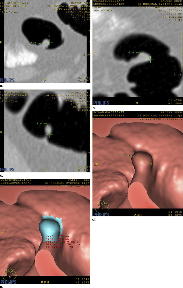
Using the same CTC system, two radiologists independently measured the maximum linear diameter of 44 polyps (reference size 3–15 mm) matched on CTC and optical colonoscopy: manual 2D optimized multiplanar reformatted planes with standard window settings (level 1500 HU, width −200 HU), manual 3D measurement with software calipers and automated 3D measurement with software. After 2 weeks, polyps were measured again. Compatibility of CTC measurement with that of optical colonoscopy and measurement reproducibility was assessed statistically.
Results
In the manual measurement, 44 polyps were analyzed and 41 in automated measurement; three polyps could not be extracted. Although the measurement difference was noted for automated, manual 3D, and manual 2D measurements, statistically supported agreement with optical colonoscopic measurement was noted only with manual 2D measurement for both observers. However, 95% limits of agreement were wide for all the measurement methods. When categorized according to the optical colonoscopic measurement, manual 2D, 3D, and automated measurements showed “good” agreement. Although intraobserver and interobserver agreement was good with manual measurement, intraobserver and interobserver agreement was excellent with automated measurement.
Conclusion
Manual 2D measurements demonstrated trends of better approximation to optical colonoscopy measurements than manual 3D or automated measurements. And automated measurement eliminated intraobserver and interobserver variability. For noninvasive CTC surveillance, manual 2D measurements are expected to measure medium-sized polyps with sufficient agreement with optical colonoscopic measurements and excellent intraobserver and interobserver variability, especially if combined with automated measurement.
It is widely believed that the majority of colorectal cancers arise from precursor adenomatous polyps and that endoscopic polypectomy is preventive ( ). In the absence of histology, maximal polyp size is the most reliable predictor of developing malignancy ( ). For polyps ≥10 mm, the risk of high-grade dysplasia or cancer ranges from 10% to 25% ( ). While there is general consensus that large, ≥10-mm polyps are excised ( ), there is a more broad range of practice management of the medium-sized (6−9 mm) and small (<6 mm) polyps, ranging from surveillance to polypectomy ( ), depending on the clinical context of the patient.
Based on this evidence, a working group has developed patient management strategies predicated by the largest polyp found at CT colonography (CTC) ( ) that are now regarded as a sensitive screening tool for colonic adenomatous polyps ( ). For this management algorithm to be useful in daily clinical practice, accurate measurement of polyps (especially for medium-sized polyps), the clinical legitimacy and the replication with optical colonoscopic measurements are essential and critical for treatment choice ( ). Moreover, for noninvasive CTC surveillance of unresected medium-sized polyps, measurements from successive follow-up examinations must be so highly repeatable and reproducible that discernment of interval polyp growth or regression can allow appropriate clinical decision making ( ). This is important because polyps left in situ require reexamination and remeasurement, perhaps after an interval of several years, which makes it less likely that the subsequent measurement will be performed by the same observer who made the first measurement. However, with CTC, a degree of intraobserver and interobserver variability is inevitable during manual measurement, which can result in misclassification and errors in the management, computer-aided automated measurement is now coming to the front ( ).
Get Radiology Tree app to read full this article<
Materials and methods
Polyp Dataset
Get Radiology Tree app to read full this article<
Get Radiology Tree app to read full this article<
CT Scanning
Get Radiology Tree app to read full this article<
Polyp Measurement
Get Radiology Tree app to read full this article<
Get Radiology Tree app to read full this article<
Get Radiology Tree app to read full this article<
Statistical Analysis
Get Radiology Tree app to read full this article<
Results
Get Radiology Tree app to read full this article<
Agreement With Optical Colonoscopic Measurement
Get Radiology Tree app to read full this article<
Table 1
Agreement Between Observer CT Measurement and Optical Colonoscopic Reference
Reader and Measurement Method Mean Measurement Difference Between Optical Colonoscopic and CTC Measurement (mm) ⁎ P Value † 95% Limits of Agreement ‡ Observer 1 Manual 2D ( n = 44) 0.39 (−0.19, 0.98) 0.182 −3.46, 4.25 Manual 3D ( n = 44) 1.15 (0.50, 1.81) 0.001 −3.18, 5.49 Automated ( n = 41) 2.21 (1.48, 2.94) 0.000 −2.41, 6.83 Observer 2 Manual 2D ( n = 44) 0.30 (−0.33, 0.93) 0.339 −3.85, 4.45 Manual 3D ( n = 44) 1.15 (0.42, 1.89) 0.003 −3.70, 6.00 Automated ( n = 41) 2.21 (1.48, 2.94) 0.000 −2.41, 6.83
Get Radiology Tree app to read full this article<
Get Radiology Tree app to read full this article<
Get Radiology Tree app to read full this article<
Get Radiology Tree app to read full this article<
Polyp Categorization
Get Radiology Tree app to read full this article<
Table 2
Polyp Size Categorization According to Observer and the Measurement Method
A. Observer 1 Polyp Size (mm) † Manual 2D Measurement <6 6−9 ≥10 <6 ( n = 20) 12 (60) 8 (40) 0 (0) 6−9 ( n = 14) 2 (14) 12 (86) 0 (0) ≥10 ( n = 10) 0 (0) 3 (30) 7 (70) κ = 0.548.
B. Observer 1 Polyp Size (mm) † Manual 3D Measurement <6 6−9 ≥10 <6 ( n = 20) 8 (40) 12 (60) 0 (0) 6−9 ( n = 14) 1 (7) 11 (79) 2 (14) ≥10 ( n = 10) 0 (0) 2 (20) 8 (80) κ = 0.427.
C. Observer 2 Polyp Size (mm) † Manual 2D Measurement <6 6−9 ≥10 <6 ( n = 20) 12 (60) 8 (40) 0 (0) 6−9 ( n = 14) 2 (14) 11 (79) 1 (7) ≥10 ( n = 10) 0 (0) 4 (40) 6 (60) κ = 0.478.
D. Observer 2 Polyp Size (mm) † Manual 3D Measurement <6 6−9 ≥10 <6 ( n = 20) 8 (40) 11 (55) 1 (5) 6−9 ( n = 14) 1 (7) 9 (64) 4 (29) ≥10 ( n = 10) 0 (0) 3 (30) 7 (70) κ = 0.330.
E. Observers 1 and 2 Polyp Size (mm) † Automated Measurement <6 6−9 ≥10 <6 ( n = 19) 5 (26) 11 (58) 3 (16) 6−9 ( n = 14) 0 (0) 9 (64) 5 (36) ≥10 ( n = 8) 0 (0) 1 (13) 7 (87) κ = 0.300.
*Values are numbers of polyps. Numbers in parentheses are percentages.
Get Radiology Tree app to read full this article<
Get Radiology Tree app to read full this article<
Intraobserver Agreement
Get Radiology Tree app to read full this article<
Table 3
Mean Difference and Bland-Altman 95% Limits of Agreement for Intraobserver Comparisons of Polyp Measurement According to Measurement Method
Observer and Measurement Method Mean Measurement Difference Between First and Second Measurement (mm) ⁎ P Value † 95% Limits of Agreement ‡ Observer 1 Manual 2D 0.07 (−0.03, 0.17) 0.19 −0.59, 0.72 Manual 3D −0.02 (−0.12, 0.07) 0.62 −0.63, 0.58 Automated 0.00 (0.00, 0.00) 1.00 0.00, 0.00 Observer 2 Manual 2D 0.25 (0.04, 0.45) 0.02 −1.08, 1.57 Manual 3D 0.30 (0.06, 0.54) 0.01 −1.27, 1.88 Automated 0.00 (0.00, 0.00) 1.00 0.00, 0.00
Get Radiology Tree app to read full this article<
Get Radiology Tree app to read full this article<
Get Radiology Tree app to read full this article<
Get Radiology Tree app to read full this article<
Interobserver Agreement
Get Radiology Tree app to read full this article<
Table 4
Mean Difference and Bland-Altman 95% Limits of Agreement for Interobserver Comparisons of Polyp Measurement According to Measurement Method
Measurement Method Mean Measurement Difference Between Observer 1 and 2 (mm) ⁎ P Value † 95% Limits of Agreement ‡ Manual 2D 0.09 (−0.15, 0.34) 0.45 −1.52, 1.70 Manual 3D 0.00 (−0.33, 0.33) 0.99 −2.16, 2.16 Automated 0.00 (0.00, 0.00) 1.00 0.00, 0.00
Get Radiology Tree app to read full this article<
Get Radiology Tree app to read full this article<
Get Radiology Tree app to read full this article<
Get Radiology Tree app to read full this article<
Discussion
Get Radiology Tree app to read full this article<
Get Radiology Tree app to read full this article<
Get Radiology Tree app to read full this article<
Get Radiology Tree app to read full this article<
Get Radiology Tree app to read full this article<
Get Radiology Tree app to read full this article<
Appendix
Get Radiology Tree app to read full this article<
References
1. Citarda F., Tomaselli G., Capocaccia R., et. al.: Efficacy in standard clinical practice of colonoscopic polypectomy in reducing colorectal cancer incidence. Gut 2001; 48: pp. 812-815.
2. Winawer S.J., Zauber A.G., Ho M.N., et. al.: Prevention of colorectal cancer by colonoscopic polypectomy. N Engl J Med 1993; 329: pp. 1977-1981.
3. Bond J.H.: Screening guidelines for colorectal cancer. Am J Med 1999; 106: pp. 7S-10S.
4. Johnson C.D., Dachman A.H.: CT colonography: The next colon screening examination?. Radiology 2000; 216: pp. 331-341.
5. Morson B.C.: Evolution of cancer of the colon and rectum. Cancer 1974; 34: pp. 9.
6. Hofstad B., Almendingen K., Vatn M., et. al.: Growth and recurrence of colorectal polyps: A double-blind 3-year intervention with calcium and antioxidants. Digestion 1998; 59: pp. 148-156.
7. Waye J.D., Lewis B.S., Frankel A., et. al.: Small colon polyps. Am J Gastroenterol 1988; 83: pp. 120-122.
8. Muto T., Bussey H.J., Morson B.C.: The evolution of cancer of the colon and rectum. Cancer 1975; 36: pp. 2251-2270.
9. Stryker S.J., Wolff B.G., Culp C.E., et. al.: Natural history of untreated colonic polyps. Gastroenterology 1987; 93: pp. 1009-1013.
10. Pickhardt P.J., Choi J.R., Hwang I., et. al.: Computed tomographic virtual colonoscopy to screen for colorectal neoplasia in asymptomatic adults. N Engl J Med 2003; 349: pp. 2191-2200.
11. Nusko G., Mansmann U., Partzsch U., et. al.: Invasive carcinoma in colorectal adenomas: Multivariate analysis of patient and adenoma characteristics. Endoscopy 1997; 29: pp. 626-631.
12. Grassi A.C.V., Fracasso P., Lapenta R., et. al.: Mediumlarge polyps of the colon: A contribution for their clinical profile and a proper surveillance. J Exp Clin Cancer Res 1997; 16: pp. 313-319.
13. Morson B.: President’s address: The polyp-cancer sequence in the large bowel. Proc R Soc Med 1974; 67: pp. 451-457.
14. Church J.M.: Clinical significance of small colorectal polyps. Dis Colon Rectum 2004; 47: pp. 481-485.
15. Pickhardt P.: By-patient performance characteristics of CT colonography: Importance of polyp size threshold data. Radiology 2003; 229: pp. 291-293.
16. Zalis M.E., Barish M.A., Choi J.R., et. al.: CT colonography reporting and data system: a consensus proposal. Radiology 2005; 236: pp. 3-9.
17. Bond J.H.: Clinical relevance of the small colorectal polyp. Endoscopy 2001; 33: pp. 454-457.
18. van Stolk R.U., Beck G.J., Baron J.A., et. al.: Adenoma characteristics at first colonoscopy as predictors of adenoma recurrence and characteristics at follow-up. Gastroenterology 1998; 115: pp. 13-18.
19. Kulling D., Christ A.D., Karaaslan N., et. al.: Is histological investigation of polyps always necessary?. Endoscopy 2001; 33: pp. 428-432.
20. Johnson C.D., Harmsen W.S., Wilson L.A., et. al.: Prospective blinded evaluation of computed tomographic colonography for screen detection of colorectal polyps. Gastroenterology 2003; 125: pp. 311-319.
21. Cotton P.B., Durvalski V.L., Pineau B.C., et. al.: Computed tomographic colonography (virtual colonoscopy): A multicenter comparison with standard colonoscopy for detection of colorectal neoplasia. JAMA 2004; 291: pp. 1713-1719.
22. Rockey D.C., Paulson E., Niedzwiecki D., et. al.: Analysis of air contrast barium enema, computed tomographic colonography, and colonoscopy: Prospective comparison. Lancet 2005; 365: pp. 305-311.
23. Taylor S., Slater A., Honeyfield L., et. al.: CT colonography: Effect of colonic distension on polyp measurement accuracy and agreement: In vitro study. Acad Radiol 2006; 13: pp. 850-859.
24. Burling D., Halligan S., Roddie M.E., et. al.: Computed tomography colonography: Automated diameter and volume measurement of colonic polyps compared with a manual technique in vitro study. J Comput Assist Tomogr 2005; 29: pp. 387-393.
25. Pickhardt P.J.: Three-dimensional endoluminal CT colonography (virtual colonoscopy): Comparison of three commercially available systems. AJR Am J Roentgenol 2003; 181: pp. 1599-1606.
26. Pickhardt P.J., Lehman V.T., Winter T.C., et. al.: Polyp volume versus linear size measurements at CT colonography: Implications for noninvasive surveillance of unresected colorectal lesions. AJR Am J Roentgenol 2006; 186: pp. 1605-1610.
27. Yeshwant S.C., Summers R.M., Yao J., et. al.: Polyps: Linear and volumetric measurement at CT colonography. Radiology 2006; 241: pp. 802-811.
28. Blake M.E., Soto J.A., Hayes R.A., et. al.: Automated volumetry at CT colonography: A phantom study. Acad Radiol 2005; 12: pp. 608-613.
29. Burling D., Halligan S., Altman D.G., et. al.: Polyp measurement and size categorisation by CT colonography: Effect of observer experience in a multi-centre setting. Eur Radiol 2006; 16: pp. 1737-1744.
30. Pickhardt P.J., Lee A.D., McFarland E.G., et. al.: Linear polyp measurement at CT colonography: In vitro and in vivo comparison of two-dimensional and three-dimensional displays. Radiology 2005; 236: pp. 872-878.
31. Bland J.M., Altman D.G.: Statistical methods for assessing agreement between two methods of clinical measurement. Lancet 1986; 1: pp. 307-310.
32. Burling D., Halligan S., Taylor S.A., et. al.: Polyp size measurement by CT colonography: Effect of viewing conditions on inter- and intraobserverobserver agreement, and agreement with colonoscopy. AJR Am J Roentgenol 2006; 186: pp. 1597-1604.
33. Sorstedt E., Persson A., Noren B., et. al.: Computed tomographic colonography: Comparison of two workstations. Acta Radiol 2005; 46: pp. 671-678.
34. Young B.M., Fletcher J.G., Paulsen S.R., et. al.: Polyp measurement with CT colonography: Multiple-reader, multiple-workstation comparison. AJR Am J Roentgenol 2007; 188: pp. 122-129.
35. Taylor S.A., Slater A., Halligan S., et. al.: CT colonography: Automated measurement of colonic polyps compared with manual techniques—Human in vitro study. Radiology 2007; 242: pp. 120-128.
36. Fenlon H.M., Nunes D.P., Schroy P.C., et. al.: A comparison of virtual and conventional colonoscopy for the detection of colorectal polyps. N Engl J Med 1999; 341: pp. 1496-1503.
37. Yee J., Akerkar G.A., Hung R.K., et. al.: Colorectal neoplasia: Performance characteristics of CT colonography for detection in 300 patients. Radiology 2001; 219: pp. 685-692.
38. Summers R.M., Beaulieu C.F., Pusanik L.M., et. al.: Automated polyp detector for CT colonography: Feasibility study. Radiology 2000; 216: pp. 284-290.
39. Summers R.M., Johnson C.D., Pusanik L.M., et. al.: Automated polyp detection at CT colonography: Feasibility assessment in a human population. Radiology 2001; 219: pp. 51-59.
40. Yoshida H., Masutani Y., MacEneaney P., et. al.: Computerized detection of colonic polyps at CT colonography on the basis of volumetric features: Pilot study. Radiology 2002; 222: pp. 327-336.
41. Näppi J., Yoshida H.: Automated detection of polyps with CT colonography: Evaluation of volumetric features for reduction of false-positive findings. Acad Radiol 2002; 9: pp. 386-397.
42. Summers R.M., Jerebko A.K., Franaszek M., et. al.: Colonic polyps: Complementary role of computer-aided detection in CT colonography. Radiology 2002; 225: pp. 391-399.
43. Yoshida H., Nappi J., MacEneaney P., et. al.: Computer-aided diagnosis scheme for detection of polyps at CT colonography. Radiographics 2002; 22: pp. 963-979.
44. Ling S.H., Summers R.M., Loew M.H., et. al.: Computer-aided detection of polyps in a colon phantom: Effect of scan orientation, polyp size, collimation, and dose. J Comput Assist Tomogr 2002; 26: pp. 1013-1018.
45. Fennerty M.B., Davidson J., Emerson S.S., et. al.: Are endoscopic measurements of colonic polyps reliable?. Am J Gastroenterol 1993; 88: pp. 496-500.
46. Schoen R.E., Gerber L.D., Margulies C.: The pathologic measurement of polyp size is preferable to the endoscopic estimate. Gastrointest Endosc 1997; 46: pp. 492-496.
47. Fennerty M.B., Davidson J., Emerson S.S., et. al.: Are endoscopic measurements of colonic polyps reliable?. Am J Gastroenterol 1993; 88: pp. 496-500.
48. Morales T.G., Sampliner R.E., Garewal H.S., et. al.: The difference in colon polyp size before and after removal. Gastrointest Endosc 1996; 43: pp. 25-28.
49. Gopalswamy N., Shenoy V.N., Choudhry U., et. al.: Is in vivo measurement of size of polyps during colonoscopy accurate?. Gastrointest Endosc 1997; 46: pp. 497-502.
50. Tsuda S., Veress B., Toth E., et. al.: Flat and depressed colorectal tumours in a southern Swedish population: A prospective chromoendoscopic and histopathological study. Gut 2002; 51: pp. 550-555.
51. Macari M., Bini E.J., Jacobs S.L., et. al.: Filling defects at CT colonography: Pseudo- and diminutive lesions (the good), polyps (the bad), flat lesions, masses, and carcinomas (the ugly). RadioGraphics 2003; 23: pp. 1073-1091.