
Rationale and Objectives
Radiologists frequently image women with the sole complaint of mastalgia (breast pain). We hypothesized that whereas the vast majority of women ultimately have no imaging explanation for their breast pain, a small percentage of patients may have a correlative imaging finding and confirm the current American College of Radiology Appropriateness Criteria recommendations.
Materials and Methods
In this Health Insurance Portability and Accountability Act (HIPAA)-compliant, institutional review board-approved retrospective review, we evaluated 236 women between the ages of 18 and 83 years who presented to our Breast Care Center in 2013 with the sole complaint of breast pain or tenderness. Patients’ clinical presentation, diagnostic imaging work-up, and clinical and radiographic follow-up were documented. Outcomes of the diagnostic work-up were compared with the American College of Radiology Appropriateness Criteria recommendations.
Results
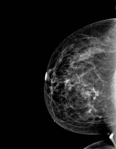
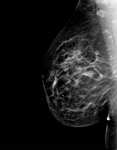
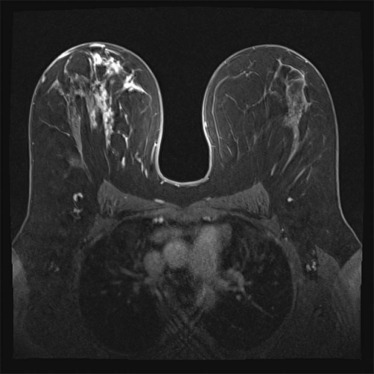
Of the 236 patients, 10 women had cyclical breast pain, 116 had noncyclical, nonfocal breast pain, and 110 had noncyclical, focal breast pain. No imaging correlates were discovered to explain the etiology of cyclical pain, supporting the American College of Radiology Appropriateness Criteria rating values. A definitive imaging correlate for breast pain was identified in seven women (3%) with noncyclical, focal pain, one of which was a cancer diagnosis (0.4%), which correlates with the American College of Radiology Appropriateness Criteria ratings. No imaging correlates were found in women with noncyclical, nonfocal pain, supporting the American College of Radiology Appropriateness Criteria ratings.
Conclusion
There was no radiological imaging finding to explain the etiology of mastalgia in most women. Diagnostic imaging may be an appropriate diagnostic evaluation in patients with noncyclical, focal breast pain, supporting the American College of Radiology Appropriateness Criteria recommendations.
Introduction
Breast pain is a common problem that affects 70–80% of women at some point in their lives , most frequently noted in premenopausal women . The incidence of cancer in patients presenting with breast pain is reported to be 0–3.2% and in one study up to 7% . Breast pain is usually self-limited and is not typically a symptom of malignant pathologic disease. Most breast pain symptomatology can be treated with reassurance, over-the-counter pain medications, or structural support .
As breast cancer awareness has increased, a concern that breast pain may indicate malignancy contributes to the trend of breast pain being the most common breast symptom causing a woman to consult her primary care physician or a breast surgeon . If patients are not treated based on symptoms and physical examination alone, they may be referred for reassurance to a breast imaging facility . These studies report that after initial imaging, most women require no intervention after reassurance that their diagnostic imaging work-up is normal. The negative predictive value of mammography and ultrasound for patients with breast pain has been reported to be 100% in three studies . However, a 2012 study showed that women who received initial imaging were more likely to have subsequent imaging, biopsies, additional visits, and higher clinical services utilization than women who did not , suggesting that these modalities should be judiciously performed.
Get Radiology Tree app to read full this article<
Get Radiology Tree app to read full this article<
Materials and Methods
Get Radiology Tree app to read full this article<
Get Radiology Tree app to read full this article<
Diagnostic Evaluation
Get Radiology Tree app to read full this article<
Get Radiology Tree app to read full this article<
Follow-up of Our Cohort
Get Radiology Tree app to read full this article<
Get Radiology Tree app to read full this article<
Get Radiology Tree app to read full this article<
Get Radiology Tree app to read full this article<
Get Radiology Tree app to read full this article<
Results
Get Radiology Tree app to read full this article<
Cyclical Breast Pain (American College of Radiology Appropriateness Criteria [ACRAC] Variants 1 and 2)
Get Radiology Tree app to read full this article<
Noncyclical, Focal Breast Pain (ACRAC Variants 3 and 4)
Get Radiology Tree app to read full this article<
Noncyclical, Nonfocal/Diffuse Breast Pain (ACRAC Variants 5 and 6)
Get Radiology Tree app to read full this article<
Discussion
Get Radiology Tree app to read full this article<
Cyclical Breast Pain (Variants 1 and 2)
Get Radiology Tree app to read full this article<
Noncyclical, Focal Breast Pain (Variants 3 and 4)
Get Radiology Tree app to read full this article<
Get Radiology Tree app to read full this article<
Get Radiology Tree app to read full this article<
Get Radiology Tree app to read full this article<
Noncyclical, Nonfocal/Diffuse Breast Pain (Variants 5 and 6)
Get Radiology Tree app to read full this article<
Limitations
Get Radiology Tree app to read full this article<
Conclusion
Get Radiology Tree app to read full this article<
Get Radiology Tree app to read full this article<
References
1. Ader D.N., Browne M.W.: Prevalence and impact of cyclic mastalgia in a United States clinic-based sample. Am J Obstet Gynecol 1997; 177: pp. 126-132.
2. Leinster S., Whitehouse G., Walsh P.: Cyclical mastalgia: clinical and mammographic observations in a screened population. Br J Surg 1987; 74: pp. 220-222.
3. Barton M.B., Elmore J.G., Fletcher S.W.: Breast symptoms among women enrolled in a health maintenance organization: frequency, evaluation, and outcome. Ann Intern Med 1999; 130: pp. 651-657.
4. Ader D.N., Shriver C.D.: Cyclical mastalgia: prevalence and impact in an outpatient breast clinic sample. J Am Coll Surg 1997; 185: pp. 466-470.
5. Goodwin P.J., Miller A., Del Giudice M.E., et. al.: Breast health and associated premenstrual symptoms in women with severe cyclic mastopathy. Am J Obstet Gynecol 1997; 176: pp. 998-1005.
6. Griffith C., Dowle C., Hinton C., et. al.: The breast pain clinic: a rational approach to classification and treatment of breast pain. Postgrad Med J 1987; 63: pp. 547-549.
7. Maddox P., Harrison B., Mansel R., et. al.: Non-cyclical mastalgia: an improved classification and treatment. Br J Surg 1989; 76: pp. 901-904.
8. Mansel R.: ABC of breast diseases. Breast pain. BMJ 1994; 309: pp. 866.
9. Morrow M.: The evaluation of common breast problems. Am Fam Physician 2000; 61: pp. 2371-2390.
10. Masroor I., Afzal S., Sakhawat S., et. al.: Negative predictive value of mammography and sonography in mastalgia with negative physical findings. J Pak Med Assoc 2009; 59: pp. 598.
11. Tumyan L., Hoyt A.C., Bassett L.W.: Negative predictive value of sonography and mammography in patients with focal breast pain. Breast J 2005; 11: pp. 333-337.
12. Loving V.A., DeMartini W.B., Eby P.R., et. al.: Targeted ultrasound in women younger than 30 years with focal breast signs or symptoms: outcomes analyses and management implications. AJR Am J Roentgenol 2010; 195: pp. 1472-1477.
13. Howard M.B., Battaglia T., Prout M., et. al.: The effect of imaging on the clinical management of breast pain. J Gen Intern Med 2012; 27: pp. 817-824.
14. Ghebrehiwet M., Paulos E., Andeberhan T.: The use of sonography and mammography in the evaluation of Eritrean women with breast pain. JEMA 2008; 3: pp. 8-11.
15. Leung J.W., Kornguth P.J., Gotway M.B.: Utility of targeted sonography in the evaluation of focal breast pain. J Ultrasound Med 2002; 21: pp. 521-526.
16. Duijm L.E., Guit G.L., Hendriks J.H., et. al.: Value of breast imaging in women with painful breasts: observational follow up study. BMJ 1998; 317: pp. 1492-1495.
17. Preece P., Baum M., Mansel R., et. al.: Importance of mastalgia in operable breast cancer. BMJ 1982; 284: pp. 1299-1300.
18. Naz N., Sohail S., Memon M.: Utility of breast imaging in mastalgia. J Liaquat Uni Med Health Sci 2010; 9: pp. 12-15.
19. Smith R.L., Pruthi S., Fitzpatrick L.A.: Evaluation and management of breast pain.2004.Elsevier, Inc.pp. 353-372.
20. Morrow M.: Management of common breast disorders: breast pain.Harris J.R. et. al.Breast diseases.1991.LippincottPhiladelphia:pp. 63-71.
21. Brand I., Sapherson D., Brown T.: Breast imaging in women under 35 with symptomatic breast disease. Br J Radiol 1993; 66: pp. 394-397.
22. Leddy R., Irshad A., Zerwas E., et. al.: Role of breast ultrasound and mammography in evaluating patients presenting with focal breast pain in the absence of a palpable lump. Breast J 2013; 19: pp. 582-589.
23. Jokich P.M., Bailey L., D’Orsi C., et. al.: ACR ® Appropriateness Criteria breast pain. American College of Radiology; June 12016. Available at: https://acsearch.acr.org/docs/3091546/Narrative/
24. Noroozian M., Stein L.F., Gaetke-Udager K., et. al.: Long-term clinical outcomes in women with breast pain in the absence of additional clinical findings: mammography remains indicated. Breast Cancer Res Treat 2015; 149: pp. 417-424.