
Rationale and Objectives
To perform magnetic resonance imaging (MRI) scans regarding material parameter and model validation in computational simulations of mechanical interaction of human soft-tissue with body-supporting devices, enhanced medical prognosis in pressure sore prophylaxis, and comfort optimization in automotive and aircraft seating.
Materials and Methods
In vivo human gluteal fat and passive muscle tissue material parameters of a volunteer evaluated via combined MRI numerical method and body-supporting foam material parameters employed in finite element (FE) simulations of tissue-support interaction were verified by a defined loading scenario using MRI. MRI of the loaded configurations were performed and compared with simulation results for displacement field information.
Results
Deformation of gluteal skin/fat and passive muscle-tissue and support material under interacting loading using numerical simulation complied with the MRI results. Accordance was found for deformed skin surface and internal fat-muscle tissue boundaries by superimposing experimental and numerical outputs. Further evidence of established through in vivo gluteal tissue parameters was thus provided and tissue material isotropy assumption was shown for use in simulated buttock loading interactions. Additionally, a new concept of FE model validation regarding non–MRI-sensitive materials such as polyurethane foam was introduced comprising peripheral surface visualization.
Conclusion
Imaging techniques are essential in biomechanical modeling and provide key information regarding model validation and validity assessment.
Pressure sores are immensely costly ($4 billion for the UK annually) and cause severe trauma for the patient. Although pressure sore development is multifactorial, it is commonly agreed that sustained mechanical tissue loading plays the most significant role during sore formation . Furthermore, persistent tissue loading also represents an essential issue regarding comfort and thrombosis etiology-related questions in automotive and aircraft seating. Analyzing mechanical stress and strain distribution inside loaded human soft tissue and optimizing medical body support devices regarding their durability and reliability in terms of tissue stress relief is gaining importance as the potential grows to numerically model detailed human anatomy and derive adequate tissue properties for use in computational simulation scenarios. Furthermore, numerical methods allow evaluation of effects of different support designs and materials on the incumbent body. They contribute to a better understanding of the etiology of stress-induced trauma such as pressure sores. Thus, body supports can be appropriately designed. In the finite element (FE) simulation process, adequate material parameters for both soft tissue and support material are indispensable to properly describe the mechanical behavior during interaction. Tissue-support interaction has been subject to several simulation studies. Because of a paucity of information on human in vivo tissue material parameters accounting for large deformations, material parameters used within these investigations have been either assumed or derived from ex vivo animal experiments . Material parameters used in the present investigation have been derived via a combined iterative numerical-experimental approach based on in vivo noninvasive testing in a human subject and magnetic resonance imaging (MRI). Following this approach, material properties of both interacting materials, specifically gluteal soft tissue and body soft foam support material, have been partially validated separately regarding distinct indenter loading experiments . With regard to the foam material, additional visual validation was performed verifying lateral straining. Validation of tissue deformation in realistic buttock loading (ie, supported gluteal tissue under body weight loading) was pending. The purpose of this study was to use the potential of MRI for providing information of the displacement field at interface boundaries of the deformed structures of both materials during interaction and thus furnish further proof of the validity of derived tissue and support material parameters by visually comparing MRI scans with FE simulation results. Interaction of gluteal soft tissue and soft foam support material was performed via a defined experimental set up and MRI scans were performed of the loaded scenario to prove material parameter validity. These images were then superimposed with simulation results and deformed interface boundaries were visually compared.
Materials and methods
The experimental protocols were approved by the ethical committee of the hospital and informed written consent was obtained from the volunteer participating in the investigations. In the following, individual material characterization of both interacting materials (ie, gluteal soft tissue and foam support material) is briefly described because it provides the basis for simulating loaded contact interactions. In addition to the assumption of isotropy, both materials were considered regarding their nonlinear long-term elastic material behavior. Therefore constitutive equations for hyperelasticity were permissible .
Gluteal Soft-tissue Characterization
Get Radiology Tree app to read full this article<
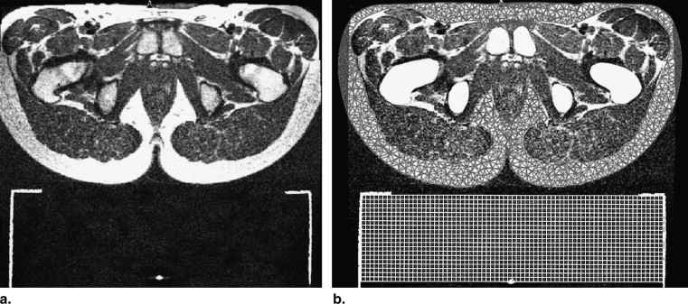
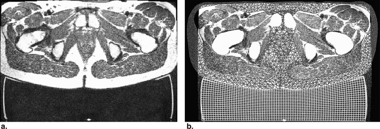
![Figure 1, Transverse magnetic resonance images of the gluteal region (a) in the initially undeformed state and (b) at maximum indenter displacement with highlight markers at [1] indicating the current indenter position.](https://storage.googleapis.com/dl.dentistrykey.com/clinical/MechanicalSoftTissuePropertyValidationinTissueEngineeringUsingMagneticResonanceImaging/0_1s20S1076633210004575.jpg)
Get Radiology Tree app to read full this article<
Get Radiology Tree app to read full this article<
Polyurethane Soft Foam Support Material Characterization
Get Radiology Tree app to read full this article<
Parameter Validation in Loaded Contact Interaction
Get Radiology Tree app to read full this article<
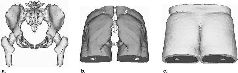
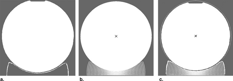
![Figure 2, (a) Test rig with foam specimen [1] and test person in face-down position (schematic). (b) Buttock loaded with foam specimen [1] in cutaway view.](https://storage.googleapis.com/dl.dentistrykey.com/clinical/MechanicalSoftTissuePropertyValidationinTissueEngineeringUsingMagneticResonanceImaging/1_1s20S1076633210004575.jpg)
Get Radiology Tree app to read full this article<
Get Radiology Tree app to read full this article<
Get Radiology Tree app to read full this article<
Get Radiology Tree app to read full this article<
Results
Gluteal Soft Tissue
Get Radiology Tree app to read full this article<
Polyurethane Soft Foam
Get Radiology Tree app to read full this article<
Get Radiology Tree app to read full this article<
Get Radiology Tree app to read full this article<
Gluteal Tissue - Soft Foam Interaction
Get Radiology Tree app to read full this article<
Get Radiology Tree app to read full this article<
Get Radiology Tree app to read full this article<
Discussion
Get Radiology Tree app to read full this article<
Get Radiology Tree app to read full this article<
References
1. Bennett G., Dealey C., Posnett J.: The cost of pressure ulcers in the UK. J Age Ageing 2004; 33: pp. 230-235.
2. Yeoman M.P., Hardy A.G.: The pathology and treatment of pressure sores in paraplegics. Br J Plast Surg 1954; 7: pp. 179-192.
3. Dansereau J.G., Conway H.: Closure of decubiti in paraplegics. Report of 2000 cases. J Plast Reconstr Surg 1964; 33: pp. 474-480.
4. Todd B.A., Thacker J.G.: Three-dimensional computer model of the human buttocks in vivo. J Rehabil Res Dev 1994; 31: pp. 111-119.
5. Oomens CWJ, Bressers OFJT, Bosboom EMH, et al. Deformation analysis of a supported buttock contact. In R.D. Kamm (ed.), Proceedings of the 2001 Bioengineering Conference, Snowbird, Utah, 2001. (BED, Vol. 50, pp. 853–854).
6. Sun Q., Lin F., Al-Saeede S., et. al.: Finite element modelling of human buttock-thigh tissue in a seated posture. Bioengin Conference, Colorado 2005; (Abstract 5–24)
7. Sun Q., Lin F., Al-Saeede S., et. al.: Soft tissue stress in buttock-thigh of a seated individual elucidated by a 3D FE Model. RESNA 28th Int Conf 2005; (Abstract 203)
8. Oomens C.W.J., Bressers O.F.J.T., Bosboom E.M.H., et. al.: Can loaded interface characteristics influence strain distributions in muscle adjacent to bony prominences?. Comput Methods Biomech Biomed Engin 2003; 6: pp. 171-180.
9. Makhsous M., Lim D., Hendrix R., et. al.: Finite element analysis for evaluation of pressure ulcer on the buttock: development and validation. J IEEE Transactions Neural Sys Rehabil Eng 2007; 15: pp. 517-525.
10. Linder-Ganz E., Gefen A.: Assessment of mechanical conditions in sub-dermal tissues during sitting: a combined experimental-MRI and finite element approach. J Biomech 2007; 40: pp. 1443-1454.
11. Then C., Menger J., Benderoth G., et. al.: A method for a mechanical characterization of human gluteal tissue. Technol Health Care 2007; 15: pp. 385-398.
12. Then C., Menger J., Benderoth G., et. al.: Analysis of mechanical interaction between human gluteal soft tissue and body supports. Technol Health Care 2008; 16: pp. 61-76.
13. James A.G., Green A., Simpson G.M.: Strain energy functions of rubber. I. Characterization of gum vulcanizates. J Appl Polym Sci 1974; 19: pp. 2033-2058.
14. Hartmann S., Tschöpe T., Schreiber L., et. al.: Large deformations of a carbon black-filled rubber. Experiment, optical measurement and parameter identification using finite elements. Euro J Mech Solid 2003; 22: pp. 309-324.
15. Lion A.: Constitutive model for carbon black filler rubber: experimental results and mathematical representation. Continuum Mech Thermodyn 1996; 6: pp. 153-169.
16. Schrodt M., Benderoth G., Kühhorn A., et. al.: Hyperelastic description of polymer soft foams at finite deformations. Tech Mech 2005; 25: pp. 162-173.
17. Hill R.: Aspects of invariance in solid mechanics. Adv Appl Mech 1978; 18: pp. 1-75.
18. Storakers B.: On the material representation and constitutive branching in finite compressible elasticity. J Mech Phy Sol 1986; 34: pp. 125-145.
19. Palevski A., Glaich I., Portnoy S., et. al.: Stress relaxation of porcine gluteus muscle subjected to sudden transverse deformation as related to pressure sore modelling. J Biomech Engin 2006; 128: pp. 782-787.
20. Gefen A., Gefen N., Linder-Ganz E., et. al.: In vivo muscle stiffening under bone compression promotes deep pressure sores. J. Biomech Engin 2005; 127: pp. 512-524.