
Rationale and Objectives
The aim of this study was to evaluate whether texture analysis (TA) can detect subtle changes in cerebral tissue caused by mild traumatic brain injury (MTBI) and to determine whether these changes correlate with neuropsychological and diffusion tensor imaging (DTI) findings.
Materials and Methods
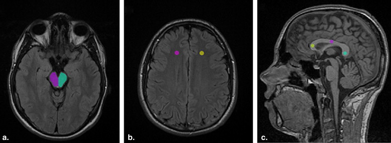
Forty-two patients with MTBIs were imaged using 1.5T magnetic resonance imaging within 3 weeks after head injury. TA was performed for the regions corresponding to the mesencephalon, centrum semiovale, and corpus callosum. Using DTI, the fractional anisotropic and apparent diffusion coefficient values for the same regions were evaluated. The same analyses were performed on a group of 10 healthy volunteers. Patients also underwent a battery of neurocognitive tests within 6 weeks after injury.
Results
TA revealed textural differences between the right and left hemispheres in patients with MTBIs, whereas differences were minimal in healthy controls. A significant correlation was found between scores on memory tests and texture parameters (sum of squares, sum entropy, inverse difference moment, and sum average) in patients in the area of the mesencephalon and the genu of the corpus callosum. Significant correlations were also found between texture parameters for the left mesencephalon and both fractional anisotropic and apparent diffusion coefficient values.
Conclusions
The data suggest that heterogeneous texture and abnormal DTI patterns in the area of the mesencephalon may be linked with verbal memory deficits among patients with MTBIs. Therefore, TA combined with DTI in patients with MTBIs may increase the ability to detect early and subtle neuropathologic changes.
Establishing a diagnosis of mild traumatic brain injury (MTBI) has proven to be challenging. Many systems have been developed to classify traumatic brain injuries (TBIs) from mild to severe. Most of these systems, such as the Glasgow Coma Scale, the duration of loss of consciousness, the presence of posttraumatic amnesia, and assorted neuroimaging techniques, rely on acute injury characteristics. The prognosis of moderate to severe traumatic brain injury is somewhat well correlated with these characteristics, but the relationship is not as clear for milder forms of traumatic brain injuries. Neuropsychological testing provides an assessment and quantification of brain function by examining brain-behavior relationships. These measurements evaluate memory recall, attention and concentration, problem-solving abilities, visual tracking, reaction time, and the speed of information processing, as well as other measures of cognitive function. Neuropsychological assessment is sensitive to the effects of traumatic brain injury and can be helpful in obtaining more information about subjective cognitive complaints in MTBI . Because brain injury does not produce a unique pattern of neuropsychological deficits, similar results may be seen that arise from the cognitive difficulties associated with other injury-related factors, such as insomnia, stress, and mood disturbances . These issues must be taken into account when neuropsychological data are analyzed.
Some victims of MTBIs may suffer from severe life-altering problems, including cognitive and emotional difficulties. Among patients with these chronic problems, the vast majority have normal findings on computed tomographic (CT) and magnetic resonance imaging (MRI) scans, the current clinical modalities of choice for the diagnosis of acute MTBI. With recent improvements, including diffusion-weighted MRI, diffusion tensor imaging (DTI), and new MRI sequences , certain MRI techniques provide even more sensitivity for MTBI than standard MRI. However, even though MRI has been found to be more sensitive for traumatic lesions than CT imaging, it still has a tendency to underestimate the condition; thus, an obvious need exists for advanced complementary techniques for imaging MTBI.
Get Radiology Tree app to read full this article<
Get Radiology Tree app to read full this article<
Get Radiology Tree app to read full this article<
Materials and methods
Study Subjects
Get Radiology Tree app to read full this article<
Neurologic and Neuropsychological Examinations
Get Radiology Tree app to read full this article<
Get Radiology Tree app to read full this article<
MRI Examinations
Get Radiology Tree app to read full this article<
Get Radiology Tree app to read full this article<
TA
Get Radiology Tree app to read full this article<
Get Radiology Tree app to read full this article<
Get Radiology Tree app to read full this article<
DTI Analysis
Get Radiology Tree app to read full this article<
Statistical Analysis
Get Radiology Tree app to read full this article<
Get Radiology Tree app to read full this article<
Results
TA Results
Get Radiology Tree app to read full this article<
Table 1
Numbers and Percentages of COM Parameters with Statistically Significant Differences ( P < .05) Between Hemispheres and Segments of the Corpus Callosum Analyzed with Wilcoxon’s Test
Differing Parameters Patient Healthy Volunteers Region of Interest COM Sequence ( n = 42) ( n = 10) Mesencephalon, right vs left_n_ = 123 T2-weighted FLAIR 55 (45%) 14 (12%) Centrum semiovale, right vs left_n_ = 123 T2-weighted FLAIR 27 (22%) 2 (2%) Corpus callosum, all segments_n_ = 123 T1-weighted MPR 30 (24%) 1 (1%)
COM, co-occurrence matrix; FLAIR, fluid-attenuated inversion recovery; MPR, magnetization prepared gradient echo.
In all, 123 texture parameters were evaluated.
Get Radiology Tree app to read full this article<
Get Radiology Tree app to read full this article<
Texture Parameter Correlation to Memory Test Scores
Get Radiology Tree app to read full this article<
Table 2
Correlations and Statistical Significance Between Texture Parameters From the Right and Left Sides of the Mesencephalon and the Memory Composite Scores ( n = 34)
Mesencephalon Right Mesencephalon Left Texture Parameter Verbal Memory Texture Parameter Verbal Memory Sum of squares (12/12 parameters)r −0.393 to −0.490 Sum of squares (6/12 parameters)r −0.341 to −0.372P .022 to .003P .049 to .030 Sum entropy (8/12 parameters)r −0.344 to −0.437 Inverse difference moment (3/12 parameters)r 0.356 to 0.385P .046 to .010P .039 to .025 Visual memory Visual memory No statistically significant correlation Sum average (5/12 parameters)r 0.387 to 0.494P .029 to .004
Table 3
Correlations and Statistical Significance Between Texture Parameters From the Genu of the Corpus Callosum and the Visual Memory Composite Scores ( n = 34)
Texture Parameter Visual Memory Entropy (4/12 parameters)r −0.390 to −0.397P .027 to .025 Angular second moment (3/12 parameters)r 0.356 to 0.385P .039 to .025
Get Radiology Tree app to read full this article<
Get Radiology Tree app to read full this article<
Get Radiology Tree app to read full this article<
Texture Correlation to DTI Results
Get Radiology Tree app to read full this article<
Get Radiology Tree app to read full this article<
Correlation of DTI to Memory Test Scores
Get Radiology Tree app to read full this article<
Get Radiology Tree app to read full this article<
Discussion
Get Radiology Tree app to read full this article<
Get Radiology Tree app to read full this article<
Get Radiology Tree app to read full this article<
Get Radiology Tree app to read full this article<
Get Radiology Tree app to read full this article<
Get Radiology Tree app to read full this article<
Conclusions
Get Radiology Tree app to read full this article<
Get Radiology Tree app to read full this article<
References
1. Alexander M.P.: Mild traumatic brain injury: a review of physiogenesis and psychogenesis. Neurology 1995; 45: pp. 1253-1260.
2. Binder L.M.: A review of mild head trauma. Part II: clinical implications. J Clin Exp Neuropsychology 1997; 19: pp. 432-457.
3. Posse S., Tedeschi R., Risinger R., et. al.: High speed 1H spectroscopic imaging in human brain by echo planar spatial-spectral encoding. Magn Reson Med 1995; 33: pp. 34-40.
4. Raucher A., Sedlacik J., Deistung A., et. al.: Susceptibility weighted imaging: data acquisition, image reconstruction and clinical applications. Z Med Phys 2006; 16: pp. 240-250.
5. Inglese M., Makani S., Johnson G., et. al.: Diffuse axonal injury in mild traumatic brain injury: a diffusion tensor imaging study. J Neurosurg 2005; 103: pp. 298-303.
6. Huisman T.A., Schwamm L.H., Schaefer P.W., et. al.: Diffusion tensor imaging as potential biomarker of white matter injury in diffuse axonal injury. AJNR Am J Neuroradiol 2004; 25: pp. 370-376.
7. Rutgers D.R., Fillard P., Paradot G., et. al.: Diffusion tensor imaging characteristics of the corpus callosum in mild, moderate, and severe traumatic brain injury. AJNR Am J Neuroradiol 2008; 29: pp. 1730-1735.
8. Kumar R., Gupta R.K., Husain M., et. al.: Comparative evaluation of corpus callosum DTI metrics in acute mild and moderate traumatic brain injury: its correlation with neuropsychometric tests. Brain Inj 2009; 23: pp. 675-685.
9. Arfanakis K., Haughton V.M., Carew J.D., et. al.: Diffusion tensor MR imaging in diffuse axonal injury. AJNR Am J Neuroradiol 2002; 23: pp. 794-802.
10. Tuceryan M., Jain A.K.: Texture analysis.Chen C.H.Pau L.F.Wang P.S.P.The handbook of pattern recognition and computer vision.1998.World Scientific PublishingRiver Edge, NJ:pp. 207-248.
11. Freeborough P.A., Fox N.C.: MR image texture analysis applied to the diagnosis and tracking of Alzheimer’s disease. IEEE Trans Med Imaging 1998; 17: pp. 475-479.
12. Bonilha L., Kobayashi E., Castellano G., et. al.: Textural analysis of hippocampal sclerosis. Epilepisa 2003; 44: pp. 1546-1550.
13. Zhang J., Tong L., Wang L., et. al.: Texture analysis of multiple sclerosis: a comparative study. Magn Reson Imaging 2008; 26: pp. 1160-1166.
14. Kjaer L., Ring P., Thomsen C., Henriksen O.: Texture analysis in quantitative MR imaging—tissue characterization of normal brain and intracranial tumours at 1.5T. Acta Radiol 1995; 36: pp. 127-135.
15. Mahmoud-Ghoneim D., Alkaabi M.K., de Certaines J.D., et. al.: The impact of image dynamic range on texture classification of brain white matter. BMC Med Imaging 2008; 23: pp. 8-18.
16. Schad L.R., Blüml S., Zuna I.: MR tissue characterization of intracranial tumors by means of texture analysis. Magn Reson Imaging 1993; 11: pp. 889-896.
17. Herlidou-Même S., Constans J.M., Carsin B., et. al.: MRI texture analysis on texture test objects, normal brain and intracranial tumours. J Magn Reson Imaging 2003; 21: pp. 989-993.
18. Jirak D., Dezortova M., Taimr P., Hajek M.: Texture analysis of human liver. J Magn Reson Imaging 2002; 15: pp. 68-74.
19. Holli K.K., Lääperi A.-L., Harrison L., et. al.: Characterization of breast cancer types by texture analysis of magnetic resonance images. Acad Radiol 2010; 17: pp. 135-141.
20. Gibbs P., Turnbull L.W.: Textural analysis of contrast-enhanced MR images of the breast. Magn Reson Med 2003; 50: pp. 92-98.
21. Harrison L., Dastidar P., Eskola H., et. al.: Texture analysis on MRI images of non-Hodgkin lymphoma. Comput Biol Med 2008; 38: pp. 519-524.
22. Harrison L., Luukkaala T., Pertovaara H., et. al.: Non-Hodgkin lymphoma response evaluation with MRI texture classification. J Exp Clin Cancer Res 2009; 28: pp. 87-100.
23. Carrol L.J., Cassidy J.D., Holm L., et. al.: Methodological issues and research recommendations for mild traumatic brain injury: the WHO Collaborating Centre Task Force on Mild Traumatic Brain Injury. J Rehabil Med Suppl 2004; 43: pp. 113-125.
24. Teasdale G., Jennett B.: Assessment of coma and impaired consciousness. A practical scale. Lancet 1974; 11: pp. 81-84.
25. Lezak M., Hovieson D., Loring D.: Neuropsychological assessment.4th ed.2004.Oxford University PressNew York
26. Morrow L.A., Ryan C.: Normative data for a working memory test: the Four Word Short-Term Memory Test. Clin Neuropsychol 2002; 16: pp. 373-380.
27. CANTAB®, the Cambridge Neuropsychological Automated Testing Battery.2004.Cambridge CognitionCambridge, UK
28. Strauss E., Sherman E.M.S., Spreen O.: A compendium of neuropsychological tests: administration, norms, and commentary.3rd ed.2006.Oxford University PressNew York
29. Mitrushina M., Boone K.B., Razani J., D’Elia L.: Normative data for neuropsychological assessment.2nd ed.2005.Oxford University PressNew York
30. Hajek M.Dezortova M.Materka A.Lerski R.Texture analysis for magnetic resonance imaging.2006.Med4PublishingPrague, Czech Republic:
31. Lo C., Shifteh K., Gold T., et. al.: Diffusion tensor imaging abnormalities in patients with mild traumatic brain injury and neurocognitive impairment. J Comput Assist Tomogr 2009; 33: pp. 293-297.
32. Mayer A.R., Ling J., Mannell M.V., et. al.: A prospective diffusion tensor imaging study in mild traumatic brain injury. Neurology 2010; 74: pp. 643-650.
33. Chu Z., Wilde E.A., Hunter J.V., et. al.: Voxel-based analysis of diffusion tensor imaging in mild traumatic brain injury in adolescents. AJNR Am J Neuroradiol 2010; 3: pp. 340-346.