
Rationale and Objectives
This study provides a systematic assessment of different methods of delivering radiologic teaching content (lecture, printed text, and digital content delivery) under standard conditions, enabling comparison of the effectiveness of these methods.
Materials and Methods
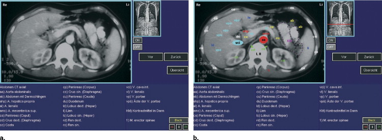
A printed atlas of sectional anatomy was used as a standard. Digital content was developed on the basis of the printed atlas. Lecturers used both the printed and the digital content to prepare lectures. Standardized teaching material thus created was presented to second-term undergraduate students who had attended the school’s anatomy course, but had not received any radiology teaching. Multiple choice examinations were used to assess the students’ ability to recognize anatomical structures in known as well as unknown images. In a survey, the students’ subjective experience of the learning process was assessed.
Results
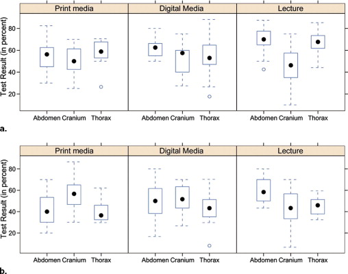
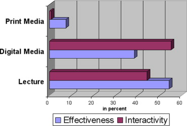
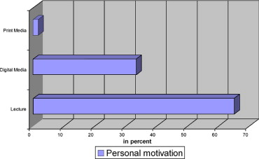
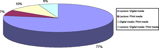
No difference was seen between the groups regarding examination results. Students preferred a combination of digital media and lectures by enthusiastic teachers.
Conclusions
The shortage of teachers requires a compromise concerning the delivery of radiologic anatomy content in a medical school setting. Based on our results, we recommend a combined approach of lecture and digital content delivery.
The use of digital media is continually extending its foothold within undergraduate, postgraduate, and continued medical education. Instructional methods that use digital methods of information delivery have been evaluated with promising results ( ). They appear to be particularly successful when the teaching content and exemplars are predominantly visual ( ). A case in point is undergraduate training in diagnostic radiology, in which image understanding and pattern recognition need to be practiced, and an essential part of teaching consists in pointing out features of an image by annotation.
That resources for teaching tend to be tightly constrained necessitates optimal use of such resources as measured by learners’ knowledge retention and development of pattern recognition skills. At the same time, the subjective learning experience should be positive and motivating. We therefore chose instructional methods based on different modes of information delivery and compared these by taking various measures that reflect the outcomes.
Get Radiology Tree app to read full this article<
Get Radiology Tree app to read full this article<
Get Radiology Tree app to read full this article<
Get Radiology Tree app to read full this article<
Get Radiology Tree app to read full this article<
Get Radiology Tree app to read full this article<
Material and methods
Teaching Groups
Get Radiology Tree app to read full this article<
Get Radiology Tree app to read full this article<
Get Radiology Tree app to read full this article<
Get Radiology Tree app to read full this article<
Table 1
Group Setup: Each Group was Taught Once in Each Topic Using a Different Instructional Method
Group No. Topic Cranium Chest Abdomen 1 Lecture Digital Book 2 Digital Lecture Book 3 Book Digital Lecture 4 Digital Book Lecture 5 Lecture Book Digital 6 Book Lecture Digital
Get Radiology Tree app to read full this article<
Testing and Teaching Procedure
Get Radiology Tree app to read full this article<
Get Radiology Tree app to read full this article<
Information Sources
Get Radiology Tree app to read full this article<
Get Radiology Tree app to read full this article<
Get Radiology Tree app to read full this article<
Get Radiology Tree app to read full this article<
Survey of Participants
Get Radiology Tree app to read full this article<
Results
Method Comparisons
Get Radiology Tree app to read full this article<
Get Radiology Tree app to read full this article<
Get Radiology Tree app to read full this article<
Table 2
Distribution of Scores Within Stratification Groups
Pattern Recognition Performance High Impact Medium Impact Low Impact Digital Print Lecture Digital Print Lecture Digital Print Lecture Good 16 23 11 11 7 16 8 3 13 Medium 12 4 9 9 11 15 18 6 14 Poor 6 10 7 13 19 7 15 15 16
Get Radiology Tree app to read full this article<
Survey
Get Radiology Tree app to read full this article<
Get Radiology Tree app to read full this article<
Get Radiology Tree app to read full this article<
Get Radiology Tree app to read full this article<
Get Radiology Tree app to read full this article<
Get Radiology Tree app to read full this article<
Discussion
Get Radiology Tree app to read full this article<
Get Radiology Tree app to read full this article<
Get Radiology Tree app to read full this article<
Get Radiology Tree app to read full this article<
Get Radiology Tree app to read full this article<
Get Radiology Tree app to read full this article<
Get Radiology Tree app to read full this article<
Get Radiology Tree app to read full this article<
Get Radiology Tree app to read full this article<
Get Radiology Tree app to read full this article<
Conclusion
Get Radiology Tree app to read full this article<
Get Radiology Tree app to read full this article<
Get Radiology Tree app to read full this article<
References
1. Mooney G.A., Bligh J.G.: Information technology in medical education: current and future applications. Postgrad Med J 1997; 73: pp. 701-704.
2. Mammone G.L., Holman B.L., Greenes R.A., et. al.: Inside BrighamRAD: providing radiology teaching cases on the Internet. Radiographics 1995; 15: pp. 1489-1498.
3. Wunderbaldinger P., Schima W., Turetschek K., et. al.: World Wide Web and Internet: applications for radiologists. Eur Radiol 1999; 9: pp. 1170-1182.
4. Grunewald M., Heckemann R.A., Gebhard H., et. al.: COMPARE radiology: creating an interactive Web-based training program for radiology with multimedia authoring software. Acad Radiol 2003; 10: pp. 543-553.
5. Grunewald M., Heckemann R.A., Wagner M., et. al.: ELERA: a WWW application for evaluating and developing radiologic skills and knowledge. Acad Radiol 2004; 11: pp. 1381-1388.
6. Grunewald M., Gebhard H., Jakob C., et. al.: Rofo 2004; 176: pp. 885-895. [Web-based training in radiology—student course in the Virtual University of Bavaria]
7. Grunewald M., Zenk J., Alibek S., et. al.: Hno 2005; 53: pp. 337-342. [Hnorad]
8. Grunewald M., Ketelsen D., Heckemann R.A., et. al.: Teach and be taught radiology: implementation of a web-based training program based on user preferences as determined by survey. Acad Radiol 2006; 13: pp. 461-468. www.tnt-radiology .de:
9. Chen M.S., Horrocks E.N., Evans R.D.: Video versus lecture: effective alternatives for orthodontic auxiliary training. Br J Orthod 1998; 25: pp. 191-195.
10. Solomon E.S., Whiton J.C.: Student evaluation of audiovisual teaching equipment. J Dent Educ 1987; 51: pp. 559-560.
11. Grunewald M., Bischoff G., Kressel M., et. al.: 2000.Urban und FischerMuenchen
12. Jaffe C.C., Lynch P.J.: Computer-aided instruction in radiology: opportunities for more effective learning. AJR Am J Roentgenol 1995; 164: pp. 463-467.
13. D’Alessandro M.P., Galvin J.R., Erkonen W.E., et. al.: The instructional effectiveness of a radiology multimedia textbook (HyperLung) versus a standard lecture. Invest Radiol 1993; 28: pp. 643-648.
14. D’Alessandro D.M., Kreiter C.D., Erkonen W.E., et. al.: Longitudinal follow-up comparison of educational interventions: multimedia textbook, traditional lecture, and printed textbook. Acad Radiol 1997; 4: pp. 719-723.
15. Erkonen W.E., D’Alessandro M.P., Galvin J.R., et. al.: Longitudinal comparison of multimedia textbook instruction with a lecture in radiology education. Acad Radiol 1994; 1: pp. 287-292.
16. Mehta A., Dreyer K.J., Montgomery M., et. al.: A World Wide Web Internet engine for collaborative entry and peer review of radiologic teaching files. AJR Am J Roentgenol 1999; 172: pp. 893-896.
17. Khorasani R., Lester J.M., Davis S.D., et. al.: Web-based digital radiology teaching file: facilitating case input at time of interpretation. AJR Am J Roentgenol 1998; 170: pp. 1165-1167.
18. Möller T.B., Reif E.: 2000.Thieme Medical PublishersStuttgart
19. McEnery K.W.: The Internet, World-Wide Web, and Mosaic: an overview. AJR Am J Roentgenol 1995; 164: pp. 469-473.
20. Richardson M.L., Norris T.E.: On-line delivery of continuing medical education over the World-Wide Web: an on-line needs assessment. AJR Am J Roentgenol 1997; 168: pp. 1161-1164.
21. Kraft S.L., Hoskinson J.J., Mussman J.M., et. al.: Development of interactive patient-based multimedia computer programs in veterinary orthopedic radiology. Vet Radiol Ultrasound 1998; 39: pp. 98-104.