
Rationale and Objectives
The aim of this study was to analyze the morphology of pancreatic cystic masses detected on multi–detector row computed tomography (MDCT) to determine whether single-dimension measurements of these masses are accurate reflections of their volumes.
Materials and Methods
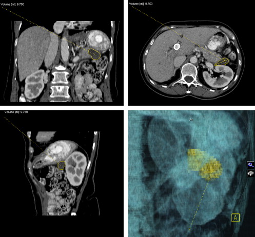
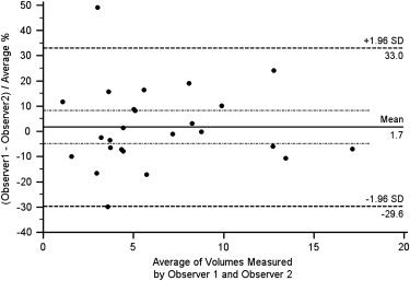
Twenty-five pancreatic cystic masses detected on MDCT in 25 patients were evaluated. Pancreatic cysts were segmented on MDCT using commercially available software. All measurements were obtained twice by two independent investigators, and the means of values for segmented cyst volume (Vs) (milliliters), maximum transaxial diameter (millimeters), and elongation value (defined as 1 − [width/length], where 1 = ellipsoid and 0 = spherical) were reported for each cystic lesion. The volume of each cyst was also calculated (Vc) using the maximum transaxial diameter, with the hypothesis that the cyst was spherical. Student’s t test was used to analyze the differences between values of Vs and Vc. Bland-Altman plots and Lin’s concordance correlation were used to assess agreement between different measurement techniques. A P value < .05 denoted statistical significance. Interobserver variability was also determined using the Bland-Altman method.
Results
There was a significant difference between Vs and Vc ( P < .0001). The elongation values ranged from 0.28 to 0.83 (mean, 0.57 ± 0.12; median, 0.56). Mean interobserver variability was 1.7% (95% confidence interval, −4.89% to 8.30%).
Conclusions
The results suggest that pancreatic cystic masses are not spherical. Therefore, a cyst’s single largest transaxial dimension is not an accurate surrogate of its actual volume.
The expanding use of cross-sectional imaging techniques (computed tomography [CT] and magnetic resonance imaging [MRI]) has increased the number of pancreatic cysts diagnosed . Because of their increasing detection rate, the management of pancreatic cystic lesions remains a challenge . Several studies have suggested a size cutoff of 3 cm as the threshold for surgical resection in patients who are asymptomatic . To our knowledge, the actual volumes of pancreatic cystic lesions have never been directly quantified with imaging modalities. As a result, their largest single diameters on computed tomographic or magnetic resonance images have traditionally been used as surrogates of their actual volumes and have been used to determine management strategies . The current literature supports follow-up of cystic masses with largest diameters of <3 cm and the resection of those masses >3 cm, depending on the clinical context .
Our objective in this study was to analyze the morphology of pancreatic cystic masses diagnosed on multi–detector row CT to test the null hypothesis that pancreatic cysts are spherical and whether the longest transaxial diameters of these masses are reliably representative of their volumes.
Materials and methods
Study Population
Get Radiology Tree app to read full this article<
Image Acquisition and Analysis
Get Radiology Tree app to read full this article<
Get Radiology Tree app to read full this article<
Get Radiology Tree app to read full this article<
Statistical Analysis
Get Radiology Tree app to read full this article<
Results
Get Radiology Tree app to read full this article<
Table 1
Quantified Parameters of Pancreatic Cysts
Cyst MTD (mm) LPD (mm) CCD (mm) Vs (mL) Vc (mL) Ve (mL) Elongation Value 1 32.6 19.1 29.75 8.08 18.14 9.70 0.6 2 22.8 19.1 27.3 5.57 6.21 6.22 0.56 3 26.85 19.95 29.6 7.16 10.13 8.30 0.42 4 16.7 10.8 14.35 1.08 2.44 1.36 0.61 5 28.6 20.25 28 8.25 12.25 8.49 0.46 6 21.9 15.05 32.3 4.35 5.50 5.57 0.71 7 25.45 20.75 26.45 4.42 8.63 7.31 0.53 8 26.5 15.4 18.35 3.70 9.74 3.92 0.805 9 30.05 27.6 26.25 8.78 14.21 11.40 0.34 10 16.45 13.55 18.35 1.56 2.33 2.14 0.51 11 23.85 15.8 30.35 5.73 7.10 5.99 0.67 12 36.5 24.85 32.3 13.44 25.46 15.34 0.595 13 20.4 14.95 19.75 2.96 4.45 3.15 0.505 14 21.55 15.95 21.2 3.57 5.24 3.82 0.655 15 24.7 18.3 23.3 5.09 7.89 5.51 0.565 16 22.5 13.05 19.5 3.20 5.96 3.00 0.54 17 22.85 17.3 22.7 4.44 6.25 4.70 0.28 18 38.4 19.5 32.9 9.89 29.65 12.90 0.835 19 33.2 22.1 38.75 12.73 19.16 14.89 0.585 20 36.15 26.65 34.2 12.79 24.73 17.25 0.54 21 22.25 16.8 23.85 3.01 5.77 4.67 0.65 22 25.5 20.3 22.8 5.02 8.68 6.18 0.545 23 25.1 17.5 21.45 3.62 8.28 4.93 0.745 24 36 29.75 38.45 17.13 24.43 21.56 0.46 25 19.6 18.2 23 3.72 3.94 4.30 0.52
CCD, craniocaudal diameter; LPD, longest perpendicular diameter; MTD, maximum transaxial diameter; Vc, calculated cyst volume; Ve, estimated cyst volume; Vs, segmented cyst volume.
Get Radiology Tree app to read full this article<
Get Radiology Tree app to read full this article<
Get Radiology Tree app to read full this article<
Get Radiology Tree app to read full this article<
Get Radiology Tree app to read full this article<
Get Radiology Tree app to read full this article<
Discussion
Get Radiology Tree app to read full this article<
Get Radiology Tree app to read full this article<
Get Radiology Tree app to read full this article<
Get Radiology Tree app to read full this article<
References
1. Laffan T.A., Horton K.M., Klein A.P., et. al.: Prevalence of unsuspected pancreatic cysts on MDCT. AJR Am J Roentgenol 2008; 191: pp. 802-807.
2. Sahani D.V., Saokar A., Hahn P.F., et. al.: Pancreatic cysts 3 cm or smaller: how aggressive should treatment be?. Radiology 2006; 238: pp. 912-919.
3. Lee C.J., Scheiman J., Anderson M.A., et. al.: Risk of malignancy in resected cystic tumors of the pancreas < or =3 cm in size: is it safe to observe asymptomatic patients? A multi-institutional report. J Gastrointest Surg 2008; 12: pp. 234-242.
4. Tanaka M., Chari S., Adsay V., et. al.: International consensus guidelines for management of intraductal papillary mucinous neoplasms and mucinous cystic neoplasms of the pancreas. Pancreatology 2006; 6: pp. 17-32.
5. Garcea G., Ong S.L., Rajesh A., et. al.: Cystic lesions of the pancreas. A diagnostic and management dilemma. Pancreatology 2008; 8: pp. 236-251.
6. Keil S., Behrendt F.F., Stanzel S., et. al.: Semi-automated measurement of hyperdense, hypodense and heterogeneous hepatic metastasis on standard MDCT slices. comparison of semi-automated and manual measurement of RECIST and WHO criteria. Eur Radiol 2008; 18: pp. 2456-2465.
7. Malvern Instruments. Particle size and shape measurement. Available at: http://www.fei.com/uploadedFiles/Documents/Content/particle_morphology.pdf . Accessed September 10, 2009.
8. Bland J.M., Altman D.G.: Statistical methods for assessing agreement between two methods of clinical measurement. Lancet 1986; 1: pp. 307-310.
9. Lin L.I.: A concordance correlation coefficient to evaluate reproducibility. Biometrics 1989; 45: pp. 255-268.
10. Walsh R.M., Vogt D.P., Henderson J.M., et. al.: Management of suspected pancreatic cystic neoplasms based on cyst size. Surgery 2008; 144: pp. 677-685.
11. Spinelli K.S., Fromwiller T.E., Daniel R.A., et. al.: Cystic pancreatic neoplasms: observe or operate. Ann Surg 2004; 239: pp. 651-659.
12. Chari S.T., Yadav D., Smyrk T.C., et. al.: Study of recurrence after surgical resection of intraductal papillary mucinous neoplasm of the pancreas. Gastroenterology 2002; 123: pp. 1500-1507.
13. Allen P.J., D’Angelica M., Gonen M., et. al.: A selective approach to the resection of cystic lesions of the pancreas: results from 539 consecutive patients. Ann Surg 2006; 244: pp. 572-582.
14. Goh B.K., Sahani D.V., Saokar A., et. al.: Pancreatic cysts 3 cm or smaller. Radiology 2007; 243: pp. 607.
15. Honda O., Sumikawa H., Johkoh T., et. al.: Computer-assisted lung nodule volumetry from multi-detector row CT: Influence of image reconstruction parameters. Eur J Radiol 2007; 62: pp. 106-113.