
Rationale and Objectives
The aims of this study were to determine agreement between clinical examination and magnetic resonance imaging (MRI) (rectal contrast and noncontrast MRI) for pelvic organ prolapse using both the pubococcygeal line (PCL) and the midpubic line (MPL) and to assess the relationship between measurements performed relative to each line.
Materials and Methods
Dynamic MRI exams in 88 women (with rectal contrast, n = 39; noncontrast, n = 49) were evaluated, followed by review of clinical exam notes. Agreement between clinical exam and MRI and the difference between PCL and MPL measurements were evaluated.
Results
Agreement of rectal contrast MRI with clinical exam was 79% for PCL and 85% for MPL ( P = .17) for cystoceles, 50% for PCL and 59% for MPL ( P = .20) for vaginal prolapse, 56% for PCL for enteroceles, and 61% for rectoceles. Agreement of noncontrast MRI with clinical exam was 67% for PCL and 78% for MPL ( P = .19) for cystoceles, 58% for PCL and 71% for MPL ( P = .10) for vaginal prolapse, 65% for enteroceles, and 40% for rectoceles. The average difference between the PCL and the MPL was 3.12 ± 0.24 cm at the bladder base and 4.88 ± 0.37 cm at the vaginal apex.
Conclusions
Agreement of MRI with clinical exam was highest for cystoceles. There was no significant difference in agreement using the MPL or PCL, suggesting that either line can be used on MRI. The average differences between the PCL and MPL at the bladder base and vaginal apex were approximately 3 and 5 cm, respectively.
Dynamic magnetic resonance imaging (MRI) of the pelvic floor can be used as an adjunct to the clinical evaluation for clarifying physical examination findings. However, the criteria for defining and grading prolapse differ between the two methods. On physical examination, the position of the pelvic viscera relative to the hymen is determined on direct visualization of the vagina, and prolapse can be quantified using the Pelvic Organ Prolapse Quantification system . There is no standardized method for evaluating prolapse on MRI, and the pubococcygeal line (PCL), HMO (H line, M line, organ prolapse) classification, and midpubic line (MPL) have been used . The PCL was initially proposed because it is easily drawn on sagittal images and approximates the pelvic floor . The MPL was subsequently proposed as a more correlative line because it approximates the hymen, which is used on clinical exam . However, two subsequent studies have found poor correlations between quantitative parameters on clinical exam and MRI using the MPL .
In an initial study to compare both the PCL and the MPL with clinical exam, Lienemann et al studied 41 asymptomatic volunteers using rectal contrast MRI. On MRI, organ descent below the MPL was used to classify prolapse. This definition differs from the typical clinical criteria of normal organ position being several centimeters above the MPL, which was also shown in an MRI study on control subjects . Differences in criteria may have accounted for the lack of prolapse in the anterior and superior compartments using the MPL on MRI, and the best correlation with clinical exam was achieved using the PCL for these compartments. A more recent patient study comparing quantitative assessment of prolapse on clinical exam, MRI, and ultrasound using both the PCL and the MPL found that only measurements in the anterior compartment showed good correlation . However, prolapse is usually evaluated qualitatively in typical clinical practice, and comparison of qualitative clinical assessment with MRI may demonstrate better agreement.
Get Radiology Tree app to read full this article<
Materials and methods
Patient Population
Get Radiology Tree app to read full this article<
Get Radiology Tree app to read full this article<
MRI Technique
Get Radiology Tree app to read full this article<
Image Analysis
Get Radiology Tree app to read full this article<
Table 1
Criteria for Diagnosing Prolapse on MRI
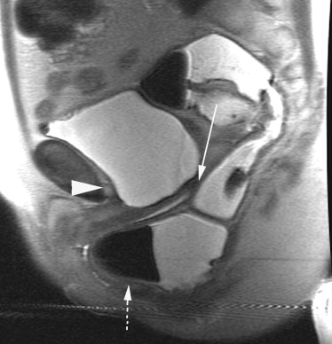
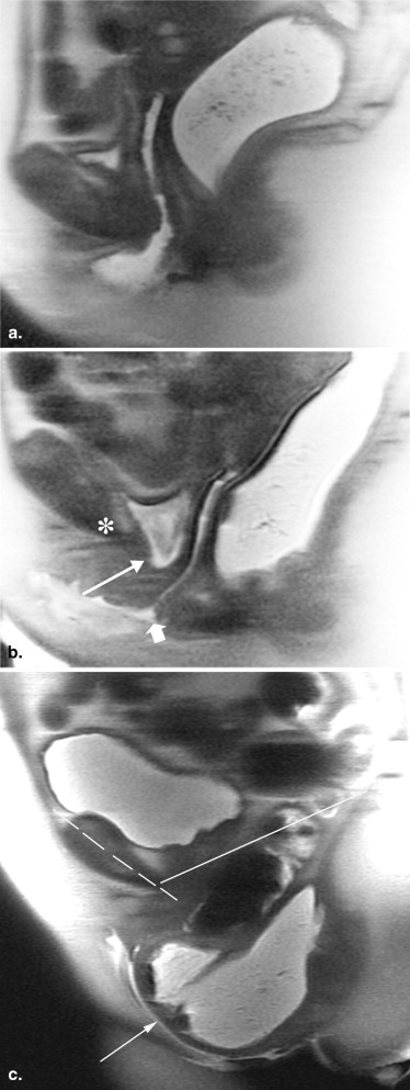
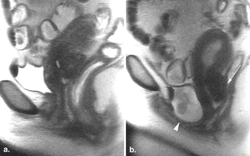
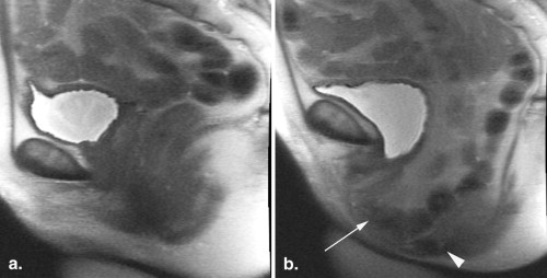
Viscera Criteria MRI diagnosis Bladder Below PCL Below MPL or ≤3 cm above MPL Cystocele Cervix/vaginal apex Below PCL Below MPL or ≤5 cm above MPL Cervical/vaginal prolapse Small bowel or fat Below apical third of vagina Enterocele Rectum ≥3 cm anterior bulge relative to anal canal Rectocele
The location of the viscera relative to the PCL and MPL was determined, and criteria listed in the table were applied. For the MPL, thresholds of 3 and 5 cm were used for the bladder and vagina, respectively, similar to clinical exam and based on a prior study in control subjects (5). On clinical exam, >2-cm descent below the total vaginal length is used to define prolapse, with 8 to 10 cm typical values for total vaginal length.
MPL, midpubic line; MRI, magnetic resonance imaging; PCL, pubococcygeal line.
Get Radiology Tree app to read full this article<
Get Radiology Tree app to read full this article<
Statistical Analysis
Get Radiology Tree app to read full this article<
Get Radiology Tree app to read full this article<
Results
Agreement of MRI with Clinical Exam
Get Radiology Tree app to read full this article<
Table 2
Frequency of Diagnosis of Cystocele and Vaginal Prolapse on MRI and Clinical Examination
Percentage of Patients with Positive Diagnoses Percentage Agreement with Clinical Diagnosis Site Patient Group (n) PCL MPL Clinical PCL MPL_P_ Bladder All patients (77) 82 99 82 72 81 .08 Rectal contrast (34) 91 100 85 79 85 .17 Noncontrast (43) 74 99 79 67 78 .19 Vagina All patients (65) 60 78 75 54 65 .04 Rectal contrast (32) 69 81 69 50 59 .20 Noncontrast (33) 52 74 82 58 71 .10
Agreement between reference line on MRI and clinical examination for presence or absence of prolapse. P values compare agreement between PCL and MPL with clinical exam.
MPL, midpubic line; MRI, magnetic resonance imaging; PCL, pubococcygeal line.
Table 3
Frequency of Diagnosis of Enterocele and Rectocele on MRI and Clinical Examination
Percentage of Patients with Positive Diagnoses Percentage Agreement with Clinical Diagnosis Site Patient Group (n) PCL Clinical PCL Bowel All patients (72) 22 42 61 Rectal contrast (33) 14 42 56 Noncontrast (39) 29 41 65 Rectum All patients (80) 38 82 49 Rectal contrast (36) 58 86 61 Noncontrast (44) 22 80 40
Agreement between reference line on MRI and clinical examination for presence or absence of prolapse.
MRI, magnetic resonance imaging; PCL, pubococcygeal line.
Get Radiology Tree app to read full this article<
Get Radiology Tree app to read full this article<
Table 4
Average Difference Between PCL and MPL Measurements at Different Sites in the Pelvis
Site Observations ∗ Difference (cm) Standard Error of the Mean (cm) Bladder 145 3.12 0.24 Vagina 107 4.88 0.37 Enterocele 43 4.46 1.01
MPL, midpubic line; PCL, pubococcygeal line.
Get Radiology Tree app to read full this article<
Get Radiology Tree app to read full this article<
Disagreement of MRI with Clinical Exam
Get Radiology Tree app to read full this article<
Get Radiology Tree app to read full this article<
Get Radiology Tree app to read full this article<
Get Radiology Tree app to read full this article<
Get Radiology Tree app to read full this article<
Get Radiology Tree app to read full this article<
Discussion
Get Radiology Tree app to read full this article<
Get Radiology Tree app to read full this article<
Get Radiology Tree app to read full this article<
Get Radiology Tree app to read full this article<
Get Radiology Tree app to read full this article<
Get Radiology Tree app to read full this article<
Get Radiology Tree app to read full this article<
Conclusions
Get Radiology Tree app to read full this article<
References
1. Bump R.C., Mattiasson A., Bø K., et. al.: The standardization of terminology of female pelvic organ prolapse and pelvic floor dysfunction. Am J Obstet Gynecol 1996; 175: pp. 10-17.
2. Yang A., Mostwin J.L., Rosenshein N.B., et. al.: Pelvic floor descent in women: dynamic evaluation with fast MR imaging and cinematic display. Radiology 1991; 179: pp. 25-33.
3. Comiter C.V., Vasavada S.P., Barbaric Z.L., et. al.: Grading pelvic prolapse and pelvic floor relaxation using dynamic magnetic resonance imaging. Urology 1999; 54: pp. 454-457.
4. Boyadzhyan L., Raman S.S., Raz S.: Role of static and dynamic MR imaging in surgical pelvic floor dysfunction. Radiographics 2008; 28: pp. 949-967.
5. Singh K., Reid W.M., Berger L.A.: Assessment and grading of pelvic organ prolapse by use of dynamic magnetic resonance imaging. Am J Obstet Gynecol 2001; 185: pp. 71-77.
6. Cortes E., Reid W.M., Singh K., et. al.: Clinical examination and dynamic magnetic resonance imaging in vaginal vault prolapse. Obstet Gynecol 2004; 103: pp. 41-46.
7. Fauconnier A., Zareski E., Abichedid J., et. al.: Dynamic magnetic resonance imaging for grading pelvic organ prolapse according to the International Continence Society classification: which line should be used?. Neurourol Urodyn 2008; 27: pp. 191-197.
8. Lienemann A., Sprenger D., Janssen U., et. al.: Assessment of pelvic organ descent by use of functional cine-MRI: which reference line should be used?. Neurourol Urodyn 2004; 23: pp. 33-37.
9. Broekhuis SR, Kluivers KB, Hendriks JC, et al. POP-Q, dynamic MR imaging, and perineal ultrasonography: do they agree in the quantification of female pelvic organ prolapse? Int Urogynecol J Pelvic Floor Dysfunct. In press.
10. Lienemann A., Anthuber C., Baron A., et. al.: Dynamic MR colpocystorectography assessing pelvic-floor descent. Eur Radiol 1997; 7: pp. 1309-1317.
11. Goh V., Halligan S., Kaplan G., et. al.: Dynamic MR imaging of the pelvic floor in asymptomatic subjects. AJR Am J Roentgenol 2000; 174: pp. 661-666.
12. Barber M.D., Lambers A., Visco A.G., et. al.: Effect of patient position on clinical evaluation of pelvic organ prolapse. Obstet Gynecol 2000; 96: pp. 18-22.
13. Silva W.A., Kleeman S., Segal J., et. al.: Effects of a full bladder and patient positioning on pelvic organ prolapse assessment. Obstet Gynecol 2004; 104: pp. 37-41.
14. Bertschinger K.M., Hetzer F.H., Roos J.E., et. al.: Dynamic MR imaging of the pelvic floor performed with patient sitting in an open-magnet unit versus with patient supine in a closed-magnet unit. Radiology 2002; 223: pp. 501-508.
15. Morren G.L., Balasingam A.G., Wells J.E., et. al.: Triphasic MRI of pelvic organ descent: sources of measurement error. Eur J Radiol 2005; 54: pp. 276-283.
16. Broekhuis S.R., Kluivers K.B., Hendriks J.C., et. al.: Dynamic magnetic resonance imaging: reliability of anatomical landmarks and reference lines used to assess pelvic organ prolapse. Int Urogynecol J Pelvic Floor Dysfunct 2009; 20: pp. 141-148.
17. Visco A.G., Wei J.T., McClure L.A., et. al.: Pelvic Floor Disorders Network. Effects of examination technique modifications on Pelvic Organ Prolapse Quantification (POP-Q) results. Int Urogynecol J Pelvic Floor Dysfunct 2003; 14: pp. 136-140.
18. Hall A.F., Theofrastous J.P., Cundiff G.W., et. al.: Interobserver and intraobserver reliability of the proposed International Continence Society, Society of Gynecologic Surgeons, and American Urogynecologic Society pelvic organ prolapse classification system. Am J Obstet Gynecol 1996; 175: pp. 1467-1470.
19. Lienemann A., Anthuber C., Baron A., et. al.: Diagnosing enteroceles using dynamic magnetic resonance imaging. Dis Colon Rectum 2000; 43: pp. 205-212.
20. Kester R.R., Leboeuf L., Amendola M.A., et. al.: Value of express T2-weighted pelvic MRI in the preoperative evaluation of severe pelvic floor prolapse: a prospective study. Urology 2003; 61: pp. 1135-1139.
21. Lockhart M.E., Fielding J.R., Richter H.E., et. al.: Reproducibility of dynamic MR imaging pelvic measurements: a multi-institutional study. Radiology 2008; 249: pp. 534-540.
22. Burrows L.J., Sewell C., Leffler K.S., et. al.: The accuracy of clinical evaluation of posterior vaginal wall defects. Int Urogynecol J Pelvic Floor Dysfunct 2003; 14: pp. 160-163.