
Rationale and Objectives
To assess the sensitivity and utility of multiphase pulsed arterial spin labeling (PASL) in detecting dynamic changes of cerebral perfusion in the presence of a middle cerebral artery (MCA) severe stenosis.
Materials and Methods
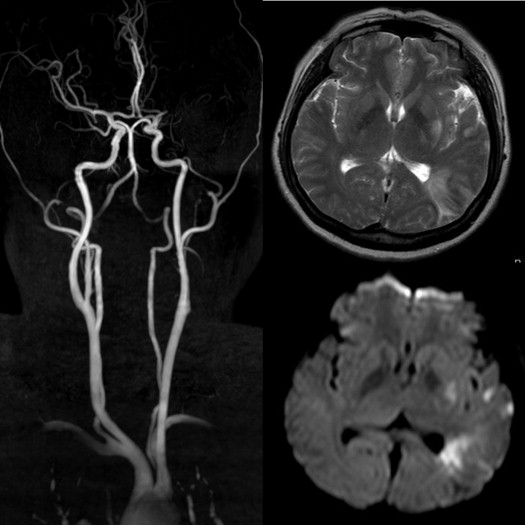
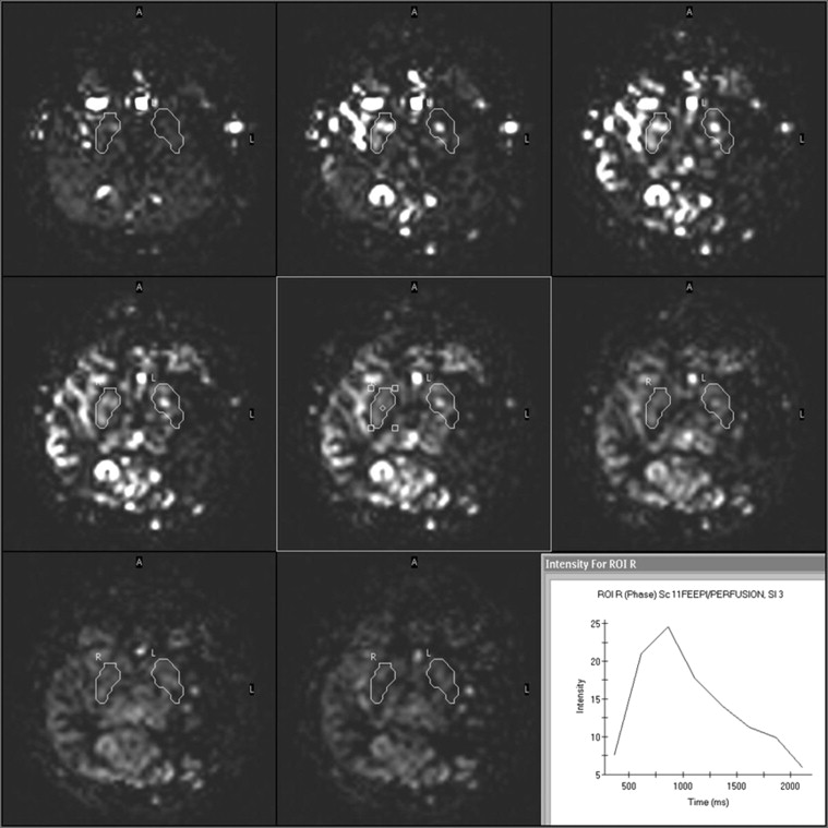
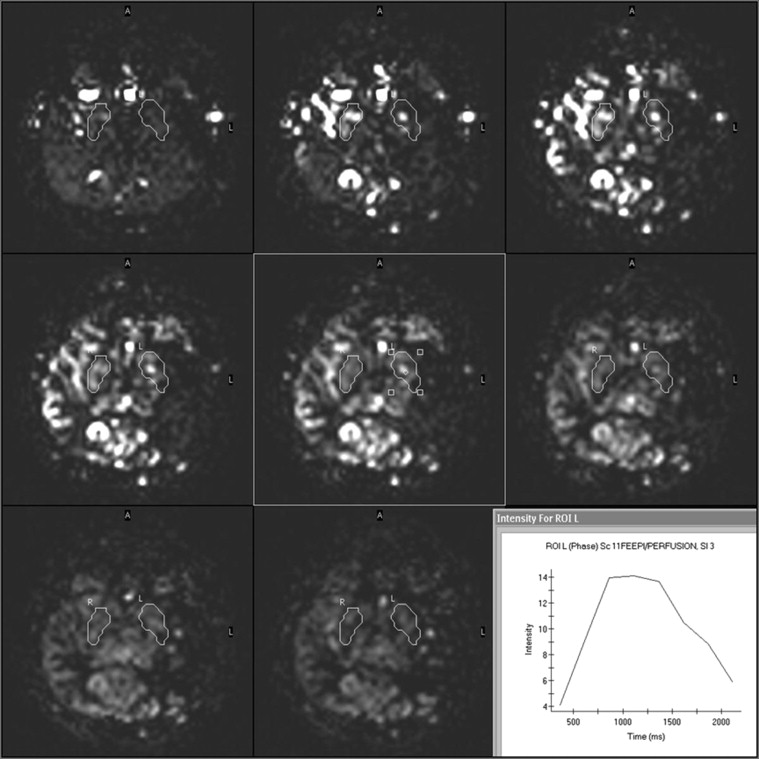
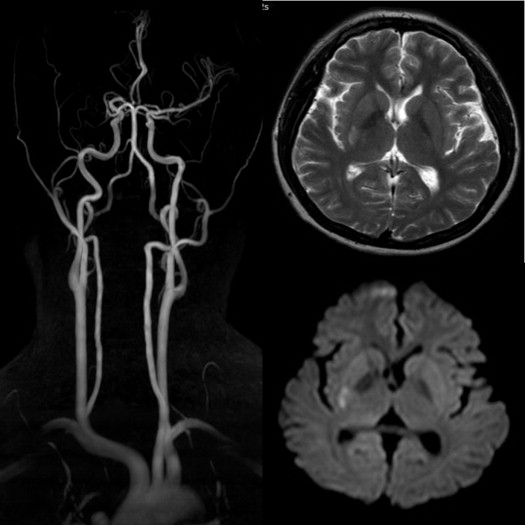
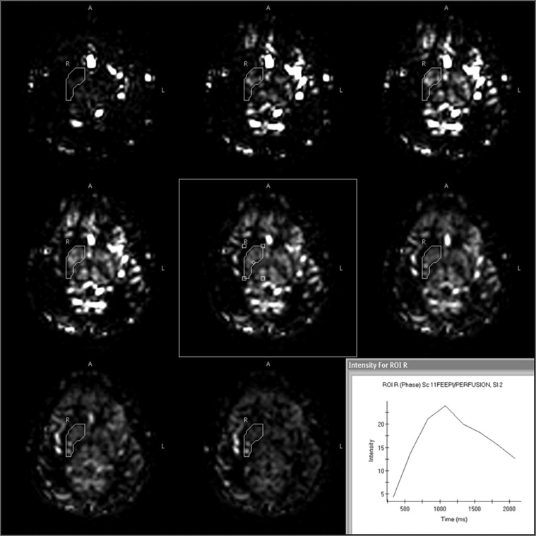
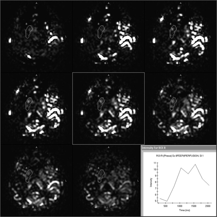
Seventeen patients with severe stenoses in unilateral M1 segment of MCA were involved in this study. All patients underwent multiphase PASL imaging. Bilateral basal ganglia were drawn by hand as regions of interest (ROIs) on eight-phase images of each patient. The signal intensities of ROIs were measured and the time–intensity curves (TICs) were acquired through postprocessing on a magnetic resonance workstation. Whether there was a significant difference in the peak signal intensities of ROIs between the narrowed and normal sides was determined by the paired samples t test.
Results
Three types of TICs were observed: eight cases with platform type, five cases with two-peak type, and four cases with single-peak type. There was a significant difference in the peak signal intensities of ROIs between the narrowed and normal sides.
Conclusions
Different types of TICs represent different cerebral hemodynamic changes. Multiphase PASL can sensitively detect the dynamic characteristics of cerebral perfusion and provide important dynamic perfusion information for clinical treatment of arterial stenosis.
In recent years, cerebral perfusion imaging has gained much attention in ischemic cerebrovascular diseases. Perfusion imaging is highly sensitive in the detection of cerebral perfusion abnormalities. A mismatch between cerebral infarction volume and the volume of hypoperfusion may respond favorably to revascularization therapies . As compared with other perfusion techniques, such as positron emission tomography, single photon emission computed tomography (CT), and CT perfusion and dynamic susceptibility contrast (DSC) magnetic resonance imaging (MRI), arterial spin labeling (ASL) is a nonionizing and completely noninvasive technique for measuring cerebral perfusion. DSC perfusion imaging can provide more hemodynamic parameters than ASL imaging, such as cerebral blood volume, mean transit time, and time to peak, but ASL imaging has its own unique advantages. A study showed that ASL techniques could identify additional and complementary hemodynamic abnormalities in about half of the patients with normal DSC perfusion imaging findings. ASL has been proved to be a useful technique for evaluating cerebral perfusion. The role of ASL in clinical practice has increased steadily in recent years. Yun et al. had quantitatively evaluated the hemodynamic changes after carotid artery stenting by measuring cerebral blood flow (CBF) using ASL. A study of Bivard et al. showed that hyperperfusion of the initially ischemic area identified on ASL at 24 hours after stroke identifies patients with better tissue and clinical outcomes. Qiao et al. found that ASL showed promise in the detection of perfusion abnormalities that correlated with clinically diagnosed transient ischemic attack in patients with otherwise normal neuroimaging results.
ASL uses magnetically labeled water protons in arterial blood as the endogenous tracer. When the labeled tracer reaches the imaging slice, it gives raise to perfusion signal. Ischemic cerebrovascular diseases frequently manifest themselves first as disturbances in cerebral hemodynamics before cerebral infarction can be observed. We often found different cerebral ischemic strokes in the patients although the same degrees of stenosis in the same cerebral arteries were detected in clinical practice, which demonstrated that there were different cerebral hemodynamic changes in the territories supplied by the narrowed arteries. In this study, multiphase pulsed ASL (PASL) technique was used to detect these hemodynamic changes. The goal of this study was to assess the sensitivity and utility of multiphase PASL in detecting dynamic changes of cerebral perfusion in the presence of a middle cerebral artery (MCA) severe stenosis.
Materials and methods
Get Radiology Tree app to read full this article<
Study Population
Get Radiology Tree app to read full this article<
MRI Protocol
Get Radiology Tree app to read full this article<
Get Radiology Tree app to read full this article<
Get Radiology Tree app to read full this article<
Image Evaluation
Get Radiology Tree app to read full this article<
Statistical Analysis
Get Radiology Tree app to read full this article<
Results
Get Radiology Tree app to read full this article<
Get Radiology Tree app to read full this article<
Get Radiology Tree app to read full this article<
Get Radiology Tree app to read full this article<
Get Radiology Tree app to read full this article<
Get Radiology Tree app to read full this article<
Get Radiology Tree app to read full this article<
Get Radiology Tree app to read full this article<
Discussion
Get Radiology Tree app to read full this article<
Get Radiology Tree app to read full this article<
Get Radiology Tree app to read full this article<
Get Radiology Tree app to read full this article<
Get Radiology Tree app to read full this article<
Get Radiology Tree app to read full this article<
Get Radiology Tree app to read full this article<
References
1. Harris A.D., Coutts S.B., Frayne R.: Diffusion and perfusion MR imaging of acute ischemic stroke. Magn Reson Imaging Clin N Am 2009; 17: pp. 291-313.
2. Zaharchuk G., Bammer R., Straka M., et. al.: Arterial spin-label imaging in patients with normal bolus perfusion-weighted MR imaging findings: pilot identification of the borderzone sign. Radiology 2009; 252: pp. 797-807.
3. Yun T.J., Sohn C.H., Han M.H., et. al.: Effect of carotid artery stenting on cerebral blood flow: evaluation of hemodynamic changes using arterial spin labeling. Neuroradiology 2013; 55: pp. 271-281.
4. Bivard A., Stanwell P., Levi C., et. al.: Arterial spin labeling identifies tissue salvage and good clinical recovery after acute ischemic stroke. Journal of Neuroimaging 2013; 23: pp. 391-396.
5. Qiao X.J., Salamon N., Wang D.J., et. al.: Perfusion deficits detected by arterial spin-labeling in patients with TIA with negative diffusion and vascular imaging. Am J Neuroradiol 2013; 34: pp. 2125-2130.
6. Samuels O.B., Joseph G.J., Lynn M.J.: A standardized method for measuring intracranial arterial stenosis. Am J Neuroradiol 2000; 21: pp. 643-646.
7. Fonda C, Ciccarone A, Mortilla M, et al. 3T arterial spin labeling (ASL) in pediatric patients: preliminary results. 6th congress and exhibition of the joint societies of paediatric radiology. 27–31 May 2011, London.
8. Williams D.S., Detre J.A., Leigh J.S., et. al.: Magnetic resonance imaging of perfusion using spin inversion of arterial water. Proc Natl Acad Sci USA 1992; 89: pp. 212-216.
9. Weber M.A., Günther M., Lichy M.P., et. al.: Comparison of arterial spin-labeling techniques and dynamic susceptibility-weighted contrast-enhanced MRI in perfusion imaging of normal brain tissue. Invest Radiol 2003; 38: pp. 712-718.
10. Detre J.A., Alsop D.C.: Perfusion magnetic resonance imaging with continuous arterial spin labeling: methods and clinical applications in the central nervous system. Eur J Radiol 1999; 30: pp. 115-124.
11. Wong E.C.: Quantifying CBF with pulsed ASL: technical and pulsed sequence factors. J Magn Reson Imaging 2005; 22: pp. 727-731.
12. Petcharunpaisan S., Ramalho J., Castillo M.: Arterial spin labeling in neuroimaging. World J Radiol 2010; 10: pp. 384-398.
13. Gregori J., Schuff N., Kern R., et. al.: T2-based arterial spin labeling measurements of blood to tissue water transfer in human brain. J Magn Reson Imaging 2013; 37: pp. 332-342.
14. Golay X., Hendrikse J., Lim T.C.: Perfusion imaging using arterial spin labeling. Top Magn Reson Imaging 2004; 15: pp. 10-27.
15. Detre J.A., Wang J., Wang Z., et. al.: Arterial spin-labeled perfusion MRI in basic and clinical neuroscience. Curr Opin Neurol 2009; 22: pp. 348-355.
16. Wolf R.L., Detre J.A.: Clinical neuroimaging using arterial spin labeled perfusion magnetic resonance imaging. Neurotherapeutics 2007; 4: pp. 346-359.
17. Petcharunpaisan S., Ramalho J., Castillo M.: Arterial spin labeling in neuroimaging. World J Radiol 2010; 2: pp. 384-398.