
Rationale and Objectives
To characterize an iodized oil emulsion for computed tomography (CT) imaging of experimental hepatic tumors in rat models.
Materials and Methods
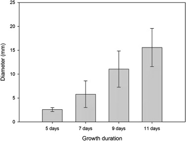
For characterizing the agents in normal rats, three rats were intravenously infused and imaged with clinical CT up to 1 week. Iopamidol solution was also used as controls ( n = 3). For evaluating the feasibility of diagnosis of hepatic tumors, 12 rats were injected with C6 glial tumor cells into the liver 11, 9, 7, and 5 days before CT ( n = 3 per day). After CT imaging, gross and histopathologic correlation of liver tumors with CT images were performed.
Results
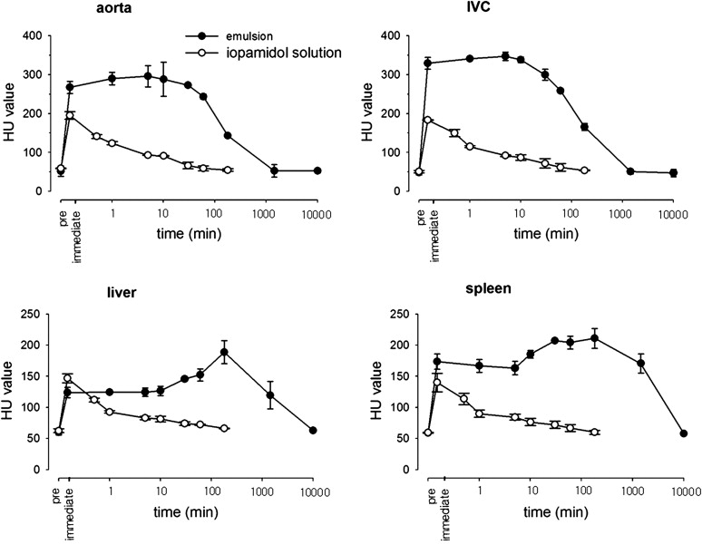
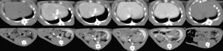
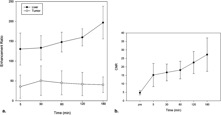
CT numbers of aorta and inferior vena cava (IVC) increased immediately after injection of the emulsion and remained above 200 Hounsfield units for 1 hour (maximum: 295.67 ± 27.65 in aorta and 347.07 ± 10.58 in IVC). The mean attenuation in liver and spleen was relatively stable between 30 and 180 minutes (maximum: 188.84 ± 18.70 in liver and 210.97 ± 15.83 in spleen). All 20 tumors later confirmed by pathology were detected as hypodense lesions on CT (sensitivity: 100%; range, 2.0–16.4 mm). The mean enhancement ratios of liver at all time points were significantly higher than those of tumors ( P < .05).
Conclusion
The hepatic enhancement achieved by the iodized oil emulsion is reticuloendothelial system–specific with the property of blood pool enhancement and longer lasting than that achievable with the current water soluble agents. Thus, this agent may offer significant advantages for diagnosis of hepatic metastases.
Computed tomography (CT) imaging enhanced with water-soluble iodinated contrast is an important diagnostic modality in the preoperative evaluation of patients with suspected hepatic metastases. The choice between surgical or palliative treatment depends on the size, number, and location of metastases as determined by imaging . However, the limitations of these contrast agents include their extravasation and accumulation in both normal hepatic parenchyma and metastatic tumors, precluding accurate diagnosis of small hepatic metastases . The lack of accurate and early detection of metastatic lesions in many cases remains the major obstacle to effective patient treatment and ultimately to survival.
In comparison to water soluble agents, contrast agents consisting of particles selectively enhance the liver and spleen because the particles are sequestered by the phagocytic reticuloendothelial system (RES) . Therefore, they may provide enhanced diagnostic accuracy for hepatic metastatic lesions, because all metastatic liver lesions do not contain Kupffer cells constituting the RES. For CT imaging, several particulate contrast agents, including perfluorooctylbromide , ethiodol oil emulsion-13 , and liposomal agents , have been investigated.
Get Radiology Tree app to read full this article<
Materials and methods
Preparation of Nanoscale Particulate Iodized Oil Emulsion
Get Radiology Tree app to read full this article<
Animal Subjects, Anesthesia, and Injection Technique
Get Radiology Tree app to read full this article<
CT Image Acquisition and Parameters
Get Radiology Tree app to read full this article<
Imaging of Normal Rats for Evaluating the Time Enhancement Curve in the Blood Pool and RES
Get Radiology Tree app to read full this article<
Imaging of Tumor Model
Tumor model
Get Radiology Tree app to read full this article<
CT imaging and analysis
Get Radiology Tree app to read full this article<
Get Radiology Tree app to read full this article<
Pathologic Evaluation
Get Radiology Tree app to read full this article<
Statistical Analysis
Get Radiology Tree app to read full this article<
Results
Get Radiology Tree app to read full this article<
Imaging of Normal Rat for Evaluation of the Time Enhancement Curve in the Blood Pool and RES
Time course of enhancement in the aorta, IVC, liver, and spleen
Get Radiology Tree app to read full this article<
Get Radiology Tree app to read full this article<
Tumor Model Evaluation
Detection of tumors and correlation with pathologic results
Get Radiology Tree app to read full this article<
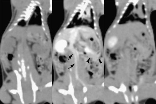
![Figure 4, Radiologic-pathologic correlation (left to right: computed tomography [CT] image, gross specimen, and pathological slide). Coronal CT images of 22-mm-sized (a) and 2-mm-sized (b) tumors are well defined in the hepatic parenchyma. The gross specimen shows the corresponding lesions ( arrows ) and C6-glial cell tumors are pathologically confirmed ( arrows ) (original magnification, ×100 (a) and ×200 (b) ; hematoxylin-eosin stain).](https://storage.googleapis.com/dl.dentistrykey.com/clinical/NanoscaledIodizedOilEmulsionasaCTContrastAgentfortheDetectionofExperimentalLiverTumorsinaRatModel/3_1s20S1076633210001777.jpg)
Get Radiology Tree app to read full this article<
Quantitative analysis of contrast enhancement on CT images
Get Radiology Tree app to read full this article<
Get Radiology Tree app to read full this article<
Discussion
Get Radiology Tree app to read full this article<
Get Radiology Tree app to read full this article<
Get Radiology Tree app to read full this article<
Get Radiology Tree app to read full this article<
Get Radiology Tree app to read full this article<
Get Radiology Tree app to read full this article<
References
1. Hughes K.S., Simon R., Songhorabodi S., et. al.: Resection of the liver for colorectal carcinoma metastases: a multi-institutional study of patterns of recurrence. Surgery 1986; 100: pp. 278-284.
2. Kao C.Y., Hoffman E.A., Beck K.C., et. al.: Long-residence-time nano-scale liposomal iohexol for X-ray-based blood pool imaging. Acad Radiol 2003; 10: pp. 475-483.
3. Rubin D.L., Desser T.S., Qing F., et. al.: Nanoparticulate contrast media. Blood-pool and liver-spleen imaging. Invest Radiol 1994; 29: pp. S280-S283.
4. Mattrey R.F.: Perfluorooctylbromide: a new contrast agent for CT, sonography, and MR imaging. AJR Am J Roentgenol 1989; 152: pp. 247-252.
5. Vermess M., Chatterji D.C., Doppman J.L., et. al.: Development and experimental evaluation of a contrast medium for computed tomographic examination of the liver and spleen. J Comput Assist Tomogr 1979; 3: pp. 25-31.
6. Seltzer S.E., Shulkin P.M., Adams D.F., et. al.: Usefulness of liposomes carrying losefamate for ct opacification of liver and spleen. Am J Roentgenol 1984; 143: pp. 575-579.
7. Chung Y.E., Hyung W.J., Kweon S., et. al.: Feasibility of interstitial CT lymphography using optimized iodized oil emulsion in rats. Invest Radiol 2010; 45: pp. 142-148.
8. Behan M., O’Connell D., Mattrey R.F., et. al.: Perfluorooctylbromide as a contrast agent for CT and sonography: preliminary clinical results. AJR Am J Roentgenol 1993; 160: pp. 399-405.
9. Sugarbaker P.H., Vermess M., Doppman J.L., et. al.: Improved detection of focal lesions with computerized tomographic examination of the liver using ethiodized oil emulsion (EOE-13) liver contrast. Cancer 1984; 54: pp. 1489-1495.
10. Blakeborough A., Ward J., Wilson D., et. al.: Hepatic lesion detection at MR imaging: a comparative study with four sequences. Radiology 1997; 203: pp. 759-765.
11. Strotzer M., Gmeinwieser J., Schmidt J., et. al.: Diagnosis of liver metastases from colorectal adenocarcinoma. Comparison of spiral-CTAP combined with intravenous contrast-enhanced spiral-CT and SPIO-enhanced MR combined with plain MR imaging. Acta Radiol 1997; 38: pp. 986-992.
12. Moss A.A., Schrumpf J., Schnyder P., et. al.: Computed tomography of focal hepatic lesions: a blind clinical evaluation of the effect of contrast enhancement. Radiology 1979; 131: pp. 427-430.
13. Tsang Y.M., Stark D.D., Chen M.C., et. al.: Hepatic micrometastases in the rat: ferrite-enhanced MR imaging. Radiology 1988; 167: pp. 21-24.
14. Miller D.L., Rosenbaum R.C., Sugarbaker P.H., et. al.: Detection of hepatic metastases: comparison of EOE-13 computed tomography and scintigraphy. AJR Am J Roentgenol 1983; 141: pp. 931-935.
15. Weichert J.P., Longino M.A., Spigarelli M.G., et. al.: Computed tomography scanning of Morris hepatomas with liver-specific polyiodinated triglycerides. Acad Radiol 1996; 3: pp. 412-417.
16. Weichert J.P., Lee F.T., Longino M.A., et. al.: Computed tomography scanning of hepatic tumors with polyiodinated triglycerides. Acad Radiol 1996; 3: pp. S229-S231.
17. Patronas N., Miller D.L., Girton M.: Experimental comparison of EOE-13 and perfluoroctylbromide for the CT detection of hepatic metastases. Invest Radiol 1984; 19: pp. 570-573.
18. Illum L., Davis S.S.: The organ uptake of intravenously administered colloidal particles can be altered using a non-ionic surfactant (Poloxamer 338). FEBS Lett 1984; 167: pp. 79-82.
19. Torchilin V.P.: Polymeric contrast agents for medical imaging. Curr Pharm Biotechnol 2000; 1: pp. 183-215.
20. Ryan P.J., Davis M.A., DeGaeta L.R., et. al.: Liposomes loaded with contrast material for image enhancement in computed tomography. Work in progress. Radiology 1984; 152: pp. 759-762.
21. Weichert J.P., Lee F.T., Longino M.A., et. al.: Lipid-based blood-pool CT imaging of the liver. Acad Radiol 1998; 5: pp. S16-S19. discussion S28–S30
22. Ivancev K., Lunderquist A., Isaksson A., et. al.: Clinical trials with a new iodinated lipid emulsion for computed tomography of the liver. Acta Radiol 1989; 30: pp. 449-457.