
Rationale and Objectives
This study is aimed at investigating neurochemical changes in scopolamine (SCP)–induced memory impairment using spatially localized in vivo magnetic resonance spectroscopy (MRS) of the hippocampus.
Materials and Methods
Four groups of mice (eight mice per group) were scanned after the injection of different SCP doses: 0, 1, 3, and 5 mg/kg (intraperitoneally). All the animals received 1 H MRS of their hippocampus at two time intervals: 30 minutes and 72 hours after SCP injection.
Results
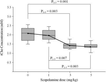
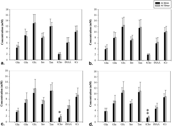
This work demonstrated that the doses of 3 mg/kg SCP or higher reduce the concentration of total choline–containing compounds, and these levels returned to baseline after 72 hours. These results are consistent with observations made by others using more invasive brain dialysis approaches. The levels of glutamate and glutamic compounds (glutamate + glutamine) were slightly changed at 3 and 5 mg/kg SCP dose, but the differences were not statistically significant ( P > .05). These findings suggest that SCP produces transient, in vivo measurable alterations in the cholinergic system in the hippocampus.
Conclusions
On this basis, we conclude that in vivo MRS is a feasible noninvasive method to probe aspects of the alterations induced by SCP in the cholinergic neurotransmission pathways in both animal models and human studies of memory impairment.
Cholinergic deficiency in human brain is the most severe and consistent biochemical change in mild cognitive impairment (MCI) and Alzheimer disease (AD) . This is seen as reduced levels of acetylcholine (ACh), choline acetyltransferase, and acetylcholinesterase reported in both necropsy brain samples and cerebrospinal fluid. In general, animal models of memory impairment have been used to understand the molecular basis of cognitive decline and identify therapeutic targets. Scopolamine (SCP), which is known as a muscarinic cholinergic receptor antagonist, has been used to induce memory deficit/amnesia such as is present in MCI and AD. SCP inhibits central cholinergic neuronal activity and impairs learning and short-term memory . Numerous studies have tried to find treatments that attenuate the effects of SCP-induced memory impairment . Several studies reported that the mechanism of memory impairment by cholinergic deficits may be through the modulation of the cerebrovasculature and decreased blood flow and blood volume may induce rapid cognitive decline . Although the precise molecular details and the influence of SCP on neurometabolic pathways have not been completely elucidated, SCP has been used widely to produce animal models of memory impairment.
The development of spatially localized in vivo proton magnetic resonance spectroscopy ( 1 H MRS) of the brain has led to both mechanistic studies of brain metabolism in animal models of disease and numerous clinical studies of human diseases and their progression . 1 H MRS is well suited for the study of cognitive disorders, because it is noninvasive, reproducible, and can sample the in vivo metabolism of many brain regions. At present, ∼20 compounds can be detected on 3T and 7T whole body magnetic resonance (MR) scanners. Some of these compounds are neurotransmitters . In particular, a resonance reflecting a composite peak representing total choline–containing compounds (tCho) is one of important metabolites in 1 H MRS studies of memory impairment. Some studies have reported that total choline signal in 1 H MRS has been changed in MCI and AD patients . This total choline peak is made up of ACh, glycerophosphocholine (GPC), phosphocholine (PC), and free choline .
Get Radiology Tree app to read full this article<
Materials and methods
Animals and Drugs
Get Radiology Tree app to read full this article<
Get Radiology Tree app to read full this article<
Proton Magnetic Resonance Imaging/Magnetic Resonance Spectroscopy
Get Radiology Tree app to read full this article<
Get Radiology Tree app to read full this article<
Quantification
Get Radiology Tree app to read full this article<
Analysis
Get Radiology Tree app to read full this article<
Results
Get Radiology Tree app to read full this article<
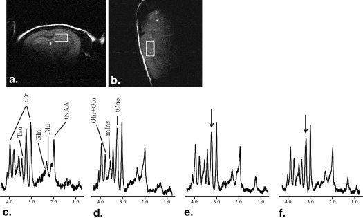
Table 1
SNR and CRLBs in Corresponding SCP Dose of Each Group
SCP Dose (mg/kg) CRLB SNR Glutamine Glutamate Amino acid Myo-inositol Taurine tCho tNAA Total creatine 0 30 m 16.88 ± 3.72 7.25 ± 1.58 7.50 ± 1.60 5.75 ± 1.16 4.63 ± 1.19 5.13 ± 0.99 6.50 ± 1.20 3.38 ± 0.74 10.13 ± 1.73 72 h 14.75 ± 3.45 7.38 ± 2.00 7.00 ± 1.41 5.38 ± 0.92 4.25 ± 0.46 5.00 ± 1.51 6.50 ± 2.00 3.13 ± 0.35 10.38 ± 1.85 1 30 m 17.88 ± 4.52 7.38 ± 2.13 7.75 ± 1.90 5.88 ± 1.13 4.50 ± 1.07 4.63 ± 0.74 6.38 ± 1.51 3.13 ± 0.35 10.50 ± 2.00 72 h 16.75 ± 5.70 7.25 ± 1.83 7.38 ± 2.00 5.63 ± 1.30 4.37 ± 1.30 5.00 ± 1.31 6.25 ± 2.05 3.00 ± 0.76 11.00 ± 2.51 3 30 m 18.50 ± 2.07 8.25 ± 2.19 8.50 ± 1.69 6.63 ± 1.85 4.50 ± 0.75 6.38 ± 2.00 7.25 ± 1.67 3.13 ± 0.35 11.63 ± 1.30 72 h 18.50 ± 5.50 8.13 ± 2.64 8.13 ± 2.64 5.75 ± 1.16 5.00 ± 1.77 5.38 ± 1.41 6.88 ± 2.42 3.50 ± 0.76 10.75 ± 1.83 5 30 m 17.63 ± 5.42 8.38 ± 2.50 8.25 ± 2.25 6.13 ± 0.99 4.75 ± 1.91 6.50 ± 1.77 7.38 ± 2.26 3.50 ± 0.76 10.13 ± 1.13 72 h 19.13 ± 6.98 8.00 ± 2.62 8.25 ± 2.71 6.25 ± 1.04 4.88 ± 1.81 5.38 ± 0.92 7.50 ± 2.14 3.38 ± 0.74 9.38 ± 1.60
CRLBs, Cramer–Rao lower bounds; h, hours; m, minutes; SCP, scopolamine; SNR, signal-to-noise ratio; tCho, total choline–containing compounds; tNAA, total N-acetylaspartate.
Get Radiology Tree app to read full this article<
Get Radiology Tree app to read full this article<
Get Radiology Tree app to read full this article<
Get Radiology Tree app to read full this article<
Get Radiology Tree app to read full this article<
Discussion
Get Radiology Tree app to read full this article<
Get Radiology Tree app to read full this article<
Get Radiology Tree app to read full this article<
Get Radiology Tree app to read full this article<
Get Radiology Tree app to read full this article<
Acknowledgments
Get Radiology Tree app to read full this article<
References
1. Saykin A.J., Wishart H.A., Rabin L.A., et. al.: Cholinergic enhancement of frontal lobe activity in mild cognitive impairment. Brain 2004; 127: pp. 1574-1583.
2. DeKosky S.T., Ikonomovic M.D., Styren S.D., et. al.: Activity in hippocampus and frontal cortex of elderly subjects with mild cognitive impairment. Ann Neurol 2002; 51: pp. 145-155.
3. Rinne J.O., Kaasinen V., Jarvenpaa T., et. al.: Brain acetylcholinesterase activity in mild cognitive impairment and early Alzheimer’s disease. J Neurol Neurosurg Psychiatry 2003; 74: pp. 113-115.
4. Pazzagli A., Pepeu G.: Amnesic properties of scopolamine and brain acetylcholine in the rat. Int J Neuropharmacol 1964; 4: pp. 291-299.
5. Huff F.J., Mickel S.F., Corkin S., et. al.: Cognitive functions affected by scopolamine in Alzheimer’s disease and normal aging. Drug Dev Res 1988; 12: pp. 271-278.
6. Tota S., Hanif K., Kamat P.K., et. al.: Role of central angiotensin receptors in scopolamine-induced impairment in memory, cerebral blood flow and cholinergic function. Psychopharmacology 2012; 222: pp. 185-202.
7. Blake M.G., Boccia M.M., Krawczyk M.C., et. al.: Choline reverses scopolamine-induced memory impairment by improving memory reconsolidation. Neurobiol Learn Mem 2012; 98: pp. 112-121.
8. Lee K.Y., Sung S.H., Kim S.H., et. al.: Cognitive-enhancing activity of loganin isolated from Cornus officinalis in scopolamine-induced amnesic mice. Arch Pharm Res 2009; 32: pp. 677-683.
9. Okaichi Y., Okaichi H.: Effects of glucose on scopolamine-induced learning deficits in rats performing the Morris water maze task. Neurobiol Learn Mem 2000; 74: pp. 65-79.
10. Claro F.T., Patti C.L., Abilio V.C., et. al.: Bovine brain phosphatidylserine attenuates scopolamine induced amnesia in mice. Prog Neuro-Psychopharmacol Biol Psychiatry 2006; 30: pp. 881-886.
11. Barber T.A., Haggarty M.K.: Memantine ameliorates scopolamine-induced amnesia in chicks trained on taste-avoidance learning. Neurobiol Learn Mem 2010; 93: pp. 540-545.
12. Saraf M.K., Anand A., Prabhakar S.: Scopolamine induced amnesia is reversed by Bacopa monniera through participation of kinase-CREB Pathway. Neurochem Res 2010; 35: pp. 279-287.
13. Estrada C., Hamel E., Krause D.N.: Biochemical evidence for cholinergic innervations of intracerebral blood vessels. Brain Res 1983; 266: pp. 261-270.
14. Lojkowska W., Ryglewicz D., Jedrzejczak T., et. al.: The effect of cholinesterase inhibitors on the regional blood flow in patients with Alzheimer’s disease and vascular dementia. J Neurol Sci 2003; 216: pp. 119-126.
15. Sato A., Sato Y.: Cholinergic neural regulation cerebral blood flow. Alzheimer Dis 1995; 9: pp. 28-38.
16. Tapp P.D., Chu Y., Araujo J.A., et. al.: Effects of scopolamine challenge on regional cerebral blood volume. A pharmacological model to validate the use of contrast enhanced magnetic resonance imaging to assess cerebral blood volume in a canine model of aging. Prog Neuro-Psychopharmacol Biol Psychiatry 2005; 29: pp. 399-406.
17. Saper C.B., Lenkinski R.E.: MR spectroscopy in translational neuroscience. J Comp Neurol 2010; 518: pp. 4089-4090.
18. Kim S.-Y., Lee H., Kim H.J., et. al.: In vivo and ex vivo evidence for ketamine-induced hyperglutamatergic activity in the cerebral cortex of the rat: potential relevance to schizophrenia. NMR Biomed 2011; 24: pp. 1235-1242.
19. Tkac I., Henry P.G., Andersen P., et. al.: Highly resolved in vivo 1H NMR spectroscopy of the mouse brain at 9.4T. Magn Reson Med 2004; 52: pp. 478-484.
20. Schepkin V.D., Brey W.W., Gorkov P.L., et. al.: Initial in vivo rodent sodium and proton MR imaging at 21.1T. Magn Reson Imaging 2010; 28: pp. 400-407.
21. Kantarci K., Weigand S.D., Petersen R.C., et. al.: Longitudinal 1H MRS changes in mild cognitive impairment and Alzheimer’s disease. Neurobiol Aging 2007; 28: pp. 1330-1339.
22. Yang Z.X., Huo S.S., Cheng X.F., et. al.: Quantitative multivoxel proton MR spectroscopy study of brain metabolites in patients with amnestic mild cognitive impairment: a pilot study. Neuroradiology 2012; 54: pp. 451-458.
23. Stadler H., Fuldner H.H.: Proton NMR detection of acetylcholine status in synaptic vesicles. Nature 1980; 286: pp. 293-294.
24. Wang X.C., Du X.X., Tian Q., et. al.: Correlation between choline signal intensity and acetylcholine level in different brain regions of rat. Neurochem Res 2008; 33: pp. 814-819.
25. Stone W.S., Croul C.E., Gold P.E., et. al.: Attenuation of scopolamine-induced amnesia in mice. Psychopharmacology 1988; 96: pp. 417-420.
26. Stone W.S., Walser B., Gold S.D., et. al.: Scopolamine and morphine-induced impairments of spontaneous alternation performance in mice: reversal with glucose and with cholinergic and adrenergic agonists. Behav Neurosci 1991; 105: pp. 264-271.
27. Konar A., Shah N., Singh R., et. al.: Protective role of Ashwagandha leaf extract and its component with an one on scopolamine-induced changes in the brain and brain-derived cells. PLoSone 2011; 6: pp. e27265.
28. Parfitt G.M., Campos R.C., Barbosa A.K., et. al.: Participation of hippocampal cholinergic system in memory persistence for inhibitory avoidance in rats. Neurobiol Learn Mem 2012; 97: pp. 183-188.
29. Tellez R., Gomez-Viquez L., Meneses A.: GABA, glutamate, dopamine and serotonin transporters expression on memory formation and amnesia. Neurobiol Learn Mem 2012; 97: pp. 189-201.
30. Sunderland T., Tariot P.N., Weingartner H., et. al.: Pharmacologic modeling of Alzheimer’s disease. Prog Neuro-Psychopharmacol Biol Psychiatry 1986; 10: pp. 599-610.
31. Klinkenberg I., Blokland A.: The validity of scopolamine as a pharmacological model for cognitive impairment: a review of animal behavioral studies. Neurosci Biobehav Rev 2010; 34: pp. 1307-1350.
32. Sunderland T., Tariot P., Mueller E.A., et. al.: Cognitive and behavioral sensitivity to scopolamine in Alzheimer patients and controls. Psychopharmacol Bull 1985; 21: pp. 676-679.
33. Sunderland T., Tariot P., Murphy D.L., et. al.: Scopolamine challenges in Alzheimer’s disease. Psychopharmacology 1985; 87: pp. 247-249.
34. Barker A., Jones R., Prior J., et. al.: Scopolamine-induced cognitive impairment as a predictor of cognitive decline in healthy elderly volunteers: a 6-year follow-up. Int J Geriatr Psychiatr 1998; 13: pp. 244-247.
35. Hasselmo M.E., McGaughy J.: High acetylcholine levels set circuit dynamics for attention and encoding and low acetylcholine levels set dynamics for consolidation. Prog Brain Res 2004; 145: pp. 207-231.
36. Toide K.: Effects of scopolamine on extracellular acetylcholine and choline levels and on spontaneous motor activity in freely moving rats measured by brain dialysis. Pharmacol Biochem Behav 1989; 33: pp. 109-113.
37. Pfister M., Boix F., Huston J.P., et. al.: Different effects of scopolamine on extracellular acetylcholine levels in neostriatum and nucleus accumbens measured in vivo: possible interaction with aversive stimulation. J Neural Trans 1994; 97: pp. 13-25.
38. McEntee W.J., Crook T.H.: Glutamate: its role in learning, memory, and the aging brain. Psychopharmacology 1993; 11: pp. 391-401.
39. Aigner T.G.: Pharmacology of memory: cholinergic–glutamatergic interactions. Curr Opin Neurobiol 1995; 5: pp. 155-160.
40. Cory-Slechta D.A.: Relationships between lead-induced learning impairments and changes in dopaminergic, cholinergic, and glutamatergic neurotransmitter system functions. Annu Rev Pharmacol Toxicol 1995; 35: pp. 391-415.
41. Riedel G., Platt B., Micheau J.: Glutamate receptor function in learning and memory. Behav Brain Res 2003; 14: pp. 1-47.