
Rationale and Objectives
Noncontrast magnetic resonance angiography (NC-MRA) of pedal artery remains challenging because of the global and regional disease load, tissue integrity, and altered microcirculation. This study aims to investigate the feasibility of the NC-MRA of pedal arteries with flow-sensitive dephasing–prepared steady-state free precession (FSD-SSFP) and to explore the effect of disease load of type II diabetes on the vessel depiction.
Materials and Methods
FSD-SSFP was performed on a 1.5-T magnetic resonance system before the contrast-enhanced MRA (CE-MRA) as a reference standard in 39 consecutive diabetic subjects (29 men and 16 women, aged 57.9 ± 11.4 years). Two experienced radiologists evaluated the overall artery visibility (VA) and the contamination from soft tissue (SC) and veins (VC) with a four-point scale. Chronic complications and measures including random blood glucose (RBG), lipid panel, body mass index, risk of diabetic foot ulcers (RDF), and glycated hemoglobin (HbA1c) by the imaging were recorded as disease load indicators. Spearman rank correlation and ordinal regression were performed to investigate the effect of disease load on the depiction of pedal arteries.
Results
The measurement of RBG and RDF were significantly correlated with the VC in CE-MRA and with the overall visibility of pedal arteries in NC-MRA ( P < .025 and P < .001, respectively). Blood pressure was the only parameter that was significantly associated with SC in NC-MRA with FSD-SSFP ( P < .025). For CE-MRA the effect of RDF on the overall VA manifested a significant linear trend ( P < .001), and the level of RBG was substantially associated with the VC ( P < .025) without significantly impacting VA and SC. Hypertension only correlated with SC in NC-MRA. VA was found independent of the presence of diabetic nephropathy, coronary artery disease, abnormal lipid panel, HbA1c (75.0%), or optimized m 1 value that ranged from 70 to 160 mT⋅ms 2 /m (mean, 125 ± 18 mT⋅ms 2 /m) in this study.
Conclusions
FSD-SSFP proved to be a useful modality of NC-MRA for pedal artery imaging in diabetic patients. The vessel depiction is subject to the local and systemic disease load of type II diabetes. Technical optimization of the flow-sensitive dephasing gradient moment and properly choosing candidate would help augment the potential of this technique in patient care of peripheral artery disease.
Peripheral artery disease (PAD) is a typical macrovascular complication that typically affects arteries below the knee in type II diabetes . The awareness and detection of PAD may be limited as a substantial proportion of patients are asymptomatic or have atypical manifestations , although significant impairment of blood flow may have already occurred . Effective screening would be useful to facilitate the early identification of PAD, thus improving prognosis and lowering the cost of the disease .
Morphological and functional parameters of peripheral arteries derived from noninvasive assessments are well accepted in clinical practice. Ankle–brachial index (ABI) is commonly used as a convenient tool in PAD screening with high sensitivity and specificity, especially for populations aged greater than 50 years with significant hemodynamic abnormalities . However, the range and severity of the diseased arteries may not be readily indicated by this index, and the reliability of ABI can be compromised by the presence of arterial calcification. Pulse wave velocity, computed tomography angiography (CTA), magnetic resonance angiography (MRA), and digital subtraction angiography (DSA) are valuable imaging modalities that provide morphological and functional details of the diseased arteries for the purposes of determining revascularization and grafting or stent patency. However, a substantial population of diabetic patients is excluded from CTA or contrast-enhanced MRA (CE-MRA) in clinical practice concerning the high dose of ionizing radiation and/or contrast agent–induced nephropathy . Therefore, the development of MRA techniques that requires no contrast medium becomes a need of significant importance for the purpose of peripheral artery imaging in diabetes.
Get Radiology Tree app to read full this article<
Get Radiology Tree app to read full this article<
Methods
Get Radiology Tree app to read full this article<
Get Radiology Tree app to read full this article<
Get Radiology Tree app to read full this article<
Get Radiology Tree app to read full this article<
![Figure 1, Illustration of normal pedal artery anatomy. (a) Anterior view. (1) Distal anterior tibial artery, (2) dorsal artery of foot, (3) lateral tarsal arteries, (4) arcuate artery, (5) deep perforating artery, (6) dorsal metatarsal artery for hallux. (b) Lateral view. (1) Distal anterior tibial artery, (2) dorsal artery of foot, (3) lateral tarsal artery, (5) deep perforating artery, (8) lateral and (9) medial plantar arteries, (10) pedal arch, (11) distal posterior tibial artery [Courtesy of Chomel et al. (16) ]. Visibility of arteries numbered 2, 3, 4, 8, 9, and 10 was assessed in this study.](https://storage.googleapis.com/dl.dentistrykey.com/clinical/NoncontrastMRAofPedalArteriesinTypeIIDiabetes/0_1s20S1076633214004565.jpg)
Get Radiology Tree app to read full this article<
Results
Get Radiology Tree app to read full this article<
Table 1
Summary of the Distribution of Abnormal Clinical Laboratory Tests, Measures, and Complications of the 39 Subjects
Measure Lipid Panel RBG HbA1c BMI LDL Triglycerides HDL TC Abnormality rate (%) 90.9 88.9 62.5 60 86.7 71.8 43.5 Complication DR Atherosclerosis Dneuro Dnephro Hypertension FW CAD % 100 84.6 69.2 56.4 53.8 30.8 23.1
BMI, body mass index; CAD, coronary artery disease; Dnephro, diabetic nephropathy; Dneuro, diabetic neuropathy; DR, diabetic retinopathy; FW, diabetic foot wound; HbA1c, glycated hemoglobin; HDL, high-density lipoprotein; LDL, low-density lipoprotein; RBG, random blood glucose; TC, total cholesterol.
Get Radiology Tree app to read full this article<
Get Radiology Tree app to read full this article<
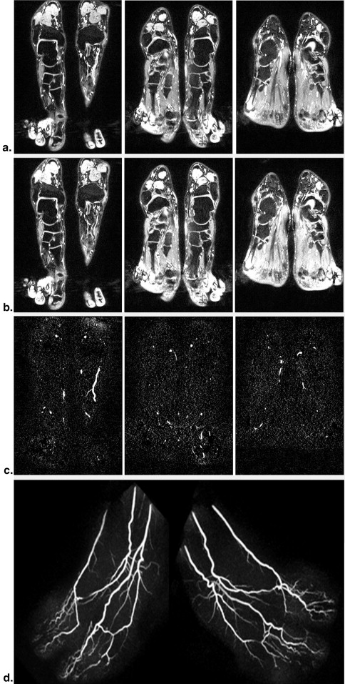
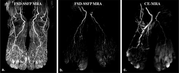
Table 2
Summary of the Scoring of the Overall VA, SC, and VC Based on the MIP FSD-SSFP Images of 234 Main Proximal Pedal Runoffs
Image Quality Scoring (Mean ± Standard Deviation) SC VC VA CE-MRA 2.4 ± 0.7 2.0 ± 0.7 2.3 ± 0.8 FSD-SSFP MRA 2.0 ± 1.2 1.4 ± 0.5 2.6 ± 0.9
CE, contrast enhanced; FSD-SSFP, flow-sensitive dephasing–prepared steady-state free precession; MIP, maximum-intensity projection; MRA, magnetic resonance angiography; SC, soft tissue contamination; VA, visibility of arteries; VC, venous contamination.
Get Radiology Tree app to read full this article<
Get Radiology Tree app to read full this article<
Table 3
Correlation Coefficient Between Disease Indicators and Image Quality for Both NC-MRA with FSD-SSFP and CE-MRA
Image Quality Indicator MRA RBG RDF Dnephro Blood Pressure HbA1c CAD m 1 VA NC 0.42* 0.39* — — — — — CE — — — — — — — VC NC — — — — — — — CE 0.44* 0.62** △△ — — — — — SC NC — 0.38* △ — 0.42* — — — CE — — — — — — —
CAD, coronary artery disease; CE, contrast enhanced; Dnephro, diabetic nephropathy; FSD-SSFP, flow-sensitive dephasing–prepared steady-state free precession; HbA1c, glycated hemoglobin; m 1 , first-order gradient moment; MRA, magnetic resonance angiography; NC, noncontrast; RBG, random blood glucose; RDF, risk of diabetic foot ulcer; SC, soft tissue contamination; VA, visibility of pedal artery; VC, vein contamination.
Bonferroni approach was applied to control the type I error.
∗ Spearman rho correlation and △ Cochran–Armitage trend test. * ,△ P < .025; ∗∗,△△ P < .001; — P > .05.
Get Radiology Tree app to read full this article<
Discussion
Get Radiology Tree app to read full this article<
Get Radiology Tree app to read full this article<
Get Radiology Tree app to read full this article<
Get Radiology Tree app to read full this article<
Get Radiology Tree app to read full this article<
Conclusions
Get Radiology Tree app to read full this article<
Get Radiology Tree app to read full this article<
References
1. Selvin E., Erlinger T.P.: Prevalence of and risk factors for peripheral arterial disease in the United States: results from the National Health and Nutrition Examination Survey, 1999–2000. Circulation 2004; 110: pp. 738-743.
2. Criqui M.H., Veronica V., Denenberg J.O., et. al.: Ethnicity and peripheral arterial disease: the San Diego Population Study. Circulation 2005; 112: pp. 2703-2707.
3. Orchard T.J., Strandness D.E.: Assessment of peripheral vascular disease in diabetes. Report and recommendations of an international workshop sponsored by the American Diabetes Association and the American Heart Association September 18–20, 1992 New Orleans, Louisiana. Circulation 1993; 88: pp. 819-828.
4. Jude E.B., Oyibo S.O., Chalmers N., et. al.: Peripheral arterial disease in diabetic and nondiabetic patients: a comparison of severity and outcome. Diabetic Care 2001; 24: pp. 1433-1437.
5. Meijer W.T., Hoes A.W., Rutgers D., et. al.: Peripheral arterial disease in the elderly: the Rotterdam Study. Arteriosclerosis, Thrombosis, and Vascular Biology 1998; 18: pp. 185-192.
6. Fowkes F.G.R., Housley E., Cawood E.H.H., et. al.: Edinburgh artery study: prevalence of asymptomatic and symptomatic peripheral arterial disease in the general population. Int J Epidemiol 1991; 20: pp. 384-392.
7. Belch W., Topol E.S., Agnelli G.M.: Critical issues in peripheral arterial disease detection and management: a call to action. Arch Intern Med 2003; 163: pp. 884-892.
8. Otah K.E., Madan A., Otah E., et. al.: Usefulness of an abnormal ankle-brachial index to predict presence of coronary artery disease in African-Americans. Am J Cardiol 2004; 93: pp. 481-483.
9. Calvin A.D., Misra S., Pflueger A.: Contrast-induced acute kidney injury and diabetic nephropathy. Nat Rev Nephrol 2010; 6: pp. 679-688.
10. Sadowski E.A., Bennett L.K., Chan M.R., et. al.: Nephrogenic systemic fibrosis: risk factors and incidence estimation. Radiology 2007; 243: pp. 148-157.
11. Miyazaki M., Lee V.S.: Nonenhanced MR angiography. Radiology 2008; 248: pp. 20-43.
12. Fan Z., Sheehan J., Bi X., et. al.: 3D noncontrast MR angiography of the distal lower extremities using flow-sensitive dephasing (FSD)-prepared balanced SSFP. Magn Reson Med 2009; 62: pp. 1523-1532.
13. Nakamura K., Miyazaki M., Kuroki K., et. al.: Noncontrast-enhanced peripheral MRA: technical optimization of flow spoiled fresh blood imaging for screening peripheral arterial diseases. MRM 2011; 65: pp. 595-602.
14. Boulton A.J.M., Armstrong D.G., Albert S.F., et. al.: Comprehensive foot examination and risk assessment. Diabetes Care 2008; 31: pp. 1679-1685.
15. Fan Z., Zhou X., Bi X., et. al.: Determination of the optimal first-order gradient moment for flow-sensitive dephasing magnetization-prepared 3D noncontrast MR angiography. Magn Reson Med 2011; 65: pp. 964-972.
16. Chomel S., Douek P., Moulin P., et. al.: Contrast-enhanced MR angiography of the foot: anatomy and clinical application in patients with diabetes. AJR 2004; 182: pp. 1435-1442.
17. Lim R.P., Hecht E.M., Xu J., et. al.: 3D nongadolinium-enhanced ECG-gated MRA of the distal lower extremities: preliminary clinical experience. J Magn Reson Imaging 2008; 28: pp. 181-189.
18. Liu X., Natasha B., Sheehan J., et. al.: Renal transplant: nonenhanced renal MR angiography with magnetization-prepared steady state free precession. Radiology 2009; 251: pp. 535-542.
19. Edmonds M.E., Roberts V.C., Watkins P.J.: Blood flow in the diabetic neuropathic foot. Diabetologia 1982; 22: pp. 9-15.
20. Uccioli L., Mancini L., Giordano A., et. al.: Lower limb arterio-venous shunts, autonomic neuropathy and diabetic foot. Diabetes Res Clin Pract 1992; 16: pp. 123-130.
21. Payne C.B.: Biomechanics of the foot in diabetes mellitus: some theoretical considerations. J Am Podiatr Med Assoc 1998; 88: pp. 285-289.
22. Zhang H.L., Kent K.C., Bush H.L., et. al.: Soft tissue enhancement on time-resolved peripheral magnetic resonance angiography. JMRI 2004; 19: pp. 590-597.
23. Dinh T., Veves A.: Microcirculation of the diabetic foot. Curr Pharm Des 2005; 11: pp. 2301-2309.
24. Fatma A., Mohammed E.: Prevalence of risk factors for diabetic foot complications. BMC family practice 2007; 8: pp. 59.
25. Le Bihan D., Breton E., Lallemand D., et. al.: MR imaging of intravoxel incoherent motions: application to diffusion and perfusion in neurologic disorders. Radiology 1986; 161: pp. 401-407.
26. Le Bihan D., Breton E., Lallemand D., et. al.: Separation of diffusion and perfusion in intravoxel incoherent motion MR imaging. Radiology 1988; 168: pp. 497-505.
27. Sun P.C., Chen C.S., Kuo C.D., et. al.: Impaired microvascular flow motion in subclinical diabetic feet with sudomotor dysfunction. Microvasc Res 2012; 83: pp. 243-248.
28. Raskin P., Pietri A., Unger R., et. al.: The effect of diabetic control on skeletal muscle capillary basement membrane width in patients with type 1 diabetes mellitus. New Engl J Med 1983; 309: pp. 1546-1550.
29. Overall W.R., Conolly S.M., Nishimura D.G., et. al.: Oscillating dual-equilibrium steady-state angiography. MRM 2002; 47: pp. 513-522.
30. Weitz J.I., Byrne J., Clagett P., et. al.: Diagnosis and treatment of chronic arterial insufficiency of the lower extremities: a critical review. Circulation 1996; 94: pp. 3026-3049.